
How Correcting Calcium Signaling Normalizes Gut Microbiota and Barrier Integrity.
This newsletter reveals how restoring ionic calcium signaling can normalize gut pH, barrier integrity, and calcium routing.
Executive Summary
Aligning gut ionized Ca2+ solubility with parathyroid hormone, calcitonin, and calcium-sensing receptor feedback reduces Ca2+ precipitation, stabilizes microbiota metabolism, and limits ectopic deposition with a mechanism-based ionic calcium approach.
Ionic Ca2+ as the Gut-to-System Calcium Control Switch
Free ionic Ca2+ in the gut lumen shapes microbial growth and biofilm behavior. When ionic Ca2+ is adequate at the mucosal surface, mucus gel structure and epithelial tight junction assembly are better supported. This improves barrier integrity and limits translocation of lipopolysaccharide and other pro-inflammatory molecules. Lower inflammatory cytokine signaling helps maintain a steadier luminal pH by normalizing bile acid handling and fermentation patterns. As pH becomes more stable, Ca2+ remains in a more soluble ionic form that can enter systemic distribution.
When ionic Ca2+ is low or repeatedly trapped in insoluble complexes, the microbiota often shifts toward increased proteolysis, higher ammonia levels, and wider pH swings. Dysbiosis → altered pH/metabolites → Ca2+ precipitation and reduced bioavailability. PTH, calcitonin, and CaSR signaling integrate these gut-level changes with renal calcium handling, bone buffering, and vascular smooth muscle responses. A practical mechanistic aim is to reduce the tendency toward ectopic calcification by directing Ca2+ toward physiologic compartments rather than soft-tissue deposition. At the cellular level, stable mitochondrial–ER Ca2+ homeostasis supports redox control and keeps oxidative stress from amplifying inflammatory signaling.
Microbiota correction, driven by a steadier ionic Ca2+ environment, restores barrier integrity, reduces inflammatory tone, and stabilizes pH. In recurrent calcium oxalate nephrolithiasis and in type 2 diabetes with arterial calcification, this sequence links gut barrier repair to more orderly ionic Ca2+ dynamics and systemic routing.
AIC Therapy fits this mechanism by restoring calcium signaling with clear endocrine integration, thereby supporting epithelial junction function and shifting the microbiota toward fewer pro-inflammatory taxa and lower proteolytic fermentation. It regulates COX-2, which reduces mucosal inflammatory drive and favors a microbiota profile with more stable short-chain fatty acid production and less pH volatility.
AIC Therapy also triggers endogenous calcitonin release, which works with CaSR and PTH feedback to guide Ca2+ distribution, so ionic Ca2+ is less available for ectopic deposition and more available for controlled physiologic use; in that setting, the microbiota tends to normalize as barrier stress and inflammatory substrate flow decrease.
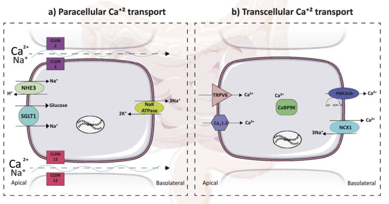
Deluque AL, Dimke H, Alexander RT. Biology of calcium homeostasis regulation in intestine and kidney. Nephrol Dial Transplant. 2025 Feb 28;40(3):435-445. doi: 10.1093/ndt/gfae204. Erratum in: Nephrol Dial Transplant. 2025 Aug 29;40(9):1810. doi: 10.1093/ndt/gfaf131. PMID: 39257024; PMCID: PMC11879016.
The figure shows that the intestine absorbs calcium in two main ways, both linked to sodium-driven water movement. One route is between neighboring cells, where calcium slips through tight junction proteins called claudin 2 and claudin 12. When the gut absorbs sodium, water follows, raising the calcium concentration in the gut fluid and creating a gradient that helps calcium move across these junctions. The other route is through the cell itself: calcium enters from the gut side through calcium channels, is held and carried across the cell by a calcium-binding protein, and then is moved out on the blood side by calcium transporters. A sodium-potassium pump on the blood side maintains the sodium gradient, supporting water flow and keeping both calcium uptake routes efficient.
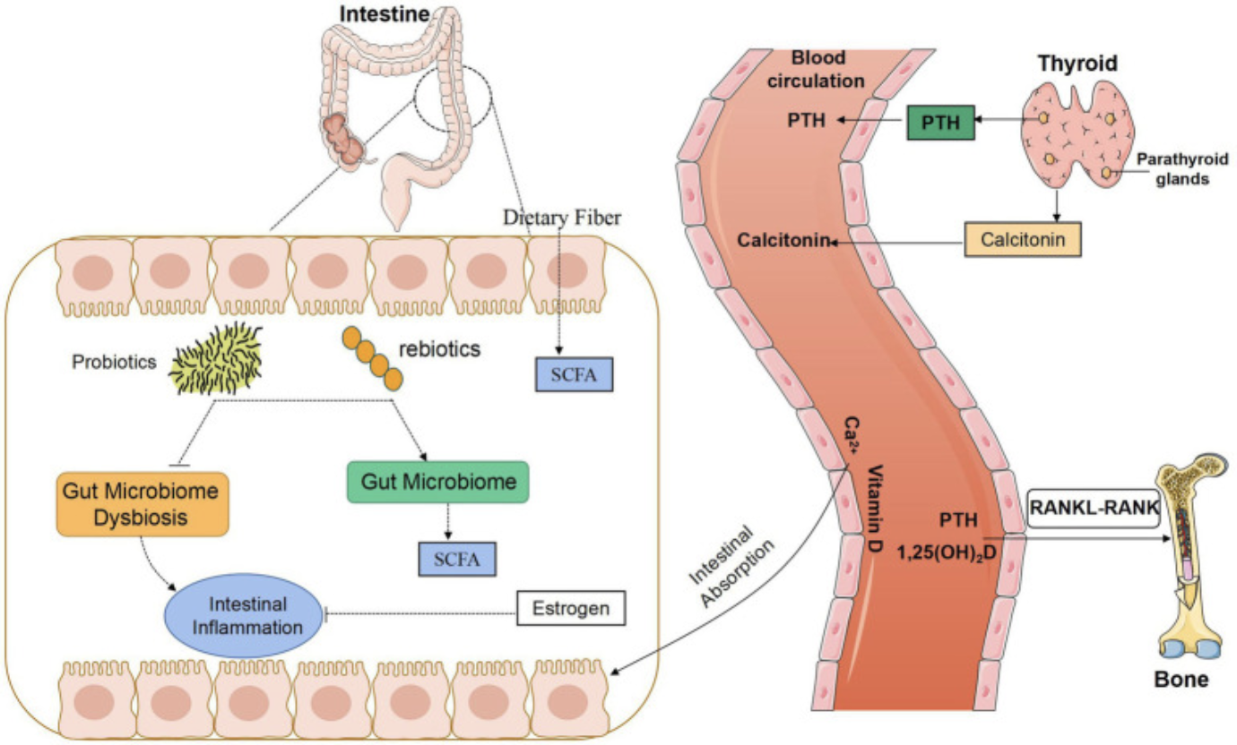
Wang J, Wu S, Zhang Y, Yang J, Hu Z. Gut microbiota and calcium balance. Front Microbiol. 2022 Dec 20;13:1033933. doi: 10.3389/fmicb.2022.1033933. PMID: 36713159; PMCID: PMC9881461.
This figure shows a possible link between gut microbiota and bone metabolism through calcium control. Gut microbes produce short-chain fatty acids that can influence calcium-regulatory factors, such as parathyroid hormone, and the overall blood calcium balance network. The diagram also highlights bone remodeling signals, including receptor activator of nuclear factor kappa B and receptor activator of nuclear factor kappa B ligand, which are key drivers of osteoclast activity. The review perspective is that gut microbiota may affect calcium metabolism across major target organs, including bone, intestine, and kidney, which together maintain blood calcium homeostasis. The clinical takeaway is that mapping these links can guide better prevention, diagnosis, and treatment approaches for calcium disorders and abnormal bone metabolism.
Mechanism-to-Clinic Trigger
If a patient has recurrent calcium oxalate stones or early vascular calcification despite a stable serum calcium, consider luminal ionic Ca2+ availability and gut pH as key variables. If bloating and loose stools improve but stone events or calcification markers do not change, consider ongoing barrier weakness and incomplete normalization of mucosal CaSR signaling. If symptoms worsen after antibiotics or bile acid disruption, consider dysbiosis that shifts metabolites and pH to promote Ca2+ precipitation and reduce functional bioavailability.
In stool chemistry, a higher pH, with more proteolytic fermentation products, suggests a microbial pattern that favors Ca2+ precipitation and ongoing mucosal irritation. In that setting, the PTH–calcitonin–CaSR feedback loop can become dysregulated, directing Ca2+ toward soft tissues and increasing the tendency for ectopic deposition.
Clinically, the key distinction is whether you only address dysbiosis or also stabilize ionic Ca2+ signaling and endocrine feedback. When ionic Ca2+ handling improves, barrier integrity tends to recover, inflammatory tone tends to fall, and the microbiota often shifts toward a more favorable profile that supports those gains.
Case Reference
A 68-year-old female with established Crohn’s disease and long-standing low day-to-day functioning started AIC Therapy as the only new change. Within weeks, a routine family visit noted a clear rise in energy, affect, and participation in normal activities compared with baseline. This rapid functional shift aligns with the sequence discussed here: stabilize luminal ionic Ca2+ and pH, support barrier integrity, reduce inflammatory signaling, and allow microbiota metabolism to shift toward a more barrier-supportive profile.
Conclusion
Stable ionic Ca2+ in the lumen supports mucus structure and tight junction assembly, thereby reducing barrier leak and lowering inflammatory signaling. With reduced inflammatory drive, bile acid handling and fermentation become steadier, luminal pH swings narrow, and Ca2+ remains soluble rather than precipitating into unavailable complexes. When Ca2+ repeatedly precipitates, dysbiosis shifts toward increased proteolysis and ammonia production, with wider pH variation; bioavailability then falls, and the cycle continues. This connects to clinical patterns such as recurrent calcium oxalate stones and arterial calcification in type 2 diabetes, in which Ca2+ distribution can shift toward soft tissues even when serum calcium appears stable.
Bone, kidney, and intestine then track the same endocrine network: parathyroid hormone, calcitonin, and calcium-sensing receptor signaling coordinate buffering, excretion, and remodeling. The above-mentioned case, a 68-year-old female with Crohn’s disease, showed a rapid functional change after a single new intervention, suggesting that barrier-linked physiology can translate into daily outcomes.
A common misconception is to treat dysbiosis in isolation, while ignoring luminal ionic Ca2+ chemistry and endocrine feedback that drive its recurrence. AIC is well-positioned here because it restores calcium signaling, regulates cyclooxygenase-2, triggers endogenous calcitonin release, and supports cellular decalcification, which lowers the tendency for ectopic deposition while the microbiota settles into a more barrier-supportive profile.
Bring calcium signaling into your practice.
Practitioners can request a consultation to discuss AIC therapy and how these mechanisms apply to specific cases.
Newsletter content is educational and contributed by participating healthcare practitioners. These statements have not been evaluated by the FDA and are not intended to diagnose, treat, cure, or prevent any disease.