
Weight-Loss Resistance in Obesity: A Calcium Signaling Disorder Inside the Adipocyte
This newsletter reveals how disordered calcium handling inside the adipocyte, not caloric failure, keeps visceral fat in storage mode and makes sustained weight loss difficult.
Executive Summary
Weight-loss resistance in obesity stems from a calcium-handling disorder within the adipocyte, in which elevated cytosolic Calcium sustains lipogenic output while suppressing lipolysis. Mitochondrial calcium overload collapses fatty acid oxidation and thermogenesis, while depleted endoplasmic reticulum calcium maintains stress signaling, keeping visceral fat inflamed and insulin-resistant. Anti-orbital Ionic Calcium Therapy( AIC) restores coordinated ionic calcium flow across the adipocyte membrane, endoplasmic reticulum, and mitochondria, reopening the pathways that caloric restriction alone cannot reach.
Disordered adipocyte calcium signaling and why visceral fat resists caloric deficit
Weight-loss resistance during caloric restriction often reflects a cellular barrier to fat mobilization, rooted in disordered calcium handling within adipocytes. Elevated cytosolic calcium concentration in fat cells stimulates lipogenic gene expression and fatty acid synthase activity while suppressing hormone-sensitive lipase-mediated lipolysis — a dual effect that locks triglyceride inside the adipocyte and makes stored fat resistant to mobilization even under sustained energy deficit.
The calcium-sensing receptor on adipocyte membranes amplifies this problem in inflamed visceral fat, where its activation increases intracellular calcium loading and drives continued adipogenesis rather than lipolytic clearance. This explains why visceral fat depots remain particularly stubborn and why lean mass is often lost before fat mass during restriction: the adipocyte itself is operating in a calcium-driven storage mode that standard caloric approaches do not correct.
This resistance to fat loss deepens as calcium dysregulation spreads across the adipocyte's internal organelles. In obesity, mitochondrial calcium uniporter expression rises in visceral adipose tissue, increasing mitochondrial calcium uptake and generating excessive reactive oxygen species. The downstream consequences are reduced fatty acid oxidation, impaired thermogenesis, and the release of proinflammatory cytokines — tumor necrosis factor-alpha and interleukin-6 — that sustain local tissue inflammation and worsen insulin resistance. Simultaneously, sarco/endoplasmic reticulum calcium-ATPase activity declines under the altered lipid environment of the obese endoplasmic reticulum, depleting luminal calcium and triggering chronic endoplasmic reticulum stress. This stress reactivates lipogenic gene programs and further blunts insulin signaling, closing a self-reinforcing loop: the more fat the adipocyte stores, the worse its calcium handling becomes, and the harder it becomes to mobilize that fat through energy restriction or increased expenditure alone.
The central challenge in sustained weight loss is not simply reducing caloric input but breaking the calcium-dependent cycle that keeps the adipocyte locked in storage mode and the surrounding tissue inflamed and insulin-resistant. When cytosolic calcium remains elevated, lipolysis stays suppressed; when mitochondrial calcium is overloaded, energy expenditure stays low; and when endoplasmic reticulum calcium is depleted, the metabolic inflammation that blocks insulin action persists.
No single dietary or pharmacologic strategy addresses all three disturbances simultaneously. AIC Therapy restores coordinated calcium signaling across the membrane, endoplasmic reticulum, and mitochondria — lowering sustained cytosolic calcium that drives lipogenesis, normalizing mitochondrial calcium loading to restore fatty acid oxidation and thermogenic capacity, and relieving endoplasmic reticulum stress that maintains insulin resistance.
Modulating calcium-dependent inflammatory signaling reduces the excess cyclooxygenase-2-driven prostaglandin pressure that sustains macrophage infiltration in obese adipose tissue. The expected results are improved fat mobilization, restored metabolic rate, and reduced insulin resistance to inflammation — the three barriers that make sustained weight loss difficult at the tissue level.
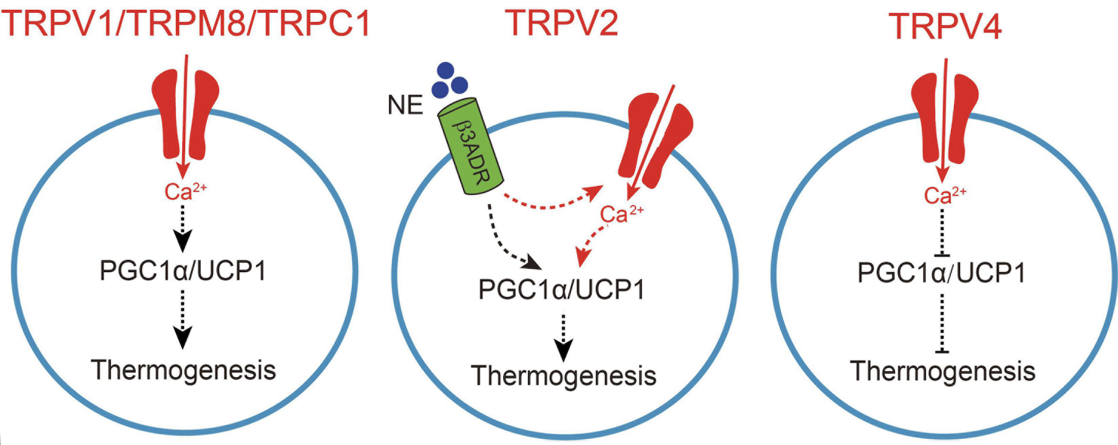
Sun W, Luo Y, Zhang F, Tang S and Zhu T (2021) Involvement of TRP Channels in Adipocyte Thermogenesis: An Update. Front. Cell Dev. Biol. 9:686173. doi: 10.3389/fcell.2021.686173.https://creativecommons.org/licenses/by/4.0/
This figure illustrates how TRP channels regulate heat production in adipocytes by modulating calcium entry. The schematic shows that calcium influx through TRPV1, TRPV2, TRPM8, and TRPC1 regulates thermogenic gene expression in adipocytes, thereby enhancing thermogenesis. When sympathetic nerve activity rises, norepinephrine is released from the sympathetic nerves and activates the β3-adrenergic receptor (β3ADR) in brown adipocytes. In this setting, TRPV2 collaborates with β3ADR to regulate peroxisome proliferator-activated receptor gamma coactivator-1 alpha (PGC1alpha) and uncoupling protein 1 (UCP1), which further enhances thermogenesis. In contrast, TRPV4-mediated calcium influx negatively regulates thermogenic gene expression in adipocytes, thereby inhibiting thermogenesis.
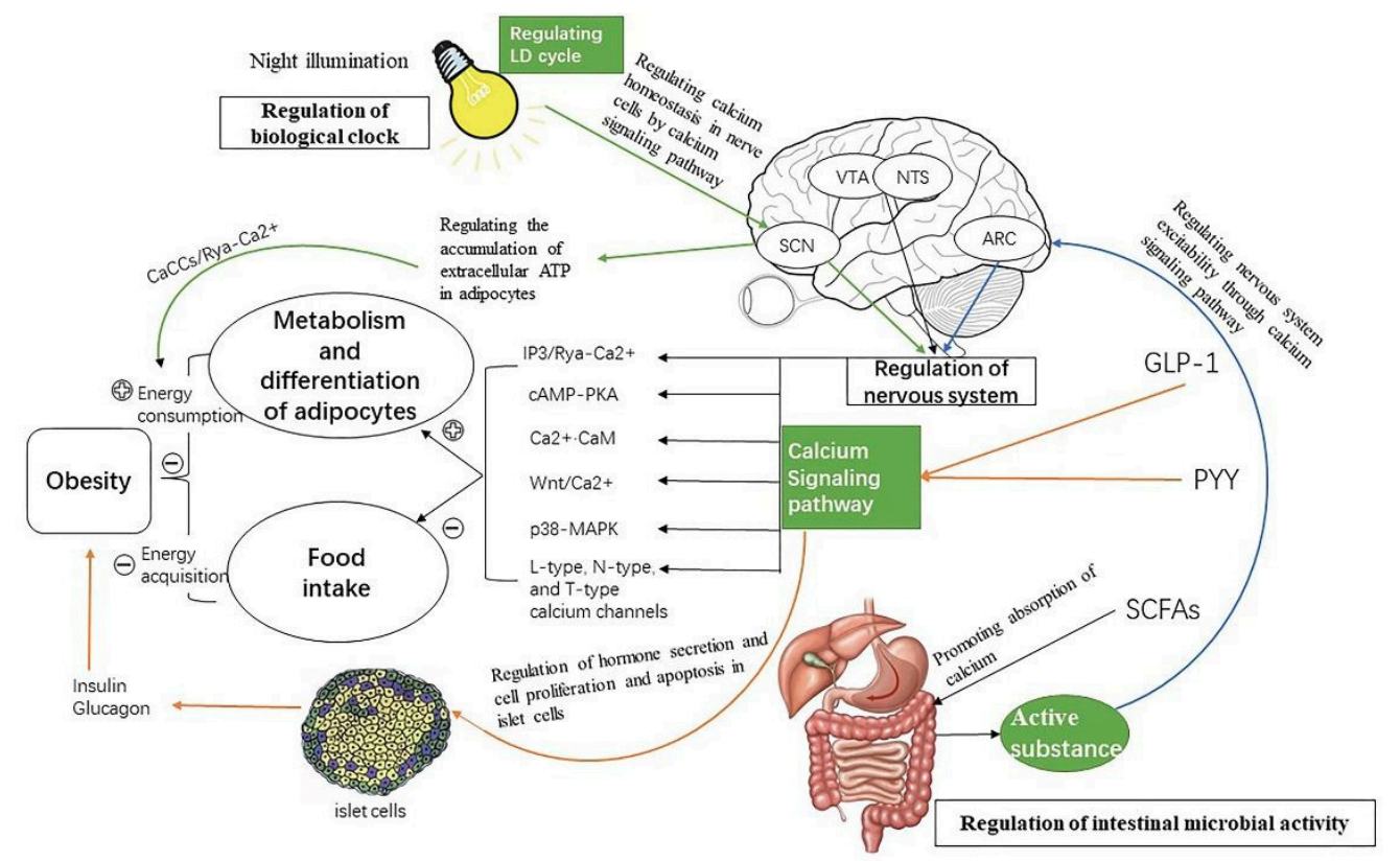
This figure illustrates how the nervous system, gut microbes, and circadian rhythms regulate obesity through calcium signaling. Light activates calcium signaling in light-responsive neurons that signal the brain to enhance satiety, and acts on adipocytes through calcium signaling pathways that promote metabolism and heat production. GLP-1, PYY, and SCFAs secreted by intestinal microorganisms act on a feeding-related brain region to enhance satiety, while other calcium signaling pathways drive lipid decomposition in adipocytes. In pancreatic tissue, calcium signaling pathways promote islet Β-cell proliferation, inhibit their apoptosis, and regulate insulin secretion, all of which are related to obesity. The signaling molecule Nesfatin-1 acts on neurons of the ventral tegmental area and other feeding-related neurons through specific calcium channels to enhance satiety, while sympathetic excitation drives brown adipocyte heat production through TRPV1 and related calcium signaling pathways.
Song, Z.; Wang, Y.; Zhang, F.; Yao, F.; Sun, C. Calcium Signaling Pathways: Key Pathways in the Regulation of Obesity. Int. J. Mol. Sci. 2019, 20, 2768. https://doi.org/10.3390/ijms20112768
https://creativecommons.org/licenses/by/4.0/
Clinical Tips
1. When caloric restriction fails to produce expected fat loss, suspect a calcium-signaling lock — elevated intracellular calcium keeps the adipocyte synthesizing fat and blocking lipolysis, even in energy deficit.
2. Visceral fat that drives disproportionate inflammation relative to total body weight points to broken ER-mitochondria calcium transfer, where the adipocyte shifts to glycolysis and amplifies oxidative stress from within.
3. Fat-loss plateaus with rising inflammatory adipokines are not just "inflammation" — they trace back to calcium-driven NF-κB activation inside hypertrophic adipocytes, recruiting immune cells into visceral fat.
4. Stalled β-oxidation from poor mitochondrial calcium uptake traps lipid intermediates inside the adipocyte, progressively damaging the electron transport chain and deepening storage dependence — a self-reinforcing metabolic block.
5. AIC therapy targets this axis directly: restoring calcium flux across the adipocyte membrane, ER, and mitochondria re-opens lipolytic signaling, recovers oxidative capacity, and quiets the ER stress driving adipocyte inflammatory output.
Clinical Snapshot
Few things frustrate a practitioner more than seeing someone do everything right — verified deficit, consistent movement, no obvious compliance gaps — and the scale stalls while abdominal fat sits untouched. The signs that set this apart from a simple plateau are worth paying attention to: cold hands and feet that worsen over weeks, energy that drops rather than stabilizes, visible loss of muscle definition while the midsection stays the same, and a metabolic rate that seems to decline faster than any textbook would predict.
This is not a willpower problem, nor a generic metabolic adaptation. It is a tissue-level shutdown in which the fat cells that are supposed to burn energy as heat — beige adipocytes in subcutaneous depots — have lost the internal calcium-driven machinery that powers this process. When the calcium cycling system inside these cells breaks down under chronic inflammatory stress, the body's secondary engine for energy expenditure goes offline at the exact moment caloric restriction demands it most. The result is a trapped state: restriction depletes muscle, fat stays locked, and the deficit that should be producing results instead deepens fatigue and cold sensitivity.
Recognizing this pattern early changes what the practitioner does next. Instead of tightening macros or adding more cardio — interventions that accelerate lean mass loss without touching the real barrier — the focus shifts to whether the adipocyte itself can still respond. The hallmark signs are all there: stalled visceral fat despite global weight change, worsening cold intolerance, inflammatory markers that creep up rather than down, and a resting metabolic rate that keeps dropping week after week. These are not vague complaints — they trace directly to the failure of calcium-dependent thermogenesis inside fat tissue, compounded by the inflammatory calcium signaling that keeps those depots insulin-resistant and metabolically inert.
AIC Therapy targets this axis by restoring coordinated calcium handling across the adipocyte membrane, endoplasmic reticulum, and mitochondria — reactivating the thermogenic machinery that caloric restriction alone cannot reach and quieting the calcium-driven inflammatory pressure that keeps beige fat shut down and visceral fat locked in storage mode.
Conclusion
Weight-loss resistance in obesity is not a behavioral or caloric problem but a tissue-level calcium disorder inside the adipocyte. Elevated cytosolic calcium keeps lipogenic programs active while blocking lipolysis; mitochondrial calcium overload shuts down fatty acid oxidation and thermogenesis; and depleted endoplasmic reticulum calcium sustains stress signaling, keeping visceral fat inflamed and insulin-resistant. Clinically, the pattern is recognizable — stalled visceral fat despite a verified deficit, worsening cold intolerance, rising inflammatory markers, and a resting metabolic rate that continues to decline faster than the restriction alone would account for.
What these patients need is not further restriction but restoration of the calcium handling that keeps the adipocyte capable of responding at all. As long as cytosolic calcium stays high, mitochondrial calcium stays overloaded, and endoplasmic reticulum calcium stays depleted, lipolysis, thermogenesis, and insulin signaling remain offline together, and each failure reinforces the next.
AIC Therapy restores coordinated calcium signaling across the adipocyte membrane, endoplasmic reticulum, and mitochondria, thereby reopening lipolytic output, restoring oxidative capacity in brown and beige fat, and restoring the thermogenic response that caloric restriction alone cannot achieve. By correcting the same calcium disturbance that drives mitochondria–endoplasmic reticulum oxidative stress and the inflammatory pressure sustaining insulin resistance, it quiets the environment, keeping visceral fat in storage mode, giving the patient's deficit a tissue that can finally respond to it.
Bring calcium signaling into your practice.
Practitioners can request a consultation to discuss AIC therapy and how these mechanisms apply to specific cases.
Newsletter content is educational and contributed by participating healthcare practitioners. These statements have not been evaluated by the FDA and are not intended to diagnose, treat, cure, or prevent any disease.