
Executive Summary
A breakdown in calcium signaling can quietly disrupt joint cell function, leading to stiffness even when scans appear normal. Chondrocytes may lose their ability to produce energy and collagen when calcium becomes trapped or misdirected. Early signs like hydration loss, fascial tension, and low-grade crystal irritation often emerge without triggering inflammatory markers.
Calcium Flow and the Hidden Roots of Joint Pain
Joint pain that lingers without a clear cause often begins where few doctors think to look: calcium flow. Before cartilage wears down, calcium signaling quietly starts to fail inside joint cells. Chondrocytes rely on this rhythmic calcium movement to generate energy, build collagen, and respond to stress. But when the system falters, joints lose their ability to recover, even if imaging looks fine and lab tests appear normal.
When calcium is misdirected into soft tissues, fascia tightens and hydration fades, well before visible degeneration. Tiny calcium clusters may accumulate in joint fluid, silently irritating the tissue. Inflammatory markers often remain low, yet stiffness and swelling persist because cells can no longer speak the language of repair. Sometimes the problem isn't inflammation at all—it’s that the cell has forgotten how to reset.
Low calcitonin levels and dysregulated calcium channel activity can make joints feel older than they are. Mitochondria inside synoviocytes begin to slow, and energy-starved cells stop repairing. Even targeted nutrition or physical therapy may fall short, not due to poor compliance, but because ionic signaling was never restored. This is where calcium signaling therapy finds its role—not by overriding biology, but by restoring the clarity it lost.
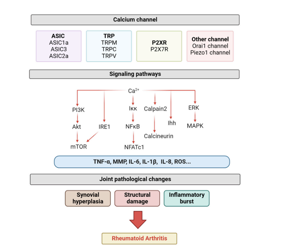
Liang HY, Yin HX, Li SF, et al. Calcium-Permeable Channels Cooperation for Rheumatoid Arthritis: Therapeutic Opportunities. Biomolecules. 2022;12(10):1383. Published 2022 Sep 27. doi:10.3390/biom12101383
Calcium channels play a central role in the development and progression of rheumatoid arthritis (RA). Channels such as ASIC, TRP, P2X7R, Orai1, and Piezo1 regulate calcium influx into joint cells, shaping how these cells respond to stress and injury. When calcium flows through these channels becomes dysregulated, it triggers inflammatory cascades that drive tissue damage and chronic synovitis. This ongoing calcium-driven activation contributes to joint deterioration, even in the early stages of disease. Targeting these specific channels may open new therapeutic avenues that go beyond conventional anti-inflammatory strategies.
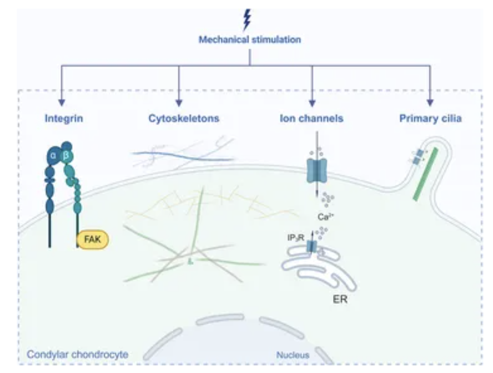
Liu Y, Jia F, Li K, et al. Critical signaling molecules in the temporomandibular joint osteoarthritis under different magnitudes of mechanical stimulation. Front Pharmacol. 2024;15:1419494. Published 2024 Jul 11. doi:10.3389/fphar.2024.1419494
As joints move or absorb pressure, the cells within them respond directly to mechanical forces. This physical stress is converted into biochemical signals—a process known as mechanotransduction, or cellular force detection. How cells perceive and interpret these signals determines how they maintain, repair, or reshape the surrounding matrix. This intricate process involves structural and signaling components such as integrins, the cytoskeleton, calcium-regulating ion channels, and primary cilia—specialized sensory hubs particularly active within the cartilage of the jaw joint.
Advanced Clinical Tips
Silent joint stiffness often stems from intracellular calcium buildup, not from visible inflammation or trauma.
ER calcium depletion reduces chondrocyte energy output, limiting recovery even after steroid or hyaluronic acid injections.
Microcalcifications in synovial fluid may provoke pain well before joint erosion becomes detectable on imaging.
TNAP overactivity breaks down protective pyrophosphate (PPi), accelerating crystal formation in chronic low-grade joint pain.
Restoring calcitonin levels can calm inflammation without relying on immunosuppressive therapies.
Case Reference
A 56-year-old woman experienced 15 years of persistent finger stiffness and pain, primarily following chronic cold exposure and repetitive strain. Conventional therapies, including topical analgesic patches, provided only short-term relief without restoring function. After initiating AIC therapy, her grip strength improved significantly, swelling resolved, and she began sleeping through the night without pain. No changes were made to her lifestyle or environment during this period. The observed recovery was attributed to AIC’s effect on calcium balance and the resolution of soft tissue calcific stress.
Conclusion
Joint pain often begins long before structural damage appears, when calcium flow inside the cell starts to break down. As seen in cellular force detection, mechanical pressure on joints isn’t just physical—it alters how ion channels and primary cilia regulate repair. Silent stiffness may emerge from intracellular calcium buildup, even when inflammatory markers remain low. In one case, chronic finger pain resolved completely after AIC therapy helped restore functional calcium balance. Rather than suppressing the body, AIC therapy corrects the miscommunication, allowing the cell to remember how to heal.
Bring calcium signaling into your practice.
Practitioners can request a consultation to discuss AIC therapy and how these mechanisms apply to specific cases.
Newsletter content is educational and contributed by participating healthcare practitioners. These statements have not been evaluated by the FDA and are not intended to diagnose, treat, cure, or prevent any disease.