Executive Summary
Chronic joint pain often persists even when inflammation is low and even when imaging studies appear normal. Beneath the surface, misdirected calcium signaling can distort cartilage response, reduce mechanical sensitivity, and block the body’s own repair commands. By restoring intracellular flow and recalibrating ionic rhythms, deeper tissue cooperation can resume—even in patients who have plateaued despite doing everything right.
The Silent Joint Shutdown
Not all cartilage damage is mechanical. In many patients, the joint fails to recover not because of inflammation or wear, but because the communication between bone and cartilage has quietly shut down. Beneath the surface, calcium signaling becomes misdirected, distorting repair timing, confusing structural cues, and slowing recovery even when nutrition, hormones, and joint protocols are in place. Osteoblasts typically send rhythmic ionic pulses that chondrocytes use to regulate gene expression and respond to stress. But when that bone rhythm fades—whether from age, overload, or ionic rigidity—the cartilage above it begins to drift, losing coordination with the very matrix that should help it regenerate.
What follows isn’t just stiffness—it’s molecular disorientation. Misallocated calcium hardens the environment, while TRPV4 signaling becomes erratic, reducing the chondrocyte’s ability to sense or respond to load. Matrix metalloproteinases remain active for too long, degrading collagen because the “off” switch isn’t received. Meanwhile, microcalcifications may begin forming—not just in bone, but inside cartilage vesicles and joint capsules. These are the patients whose joints flare with movement but don’t respond to anti-inflammatory diets or injections. What’s more? Calcitonin signaling, meant to buffer immune tension in the joint, often fades early, creating an environment where repair never catches up. And ER-mitochondria disconnection inside cartilage cells quietly limits ATP, making physical rehab feel harder than it should.
This isn’t cartilage degeneration—it’s a failed dialogue. Chondrocytes aren’t misbehaving—they’re cut off. Nociceptors become hypersensitive, not from trauma, but from calcium trapped in sensory endings. Even in autoimmune cases, flare-ups often follow ionic shifts before cytokines ever rise.
Subchondral edema? If is often just a symptom of ionic traffic jams below the cartilage. What changes things isn't adding more inputs—it’s restoring the signal clarity that once kept this joint adaptive. AIC therapy may support this by correcting the calcium flow that enables cells to receive, respond to, and repair. For patients who are doing everything right but still plateau, this may be the hidden axis holding the joint in limbo.
Savadipour A, Nims RJ, Rashidi N, et al. Membrane stretch as the mechanism of activation of PIEZO1 ion channels in chondrocytes. Proc Natl Acad Sci U S A. 2023;120(30):e2221958120. doi:10.1073/pnas.2221958120
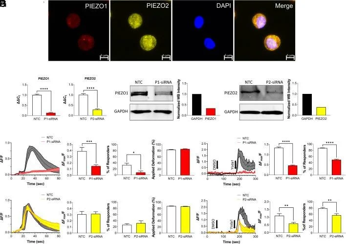
This figure illustrates how PIEZO1 and PIEZO2 channels regulate calcium signaling in cartilage cells in response to mechanical or chemical stimulation.
Panel A shows that both PIEZO1 and PIEZO2 are present in porcine chondrocytes, confirmed through immunostaining.
Panels B and C demonstrate that targeted gene silencing effectively reduces the mRNA and protein levels of each channel.
Panels D through G reveal that when either PIEZO1 or PIEZO2 is silenced, chondrocytes show a weaker calcium response and reduced sensitivity to mechanical loading or pharmacologic activation.
Together, these results suggest that healthy cartilage function depends on intact PIEZO signaling to trigger proper calcium influx during mechanical stress and repair.
Amin AK, Huntley JS, Bush PG, Simpson AH, Hall AC. Chondrocyte death in mechanically injured articular cartilage--the influence of extracellular calcium. J Orthop Res. 2009;27(6):778-784. doi:10.1002/jor.20809
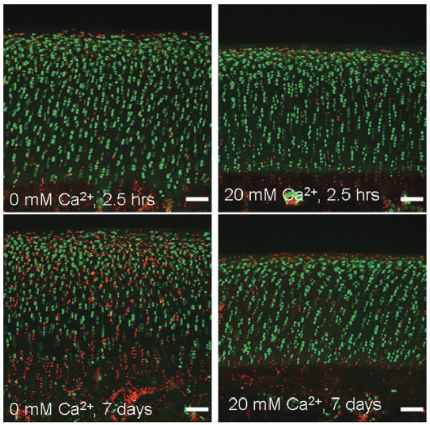
This Figure shows how extracellular calcium levels affect the pattern of cartilage cell death after injury.
In the short term (2.5 hours), both calcium-rich and calcium-free conditions result in cell loss near the joint surface.
But over 7 days, calcium-free conditions lead to deeper tissue damage, spreading cell death into the middle and deep cartilage layers.
In contrast, when calcium is present, the damage stays localized near the surface, suggesting a stabilizing effect on deeper tissue.
Clinically, this highlights that while reducing calcium may lower immediate damage, it may compromise long-term joint integrity unless carefully managed.
Where AIC Fits Clinically
Most protocols for chronic or immune-driven bone disorders still treat the skeleton as a structural endpoint. But AIC therapy shifts the starting point entirely, reframing calcium not as a static nutrient, but as a mobile signal that calibrates tissue behavior across systems. It engages where conventional interventions taper off: at the intracellular level, where calcium dynamics regulate osteoblast output, marrow signaling, and vascular responsiveness in the surrounding environment.
Instead of pushing or replacing what the body lacks, AIC works by restoring directional calcium flow. It helps shift calcium away from congested soft tissues and back into active skeletal cycling. This transition clears local calcifications and reopens cellular communication between bone, nerve, and immune cells, subtly improving how each responds under stress.
In cases where patients don’t respond to nutrient support, minerals, or even well-designed protocols, the barrier may not be input—it may be flow. AIC restores the rhythm that governs intracellular calcium movement, including the signals that help regulate structural repair and local inflammatory tone.
Even mild reactivation of physiological calcium-based signaling, such as the calcitonin axis, can be sufficient to reset the repair process from within. This opens up a different kind of intervention—not one that suppresses or overrides, but one that removes the silence that kept tissues from responding in the first place. AIC doesn’t rush recovery; it reminds the system how to listen.
Overlooked Clinical Signals in Joint Pain
Clinical Insight #1
Cartilage isn’t always the first to break down in chronic joint pain.
Subchondral bone signaling loss and marrow dysfunction often begin long before cartilage frays.
Clinical Insight #2
Meniscal pain may reflect fluid stagnation, not just structural wear.
Impaired lymphatic flow can amplify mechanical stress and mimic symptoms of a meniscus tear.
Clinical Insight #3
Post-detox joint flares don’t always mean toxin rebound.
Misplaced intracellular minerals, especially calcium and phosphate, can drive flare-like responses.
Clinical Insight #4
Joint clicking without pain can still indicate early dysfunction, especially in conjunction with fatigue.
Micro-instability and delayed ligament signaling may manifest long before imaging changes become apparent.
Case Reference
A 42-year-old woman with longstanding hand arthritis experienced pain unresponsive to prescription medications, and daily activities became difficult. Despite normal labs, joint stiffness and inflammatory swelling persisted, limiting her grip strength and quality of life. After introducing AIC therapy, symptoms began to reverse within weeks—swelling reduced, finger flexibility returned, and energy levels noticeably improved. What changed was not the visible inflammation but the internal signal flow; ionic rigidity was cleared, restoring the tissue’s ability to respond. This case illustrates that for patients plateauing despite standard protocols, restoring calcium signaling may reactivate dormant repair capacity.
Conclusion
Some patients experience pain and stiffness despite having good laboratory results, no obvious inflammation, and consistent care. As the data shows, calcium doesn’t just strengthen tissue—it instructs it. When that instruction is disrupted, cellular repair slows, even if mechanical input is ideal. Recent findings demonstrate that cartilage loses its direction when PIEZO channels are silenced or calcium buffering is unstable, which can shift the tissue from an active state of adaptation to a state of silent degradation. What we’ve also observed clinically is that restoring intracellular calcium rhythm, not just raising levels, can reset these signals. This is where AIC therapy stands apart: it doesn’t override the system—it gives the system back its voice.
Bring calcium signaling into your practice.
Practitioners can request a consultation to discuss AIC therapy and how these mechanisms apply to specific cases.
Newsletter content is educational and contributed by participating healthcare practitioners. These statements have not been evaluated by the FDA and are not intended to diagnose, treat, cure, or prevent any disease.