Rebuilding Immune Power Through Targeted Calcium Signaling
This newsletter explores how restoring precise intracellular calcium flow can energize immune cells, strengthen pathogen defense, and improve infection outcomes.
Executive Summary
Calcium microdomains at the ER-mitochondria interface control the metabolic tempo of immune cells during activation. Disruption in this calcium transfer can weaken T-cell responses, blunt NK cell function, and heighten inflammatory stress, even when standard blood calcium levels look normal. AIC therapy delivers ionized calcium directly to support mitochondrial ATP production, stabilize immune activation, and reduce harmful inflammatory amplification.
The Hidden Driver of Immune Energy
Calcium microdomains at endoplasmic reticulum–mitochondria junctions set the tempo for ATP production during immune activation. In patients with recurrent infections, serum values may appear normal, while intracellular Ca²⁺ handling is impaired. During T-cell receptor signaling, mitochondrial Ca²⁺ entry through the uniporter drives TCA flux and oxidative phosphorylation, and weak Ca²⁺ transfer can coincide with low IL-2 after stimulation assays. Vaccine responses often correlate with this bioenergetic reserve, so fatigue states with poor mitochondrial function may exhibit blunted antibody and T-cell responses.
Natural killer cell performance also depends on calcium-tuned metabolism. Low CD107a degranulation or weak cytotoxicity can reflect limited mitochondrial Ca²⁺ support even when glucose availability seems adequate. In inflamed tissue, extracellular ATP released from immune and stromal cells opens purinergic Ca²⁺ channels on macrophages, creating intercellular Ca²⁺ waves that amplify local inflammation. Gout, burns, or trauma create ATP-rich microenvironments that heighten these signals, which explains why simple measures that lower local metabolic demand and protect tissue can help alleviate the condition.
Inflammasome assembly, particularly NLRP3, requires calcium mobilization, and mitochondrial stress can add fuel by releasing oxidized mtDNA. Sleep disruption, hypoxia, and metabolic imbalance frequently aggravate this Ca²⁺-linked stress. Because intracellular Ca²⁺ dynamics can be abnormal despite a normal basic metabolic panel, the interpretation of “normal labs” benefits from pairing clinical context with functional immune readouts rather than relying solely on serum calcium levels.
AIC therapy delivers ionized calcium in a form that efficiently reaches the microdomains where the endoplasmic reticulum and mitochondria interface, sustaining the calcium signals that drive mitochondrial ATP production during immune activation. In clinical settings, stable microdomain calcium flow supports robust T-cell cytokine release, NK cell degranulation, and macrophage coordination, even in patients whose standard serum calcium levels appear normal. By promoting calcitonin-driven redistribution of calcium toward bone and away from soft tissues, AIC can help reduce ectopic calcium stress on the microcirculation, thereby tempering excessive inflammatory amplification.
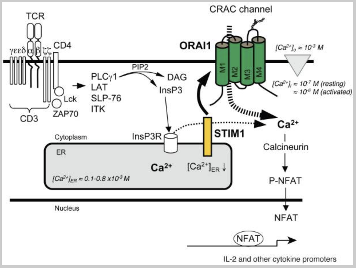
Feske S, Picard C, Fischer A. Immunodeficiency due to mutations in ORAI1 and STIM1. Clin Immunol. 2010;135(2):169-182. doi:10.1016/j.clim.2010.01.011
Some patients have a normal number of lymphocytes, yet their T cells are unable to activate properly. In some cases, this is due to reduced calcium entry into the cell. Normally, when the T-cell receptor is stimulated, it activates two enzymes called tyrosine kinases, which assemble a signaling complex and activate an enzyme called phospholipase C gamma. This enzyme splits a membrane molecule into two messengers, one of which releases calcium from storage inside the endoplasmic reticulum. As these stores empty, a sensor protein called STIM1 detects the drop and activates another protein, ORAI1, to open a channel in the cell membrane. This allows calcium from outside the cell to flow in, raising the concentration inside from about 100 nanomoles to about 1 micromole. This sustained calcium signal is crucial for activating calcineurin, which in turn translocates the transcription factor NFAT into the nucleus, thereby regulating genes that control immune responses.
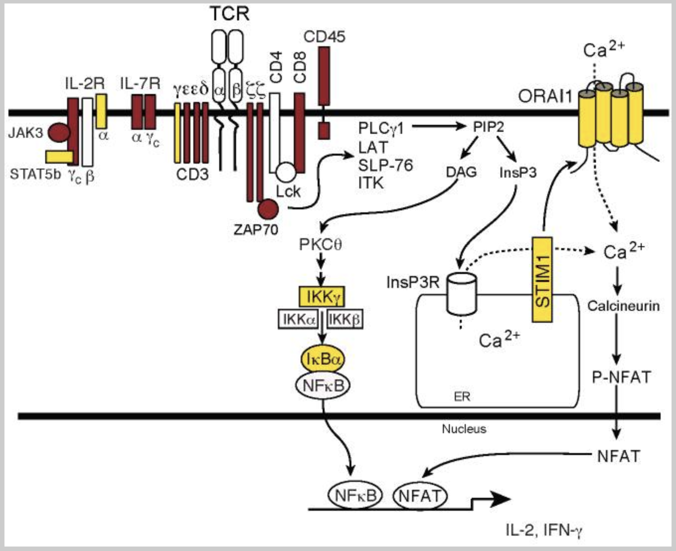
Feske S, Picard C, Fischer A. Immunodeficiency due to mutations in ORAI1 and STIM1. Clin Immunol. 2010;135(2):169-182. doi:10.1016/j.clim.2010.01.011
Severe combined immunodeficiency is a serious condition where the immune system cannot develop or activate key defense cells. Around half of the cases are caused by changes in a single gene essential for several immune cell communication receptors. Other cases result from defects affecting the processing of DNA building blocks, DNA repair, mitochondrial energy production, thymus development, or the transfer of signals in immune cells. Some defects prevent T lymphocytes from developing at all, while others allow normal numbers but block their activation. In these activation defects, patients may have normal blood counts yet remain highly prone to infections because their T cells cannot respond to threats.
Five Common Clinical Misconceptions
1. Misconception: Serum calcium is normal, so immune Ca²⁺ is fine.
Clinical move: treat persistent infections or vaccine nonresponse as a possible intracellular Ca²⁺ problem, and use stimulated lymphocyte assays or NK functional tests rather than stopping at the chemistry panel.
2. Misconception: T-cell calcium entry is only plasma-membrane store-operated flow.”
Clinical move: remember the MCU and mitochondrial Ca²⁺ uptake in T-cell activation, and anticipate weak responses in patients with mitochondrial stress, malnutrition, or severe sleep apnea that disturbs Ca²-linked energy supply.
3. Misconception: NK cells run on glycolysis, so mitochondria and Ca²⁺ matter less.
Clinical move: when CD107a or killing assays are low, address Ca²-dependent oxidative phosphorylation as well as glucose control, hydration, and hypoxia, and recheck function after correcting those stressors.
4. Misconception: Inflammation spreads only via cytokines.
Clinical move: in hot, ATP-rich tissue, purinergic receptors open Ca²⁺ channels that amplify local inflammation, so emphasize gentle wound care, edema control, and avoidance of unnecessary manipulation that spills more ATP.
5. Misconception: Inflammasomes ignore calcium.
Clinical move: in autoinflammatory flares or severe sepsis trajectories, reduce upstream Ca²⁺ stressors where possible, watch drugs that disturb Ca²⁺ movement, and pair anti-cytokine decisions with steps that normalize Ca²⁺ signaling and mitochondrial load.
Case Reference 1
A 64-year-old man from Tanzania with HIV started AIC therapy twice to three times daily. In just six days, his viral load fell from 2,380 to 75 copies/ml. This rapid drop demonstrates the strong immune-boosting effect of AIC therapy, enabling the body to suppress the virus with exceptional speed. Even without a recorded CD4 count, the outcome shows clear immune activation and control.
Case Reference 2
a 62-year-old man from Canada with HIV, began AIC therapy two to three times daily. Over a period of just ten weeks, his viral load dropped from 2,300 to 50 copies/ml. This remarkable decline highlights the strong immune-supportive action of AIC therapy, empowering the body to suppress viral replication to near-undetectable levels. Even without CD4 data, the result demonstrates clear, rapid immune activation.
Conclusion
AIC therapy directly addresses a hidden weakness in immune performance: impaired calcium delivery to mitochondrial microdomains. By stabilizing these signals, it supports the ATP production needed for strong T-cell activation and NK cell cytotoxicity. The result is a more coordinated immune response, with faster viral control and better pathogen clearance. Clinical cases show rapid viral load reductions in HIV patients, highlighting real-world immune activation. Beyond infections, this approach also helps temper excessive inflammation by guiding calcium away from soft tissues and into the bone. In both prevention and treatment, AIC therapy stands out as a precise, clinically relevant immune support strategy.
Bring calcium signaling into your practice.
Practitioners can request a consultation to discuss AIC therapy and how these mechanisms apply to specific cases.
Newsletter content is educational and contributed by participating healthcare practitioners. These statements have not been evaluated by the FDA and are not intended to diagnose, treat, cure, or prevent any disease.