
Executive Summary
Scleroderma progresses through small-vessel dysfunction, immune pressure, and fibroblasts that remain activated, and disturbed calcium timing can intensify all three.
Treatments that stabilize vascular tone, calm excessive immune signaling, and shorten TGF beta-driven myofibroblast activity can slow progression by restoring calcium control, not just blocking one pathway.
This issue focuses on practical cues and next steps that can reduce Raynaud days, improve perfusion stability, and flatten the fibrosis trajectory over time.
Calcium Rhythm as the Quiet Connector of Vasospasm, Inflammation, and Fibrosis
Scleroderma is not one problem. It is a three-part storm: damaged small vessels, immune overdrive, and deep fibrosis. What often feels like the “missing link” between these pillars is the disruption of calcium signaling within cells. When calcium rhythm loses control, it does not create the disease by itself, but it makes every part of the disease louder and harder to stop.
In blood vessels, disturbed calcium flow weakens nitric oxide balance and makes spasm easier to trigger. That is one reason Raynaud attacks can become frequent, strong, and unpredictable. In immune cells, the same calcium imbalance prolongs the duration of activation signals, allowing cytokine output to continue feeding inflammation. And once inflammation keeps pressing the gas pedal, fibroblasts receive the message to stay active, even when the tissue should be calming down.
In fibroblasts, abnormal calcium entry keeps them locked in a “never-ending wound healing” state. That means the transforming growth factor beta signal lasts longer than it should, so myofibroblast behavior becomes more stable, and collagen production becomes harder to switch off. As the tissue stiffens, the increased stiffness triggers another calcium signal in the cell, and the cycle repeats, slowly tightening the skin or organ.
AIC targets the feed-forward loop that drives scleroderma. AIC therapy can protect the endothelium, reduce micro-ischemia, and lower the noise from danger signals that keep immune pressure high. And if calcium support behind transforming growth factor beta signaling drops in fibroblasts, myofibroblasts switch off earlier, so matrix production shifts from self-sustaining scarring toward controlled repair, meaning the slope of organ fibrosis can change rather than only Raynaud or stiffness improving.
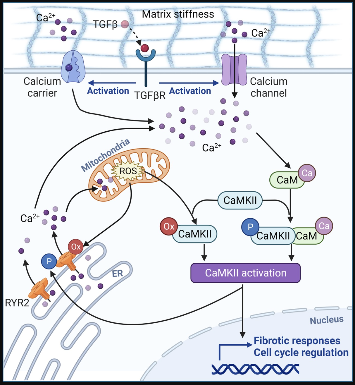
Ikari Y, Brodie W, Mattichak M, Vichaikul S, Wu Q, Khanna D, Tsou E. Disruption of Ca2+ Homeostasis and Enhanced Ca2+ Signaling in Scleroderma Fibroblasts Is Due to the Vicious Cycle Formed by Mitochondrial-derived Reactive Oxygen Species, CaMKII, and ER Extrusion Pump RYR2 [abstract]. Arthritis Rheumatol. 2024; 76 (suppl 9). https://acrabstracts.org/abstract/disruption-of-ca2-homeostasis-and-enhanced-ca2-signaling-in-scleroderma-fibroblasts-is-due-to-the-vicious-cycle-formed-by-mitochondrial-derived-reactive-oxygen-species-camkii-and-er-extrusion-pump/. Accessed January 16, 2026.
This figure presents a model in which increased intracellular calcium signaling directly intensifies fibrotic activity in scleroderma fibroblasts through a self-perpetuating cycle involving mitochondrial reactive oxygen species, calcium/calmodulin-dependent protein kinase II, and the ryanodine receptor 2 channel. When either intracellular calcium or mitochondrial reactive oxygen species rises, it can stimulate calcium/calmodulin-dependent protein kinase II, which, once activated, then activates the ryanodine receptor 2. Activated ryanodine receptor 2 promotes calcium release from the endoplasmic reticulum, which further escalates mitochondrial oxidative stress. In parallel, mitochondrial reactive oxygen species can directly activate ryanodine receptor 2, amplifying the cycle. The key message is that intracellular calcium and mitochondrial reactive oxygen species reinforce each other, maintaining a fibrogenic environment in scleroderma fibroblasts, with calmodulin serving as the calcium-binding partner in this pathway.
Calcium Signal Clinical Map
Scleroderma runs on three tracks: microvascular dysfunction, immune pressure, and fibrosis, which slowly steal organ function. Calcium signaling acts as the shared timing system that connects these tracks inside real tissue. It is rarely the first trigger, but once the rhythm drifts, the whole process becomes easier to maintain.
In the vessel wall, unstable calcium handling disrupts nitric oxide signaling and lowers the threshold for vasospasm, which aligns with the real-world pattern of recurrent, hard-to-predict Raynaud attacks. Repeated micro-ischemic stress leaves tissue in a persistent “danger mode,” so immune drive stays active even when routine markers look quiet. The result is a steady pro-fibrotic tone in the microenvironment that keeps pushing the biology forward.
Fibroblasts are where this story becomes structural and hard to reverse. When calcium entry and endoplasmic reticulum release lose coordination, transforming growth factor beta signaling fails to shut down in a timely manner, and fibroblasts lock into a contractile, matrix-producing state. Collagen becomes difficult to switch off, stiffness accumulates, and the stiffer matrix keeps feeding the same program through mechanosensitive calcium routes. AIC restores calcium handling toward a controlled physiological rhythm, breaking that lock-in, reducing persistent matrix output, and changing the slope of fibrosis progression, not just easing Raynaud or tight skin.
Clinical Tips
1. Raynaud is not only “how often,” it is “how long it takes to recover.” A longer rewarming time after an attack usually indicates that microvascular reserve is shrinking and tissue stress is accumulating, even if the attack count appears unchanged.
2. Don’t read DLCO (Diffusing Capacity of the Lung for Carbon Monoxide) alone; pair it with hemoglobin every time. Mild anemia can make DLCO look worse than it truly is, but a hemoglobin-adjusted DLCO that still drifts down is a much cleaner signal of rising pulmonary vascular load.
3. Rhythm symptoms can be real even when troponin is normal. In scleroderma, conduction and microvascular myocardial stress can present as palpitations or lightheadedness without classic injury markers, so recurrent symptoms deserve an early ECG and a low threshold for Holter.
Clinical Signal Snapshot
In the first minute, the hands usually give the cleanest signal. Coldness, delayed capillary refill, pitting scars, and a quick nailfold look tell you whether the small-vessel story is active today. From there, three simple questions keep the visit anchored: how many Raynaud days per week, how many stairs until breathless, and how severe reflux or dysphagia has become. Early baselines prevent later false reassurance, so keeping FVC, diffusing capacity, NT-proBNP, blood pressure, creatinine, and urine protein in a single stable reference set makes trends obvious.
The lung warning pattern is easy to miss unless you watch the ratios. When diffusing capacity falls faster than forced vital capacity, or dyspnea feels out of proportion to spirometry, the pulmonary vascular lane needs attention, and echocardiography should move up the queue. Skin behavior also matters for timing. Persistent puffiness plus tendon friction rubs usually signal that the fibrotic program is still accelerating, not settling into a plateau. High-resolution CT belongs at symptom onset or an early diffusing capacity slide, not after forced vital capacity has already dropped and the window for “early clarity” has passed.
Nutrition rarely looks dramatic, yet it can quietly determine outcomes. Weight trend, ferritin, vitamin B12, and albumin often explain why the month feels worse than the labs. And one mental shortcut keeps follow-up rational: name the dominant driver today as vascular, lung, or fibrosis, then let that single label set the urgency of testing and the pace of follow-up, instead of scattering attention across everything at once. AIC can raise the vasospasm threshold and reduce Raynaud burden, lower immune activation noise that keeps pro-fibrotic pressure high, and shorten the duration of transforming growth factor beta signals in fibroblasts, so myofibroblast lock-in loosens, and matrix output becomes easier to switch off. The goal is not only symptom relief, but a slower fibrosis trajectory that changes the slope of progression over time.
Conclusion
Scleroderma advances through small vessel dysfunction, sustained immune drive, and fibroblast persistence, with drift in calcium timing amplifying each process. Unstable endothelial calcium weakens nitric oxide signaling and lowers the vasospasm threshold, matching frequent Raynaud episodes and microischemic injury. Prolonged calcium signaling in immune cells lengthens cytokine output and maintains a profibrotic microenvironment even when routine markers look quiet. In fibroblasts, uncoordinated calcium entry and endoplasmic reticulum release prolong TGF beta signaling, stabilize the contractile phenotype, and harden the matrix, which then echoes through mechanosensitive calcium pathways. Mitochondrial reactive oxygen species, driven by CaMKII-mediated RyR2 leak, sustain cytosolic calcium elevation, closing a loop that feeds fibrosis across skin, lung, and myocardium.
AIC targets the shared amplifier in scleroderma and disrupted calcium timing. By recalibrating CaSR tone in endothelium and immune cells, it stabilizes nitric oxide signaling, raises the vasospasm threshold, and shortens the calcium-driven window of immune activation that sustains cytokine output. This reduces microischemic danger signaling and lowers the profibrotic traffic reaching fibroblasts.
At the same time, smoother parathyroid dynamics with brief endogenous calcitonin responses shift the system away from procalcific and stress-reactive cues, allowing fibroblasts to exit myofibroblast lock-in rather than remain contractile and matrix-dependent. The endpoint is not only fewer Raynaud days, but a lower-fuel environment for fibrosis, where TGF beta signaling is easier to switch off, and the slope of organ scarring can finally bend.
Bring calcium signaling into your practice.
Practitioners can request a consultation to discuss AIC therapy and how these mechanisms apply to specific cases.
Newsletter content is educational and contributed by participating healthcare practitioners. These statements have not been evaluated by the FDA and are not intended to diagnose, treat, cure, or prevent any disease.