Calcium Signaling in Inflammation: Translating Molecular Insight Into Clinical Strategy
This newsletter explores how calcium misregulation drives inflammation and examines therapeutic approaches that reestablish physiological calcium signaling in chronic disease.
Executive Summary
Persistent inflammation is now understood to involve more than immune activation—it reflects disrupted calcium regulation at the mitochondrial, endoplasmic reticulum, and membrane channel levels. These intracellular imbalances contribute to chronic disease patterns even when systemic markers such as CRP or WBC appear normal. From metabolic dysfunction to neuro-inflammation and joint degradation, calcium signaling plays an active role in sustaining inflammation and tissue damage. Therapeutic approaches that restore proper calcium flow and receptor sensitivity, including AIC therapy, represent a clinically meaningful strategy to support resolution without broad immunosuppression.
How Calcium Shapes the Inflammatory Cascade
Inflammation inside blood vessels silently weakens the endothelial barrier, allowing calcium to enter where it shouldn’t and activate destructive signaling like the NLRP3 inflammasome. This process plays a key role in making plaques unstable, especially in early atherosclerosis, but can often be modulated by targeting calcium-sensing receptors and improving nitric oxide support.
In osteoarthritis, inflammatory signals raise calcium levels inside chondrocytes, pushing them into a breakdown mode that wears away cartilage. Meanwhile, inflamed joint linings release calcium-loaded particles that further irritate the area, making pain and stiffness worse over time.
Autoimmune diseases often flare up when T cells become overly sensitive to calcium, especially through a signaling axis called calcineurin–NFAT. With the right combination of natural modulators and immune-balancing nutrients, it may be possible to ease this calcium-driven overreaction without suppressing immunity.
In metabolic conditions like insulin resistance, excess calcium inside fat cells can block healthy energy regulation and worsen inflammation. Restoring proper calcium flow in these cells has been shown to improve glucose handling and reduce inflammatory stress on blood vessels.
In the brain, chronic inflammation disrupts how microglia handle calcium, leading to oxidative stress and faster buildup of toxic proteins like amyloid. When this calcium misfiring is corrected, even in its early stages, brain cells exhibit improved energy production and healthier synaptic communication.
During infections, immune cells such as neutrophils and macrophages rely on brief calcium bursts to combat pathogens. But when calcium stays elevated too long, it can lock these cells into an aggressive state, causing long-term tissue damage even after the infection has passed.
Across nearly every chronic disease, calcium signaling becomes distorted by inflammation, turning a vital messenger into a driver of dysfunction. This is where AIC shows promise—not as just another supplement, but as a tool that encourages the body to restore balance from within. By gently activating calcitonin and helping shift calcium away from inflamed tissues, AIC may support both inflammation resolution and long-term cellular health.
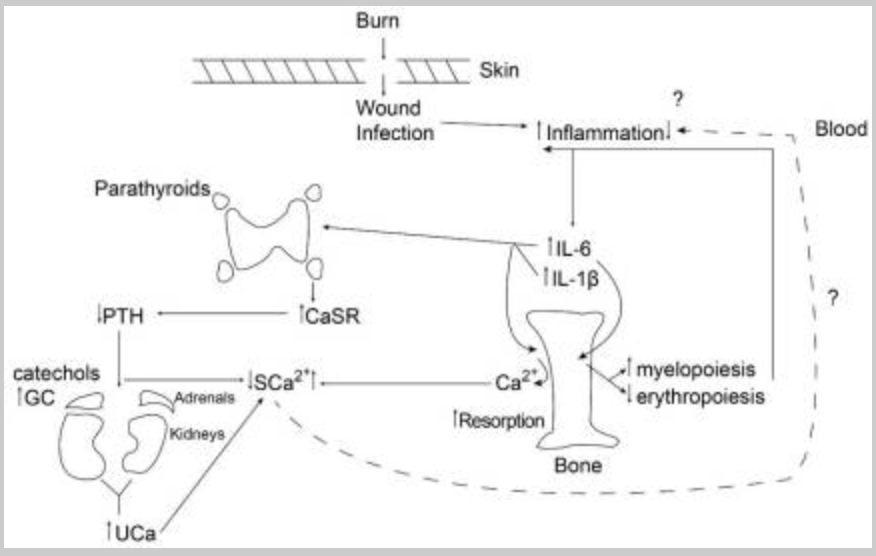
Klein GL. The Role of Calcium in Inflammation-Associated Bone Resorption. Biomolecules. 2018;8(3):69. Published 2018 Aug 1. doi:10.3390/biom8030069
This figure illustrates the events that occur in the body within 24 hours after a severe burn in children. The injury triggers inflammation, which increases certain immune signals, such as IL-6 and IL-1β, leading to the release of calcium from the bone. At the same time, the parathyroid’s calcium-sensing receptor (CaSR) becomes more active, which lowers parathyroid hormone (PTH) and causes the kidneys to lose more calcium in the urine. Stress hormones from the adrenal glands also support this calcium loss through urine. This response strikes a balance between releasing calcium from the bones and removing it through the kidneys, which may help control inflammation in the early stages after injury.
Cellular Realities That Shift How We Understand Inflammation
Let’s revisit five widely accepted clinical ideas—this time through the lens of molecular signaling and emerging translational insights.
Prevailing View 1: Mitochondria are primarily energy producers, not key inflammatory drivers. Deeper Layer: Mitochondrial damage leads to the release of mtDNA and cardiolipin, activating NLRP3. Supporting mitochondrial integrity helps reduce sterile inflammation in fatigue and autoimmunity.
Prevailing View 2: Protein misfolding is a neurological concern, not a systemic inflammatory trigger. Deeper Layer: ER stress from misfolded proteins activates the unfolded protein response and NF-κB. Enhancing proteostasis may help reduce chronic inflammation in conditions such as diabetes, cancer, and aging.
Prevailing View 3: Normal CRP and WBC values rule out active inflammation. Deeper Layer: Neuroinflammation, adipose cytokine activity, and perivascular signaling often occur without systemic laboratory changes—normal labs do not exclude underlying inflammatory activity.
Prevailing View 4: Tissue calcification is a passive consequence of aging. Deeper Layer: Calcium deposition reflects the reprogramming of soft tissues through chronic inflammation, driven by cytokine-mediated osteogenic pathways—this is an active, immune-mediated process, not a passive accumulation.
Prevailing View 5: Calcium plays no significant role in inflammatory signaling. Deeper Layer: Intracellular calcium is essential for initiating and sustaining immune activation. Dysregulated calcium dynamics can perpetuate chronic inflammatory states.
Case Reference
A 66-year-old female patient with a 10-year history of rheumatoid arthritis reported severe joint pain, stiffness, and difficulty walking, unresponsive to multiple conventional interventions including gold injections. Her symptoms included extreme tenderness in the toes, limited mobility in the knees, and deformity in the fingers.
After initiating AIC therapy under peer guidance, she experienced a marked reduction in pain within two weeks. She regained full toe-to-ground mobility and reported significant improvements in daily function and ambulation. This case illustrates the potential of targeted AIC therapy in modulating inflammatory joint conditions and supporting structural recovery in chronic autoimmune settings.
Conclusion
Inflammation destabilizes calcium-dependent pathways in endothelial, cartilage, and neural tissues, thereby setting the stage for progressive damage even in the early stages of disease. As seen in acute injuries, such as pediatric burns, calcium homeostasis is tightly regulated through bone, renal, and hormonal responses within hours of onset.
Molecular-level findings now reveal that unresolved inflammation can persist beneath normal labs, driven by mitochondrial signals, ER stress, and misdirected calcium flux. Clinically, a patient with treatment-resistant rheumatoid arthritis experienced rapid improvement in mobility and pain after starting AIC therapy.
Taken together, these insights suggest AIC therapy may restore intracellular calcium dynamics, promote calcitonin activity, and interrupt chronic inflammatory cycles without blocking natural immune resolution.
Bring calcium signaling into your practice.
Practitioners can request a consultation to discuss AIC therapy and how these mechanisms apply to specific cases.
Newsletter content is educational and contributed by participating healthcare practitioners. These statements have not been evaluated by the FDA and are not intended to diagnose, treat, cure, or prevent any disease.