Executive Summary
Chronic back pain often reflects intracellular calcium collapse long before structural degeneration appears on imaging. Misplaced calcium in fascia and discs silences repair, stiffens tissues, and blocks metabolic recovery. Restoring ionic signaling awakens mitochondrial energy flow and reactivates tissue responsiveness where other therapies fail.
Healing Signal
Chronic back pain often begins where we don't look—deep in the tissue matrix, where misallocated calcium reduces fascial glide and microvascular flow long before structural changes appear. When spinal muscles fatigue despite clean labs and optimal nutrition, the missing signal is frequently mitochondrial calcium, not a lack of ATP. Stubborn soft tissue injuries that don’t heal are often due to impaired ER calcium refilling, not just oxidative load. Pain that lingers in low-inflammatory states may stem from stiffened calcium channels unable to conduct proper intracellular communication. Intervertebral discs lose hydration not only from pressure or aging, but also when chondrocyte calcium signaling falters. Vertebral inflammation and marrow hypersensitivity often reflect disrupted calcitonin rhythms more than overt bone loss. Dense, fibrotic-feeling fascia can conceal microcalcifications that resist manual release but respond to ionic rebalancing. In dorsal root neurons, calcium overload drives hyperexcitability and sustained nociceptive output. Even minor imbalances in calcium flow can disconnect the signaling between bone, muscle, and nerve, leading to neuromuscular misfiring. Normal serum calcium masks dysfunctional distribution, especially in patients presenting with stiffness and tightness unexplained by imaging. Local oxygen deprivation may be traced back to microvascular calcification, which impairs delivery at the capillary level. In patients who don’t respond to magnesium therapy, the problem is often intracellular calcium is still locked in the wrong place. Recurring pain loops in the spinal cord may stem from disrupted calcium buffering in interneurons, not simply central sensitization. Back pain with clean imaging often reflects ionic rigidity at the cellular level rather than visible degeneration. And until intracellular calcium flow is reestablished, therapeutic input remains limited—because the tissue, quite literally, can’t hear the signal.
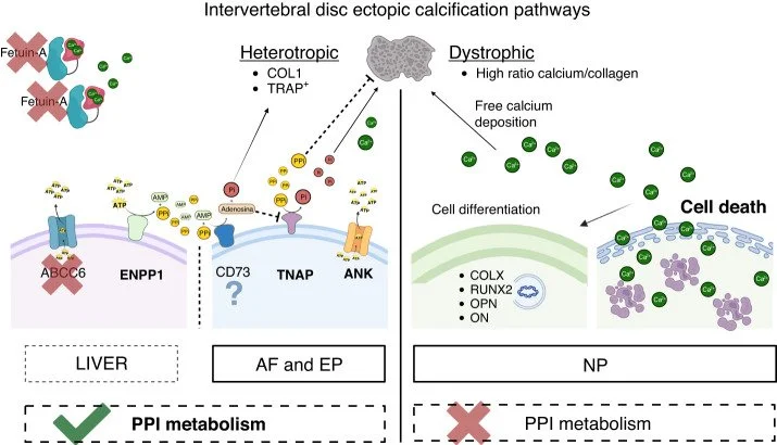
Novais EJ, Narayanan R, Canseco JA, et al. A new perspective on intervertebral disc calcification-from bench to bedside. Bone Res. 2024;12(1):3. Published 2024 Jan 22. doi:10.1038/s41413-023-00307-3
This figure shows how different regions of the intervertebral disc follow different calcification pathways. The annulus fibrosus (AF) and endplate (EP) respond to extracellular pyrophosphate (PPi) signaling and follow a regulated, ectopic calcification process. In contrast, the nucleus pulposus (NP) undergoes dystrophic calcification that appears unaffected by changes in PPi metabolism. Key proteins like ENPP1, ABCC6, and ANK regulate PPi availability by converting extracellular ATP and releasing it into circulation. When PPi is reduced, either through overactive TNAP or insufficient ENPP1 signaling, calcification-promoting pathways dominate, especially where protective mechanisms are absent or inactive.
Where AIC Fits Clinically
The mechanism behind this dysfunction isn’t just known, it’s addressable. AIC therapy restores intracellular calcium rhythms by resolving ionic congestion and reopening ER-mitochondria communication. As soft tissue calcium is redirected, stiffness lifts and ATP generation in stabilizer muscles reactivates. This allows the body’s existing repair systems to work again, not by force, but by removing the silent block.
Achieving Clinical Turnaround Through Calcium Signaling Restoration
Chronic back pain that resists structural or anti-inflammatory interventions often reflects disrupted calcium flow at the cellular level, particularly between the endoplasmic reticulum and mitochondria. This loss of intracellular rhythm impairs ATP production in postural stabilizers and deep fascial tissues, even when nutrients are sufficient. Calcium misallocation into soft tissue stiffens the local matrix and silences repair signaling, contributing to pain persistence and reduced flexibility. By restoring intracellular calcium oscillation and directing ionic flow back toward skeletal compartments, tissue responsiveness and recovery pathways begin to reengage. This shift doesn't block pain—it removes the biochemical rigidity that kept the system from resolving it.
Advanced Clinical Tips
Calcium buildup in fascia reduces glide and stiffness long before imaging shows degeneration.
Paraspinal mitochondria underperform when calcium entry through MCU is blocked, not from nutrient lack.
Dystrophic disc calcification bypasses systemic calcium control and resists standard chelation efforts.
Excess TNAP activity depletes PPi, accelerating ectopic mineralization in disc and endplate tissue.
Low calcitonin signaling weakens vertebral immune tone and slows down repair under load.
Clinical Pattern We’ve All Seen
The patient has clean imaging, good compliance, and no clear reason for persistent back pain, but nothing has improved. Manual therapies bring temporary relief, but stiffness always returns, and fatigue sets into the deep stabilizers. Labs show normal calcium, yet tissue feels dense, unresponsive, and inflamed without heat. These aren’t complicated cases, they’re misread ones. What’s failing isn’t the structure, it’s the signaling that is used to tell those tissues how to recover.
Conclusion
Calcium signaling collapse begins long before discs degenerate or inflammation shows up,it starts silently, in the fascia. In the disc, when PPi metabolism fails, protective regulation gives way to calcification that resists both chelation and physical therapy. Without calcium flow between the ER and mitochondria, cellular recovery stalls, even when all nutrients are present. Patients who look fine on imaging but never improve often suffer from this invisible ionic block. AIC therapy addresses the cause at its core—by restoring intracellular rhythm, it gives tissue back its ability to respond.
Bring calcium signaling into your practice.
Practitioners can request a consultation to discuss AIC therapy and how these mechanisms apply to specific cases.
Newsletter content is educational and contributed by participating healthcare practitioners. These statements have not been evaluated by the FDA and are not intended to diagnose, treat, cure, or prevent any disease.