When Calcium Leaks, Myalgia Lingers
This newsletter reveals how disturbed calcium handling drives stubborn myalgia and how restoring rhythms improves function.
Executive Summary
Persistent myalgia often reflects calcium leak physiology, not simple deconditioning or lactic acid buildup alone.
Oxidative stress loosens channel control, raises resting calcium, and lowers mitochondrial efficiency, amplifying pain.
Stabilizing calcium rhythms correlates with steadier mobility, quieter nights, and fewer flares in practice.
Why calcium handling matters in stubborn myalgia
In stubborn myalgia, calcium handling deserves attention. Oxidative stress can destabilize the ryanodine receptor complex, detach calstabin1 from the channel, and create a slow sarcoplasmic leak, increasing resting cytosolic calcium. Patients often describe low-load fatigue, nocturnal cramps, and poor recovery despite routine labs appearing normal, which corresponds with this physiology. Mitochondria sense that leak, shift toward higher ROS, and contractile efficiency declines; as a result, even simple activity now feels heavier.
Clinical cues include disproportionate fatigue after mild exertion, muscle tenderness without inflammatory markers, and sleep that does not restore strength. Approaches that reduce oxidative burden, improve sleep architecture, and guide paced activity can lower this leak pressure and raise thresholds in daily practice.
Pain pathways reflect these calcium shifts clearly. Within the dorsal horn, CaMKII translates abnormal calcium transients into persistent excitability, maintaining central gain even when peripheral input is modest. At the sensory terminal, CaMKII phosphorylation modulates TRPV1, explaining why heat and inflammatory flares are perceived as intensified. Such patients may describe a warm shower as burning or a light touch as stinging, while imaging remains unremarkable. When mechanical allodynia appears with little swelling, CaMKII-driven signaling is a more likely contributor than unresolved inflammation. In practice, calming oxidative tone, stabilizing sleep, and restoring graded movement often bring better relief than escalating analgesics.
Joints provide the same calcium narrative in a slower progression. Chondrocytes rely on TRPV4 to register physiologic load; under abnormal strain, they drift toward hypertrophy, RUNX2 activation, and MMP-13–mediated degradation. This explains cartilage decline even in the presence of minimal systemic inflammation. Clinical reading should extend beyond pain scores to function under normal loading, stiffness after rest, and weekly activity tolerance. Strategies that reduce load peaks, smooth daily mechanical input, and support mitochondrial resilience foster steadier function rather than just short-term pain relief. Patients frequently report fewer flares and longer usable hours when calcium signaling returns to rhythm.
AIC therapy acts on the calcium-sensing receptor, enhancing endogenous calcitonin, calming osteoclast activity, and gradually producing steadier bones with fewer nocturnal cramps. By strengthening anti-calcification defenses, it shifts calcium away from soft tissues, easing vascular stiffness and muscle irritability so circulation feels smoother and trigger points resolve. As intracellular oscillations normalize and CaMKII reactivity is moderated, nociceptive gain diminishes and sleep continuity strengthens, which patients notice as smaller pain spikes and more restorative mornings. At the joint level, by sustaining mitochondrial redox balance and physiologic load sensing, AIC contributes to cartilage preservation, reflected in easier stair climbing, extended walking windows, and less post-activity soreness.
Despite the varied clinical details, they converge on a shared outcome: steadier calcium rhythms. Attention then shifts to skeletal muscle and the release channel that governs calcium flow from the sarcoplasmic reticulum, the ryanodine receptor type 1 complex. Under oxidative or adrenergic stress, and in specific variants, channel organization becomes less organized, resting calcium levels drift upward, mitochondrial efficiency declines, and low-load fatigue, accompanied by night cramps, becomes more likely.
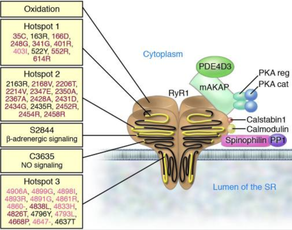
Bellinger AM, Mongillo M, Marks AR. Stressed out: the skeletal muscle ryanodine receptor as a target of stress. J Clin Invest. 2008;118(2):445-453. doi:10.1172/JCI34006
In skeletal muscle, the sarcoplasmic reticulum calcium-release channel is depicted as a large, four-part receptor composed of four identical subunits.
Anchoring proteins position key regulators on their cytoplasmic side, including a kinase, a phosphatase, and a phosphodiesterase, creating a local signaling complex that controls channel behavior.
The components are illustrated as bound to one subunit of the four-part receptor.
The figure also depicts stress inputs and three regions where disease-causing mutations cluster, with malignant hyperthermia in red, central core disease in pink, and mixed or multiminicore or nemaline rod disease in black, and a minus sign indicating a deletion.
Five Common Clinical Misconceptions
Misconception 1: Muscle fatigue is mainly explained by lactic acid buildup.
Clinical move: Examine calcium leaks from the ryanodine receptor more closely. Even mild oxidative stress can destabilize the channel, raise resting calcium, and blunt mitochondrial efficiency. Patients with “normal labs” but poor recovery often fit this profile.
Misconception 2: Pain intensity correlates with the amount of visible inflammation.
Clinical move: Central gain through calcium-sensitive kinases, such as CaMKII, can amplify sensory input without causing swelling. When a light touch feels painful or a warm shower burns, targeting redox tone and sleep can calm these circuits more effectively than escalating anti-inflammatories.
Misconception 3: Cartilage loss always follows systemic inflammation.
Clinical move: Chondrocytes sense local load through calcium channels such as TRPV4. Abnormal mechanical strain drives hypertrophy and matrix breakdown, even when systemic markers stay low. It may be worth including questions on stiffness after rest and weekly function, in addition to inflammatory labs.
Misconception 4: Night cramps are simply electrolyte depletion.
Clinical move: Many cramps arise from altered calcium signaling rather than sodium or magnesium deficiency. Stabilizing calcium handling—through improved sleep architecture, reduced oxidative load, and pacing activity—often relieves cramps that don’t respond to supplements alone.
Misconception 5: Bone strength reflects only mineral density.
Clinical move: Endogenous calcitonin tone, regulated through calcium-sensing pathways, influences both osteoclast quieting and soft-tissue decalcification. Supporting this balance can help reduce vascular stiffness and improve muscle irritability, resulting in fewer cramps and more stable mobility.
Case Reference
A 53-year-old woman with long-standing knuckle and knee arthritis described years of nightly pain that forced her to rely on heating pads and left her exhausted and depressed despite multiple trials of medicines and supplements. On starting therapy, she noticed a marked change within just ten days, with swelling and pain easing to the point that she no longer needed nightly heating pads. Her husband observed that she moved more freely and seemed brighter, and she herself reported feeling finally released from the cycle of nightly suffering and able to return to daily tasks with greater comfort.
Conclusion
Myalgia that lingers often hides a calcium story. When oxidative stress loosens control at the muscle release channel, resting calcium creeps up, mitochondria work harder, and the same walk hurts more. Patients describe low-load fatigue, night cramps, and sleep that fails to restore strength, even when routine labs look normal. Reading function through a calcium lens suggests practical steps.
Reduce oxidative stress, protect sleep architecture, and regulate activity to allow channels to regain stability. As rhythms settle, pain spikes decrease, mobility steadies, and flares occur less frequently. Supporting the calcium-sensing axis further helps by easing soft-tissue calcium pressure, quieting osteoclast activity, and improving circulation comfort. For myalgia marked by light-load pain and night cramps, restoring calcium rhythm is the pivot.
AIC therapy supports this shift by triggering endogenous calcitonin, reinforcing decalcification defenses, and tempering CaMKII-driven gain, resulting in steadier function and longer, easier walks.
Bring calcium signaling into your practice.
Practitioners can request a consultation to discuss AIC therapy and how these mechanisms apply to specific cases.
Newsletter content is educational and contributed by participating healthcare practitioners. These statements have not been evaluated by the FDA and are not intended to diagnose, treat, cure, or prevent any disease.