Notice. The following information is provided for your convenience. ACRI is not directly involved in this research and does not make any medical claims.

AIC THERAPY in Osteoporosis
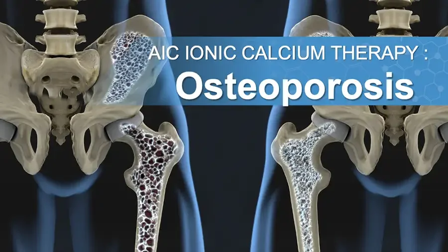
3D illustration: pelvis and femurs, porous bone vs dense bone. AIC IONIC CALCIUM THERAPY : Osteoporosis
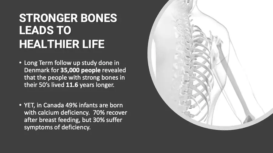
STRONGER BONES LEADS TO HEALTHIER LIFE Long Term follow up study done in Denmark for 35,000 people revealed that the people with strong bones in their 50’s lived 11.6 years longer. YET, in Canada 49% infants are born with calcium deficiency. 70% recover after breast feeding, but 30% suffer symptoms of deficiency. Illustration: grayscale skeleton of upper torso in a circle.
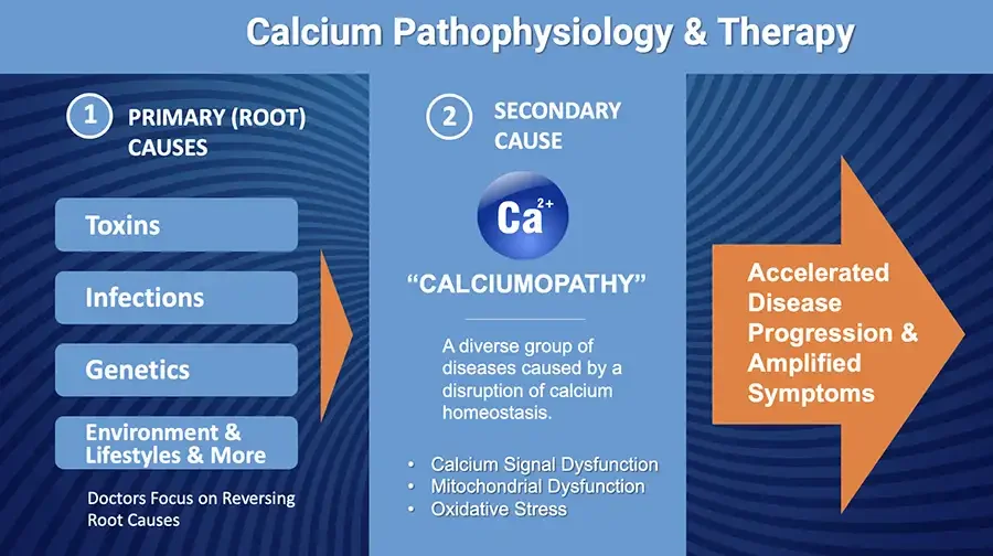
Calcium Pathophysiology & Therapy 1 PRIMARY (ROOT) CAUSES Toxins Infections Genetics Environment & Lifestyles & More Doctors Focus on Reversing Root Causes Orange arrow pointing right. 2 SECONDARY CAUSE Ca²⁺ “CALCIUMOPATHY” A diverse group of diseases caused by a disruption of calcium homeostasis. Calcium Signal Dysfunction Mitochondrial Dysfunction Oxidative Stress Large orange arrow pointing right, containing text. Accelerated Disease Progression & Amplified Symptoms
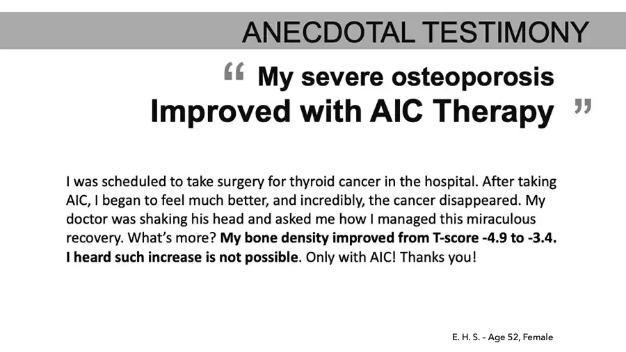
ANECDOTAL TESTIMONY “My severe osteoporosis Improved with AIC Therapy” I was scheduled to take surgery for thyroid cancer in the hospital. After taking AIC, I began to feel much better, and incredibly, the cancer disappeared. My doctor was shaking his head and asked me how I managed this miraculous recovery. What’s more? My bone density improved from T-score -4.9 to -3.4. I heard such increase is not possible. Only with AIC! Thanks you! E. H. S. – Age 52, Female
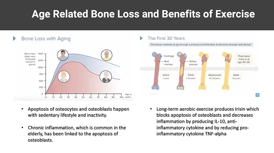
Age Related Bone Loss and Benefits of Exercise Bone Loss with Aging Area chart: bone mass vs age, blue and red curves, face icons. Bone mass (total mass of skeletal calcium in grams) 1500 1250 1000 750 500 250 0 0 10 20 30 40 50 60 70 80 90 100 Age in years Faint cut-off watermark. Apoptosis of osteocytes and osteoblasts happen with sedentary lifestyle and inactivity. Chronic inflammation, which is common in the elderly, has been linked to the apoptosis of osteoblasts. The First 30 Years The bones continue to go through a process of ossification to become stronger and denser. Diagram: four femurs showing bone development stages. Cartilage Red marrow Yellow marrow Peak bone mass is at age 30–35. Infant <1 year Child 1–10 years Adolescent 10–20 years Adult 25 years Faint watermark. Small blue icon. Long-term aerobic exercise produces Irisin which blocks apoptosis of osteoblasts and decreases inflammation by producing IL-10, anti-inflammatory cytokine and by reducing pro-inflammatory cytokine TNF-alpha
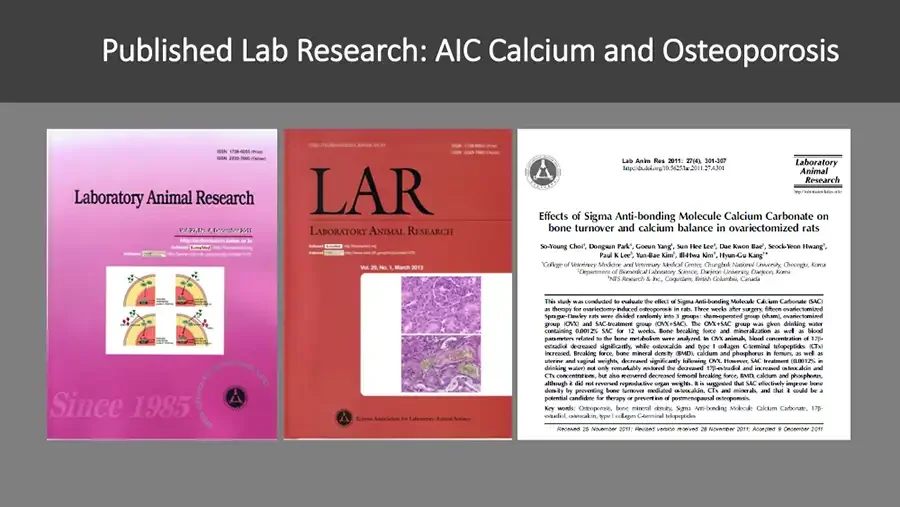
Published Lab Research: AIC Calcium and Osteoporosis Journal cover, pink, with cell diagram. ISSN 1738-6055 (Print) ISSN 2233-7660 (Online) Laboratory Animal Research Indexed KoreaMed Indexed Logo. Since 1985 Circular text around logo. Logo. Journal cover, red, with histology images. ISSN 1738-6055 (Print) ISSN 2233-7660 (Online) LAR LABORATORY ANIMAL RESEARCH Indexed KoreaMed Indexed Logo. Vol. 29, No. 1, March 2013 Histology micrographs. Logo. Korean Association for Laboratory Animal Science First page of journal article. Lab Anim Res 2011: 27(4), 301-307 http://dx.doi.org/10.5625/lar.2011.27.4.301 Laboratory Animal Research Effects of Sigma Anti-bonding Molecule Calcium Carbonate on bone turnover and calcium balance in ovariectomized rats So-Young Choi¹, Dongsun Park¹, Goeun Yang¹, Sun Hee Lee¹, Dae Kwon Bae¹, Seock-Yeon Hwang², Paul K Lee³, Yun-Bae Kim¹, Ill-Hwa Kim¹, Hyun-Gu Kang¹* Superscript numbers in author line uncertain. ¹College of Veterinary Medicine and Veterinary Medical Centre , Chungbuk National University, Cheongju, Korea ²Department of Biomedical Laboratory Science, Daejeon University, Daejeon, Korea ³NES Research & Inc., Coquitlam, British Columbia, Canada This study was conducted to evaluate the effect of Sigma Anti-bonding Molecule Calcium Carbonate (SAC) as therapy for ovariectomy-induced osteoporosis in rats. Three weeks after surgery, fifteen ovariectomized Sprague-Dawley rats were divided randomly into 3 groups: sham-operated group (sham), ovariectomized group (OVX) and SAC-treatment group (OVX+SAC). The OVX+SAC group was given drinking water containing 0.0012% SAC for 12 weeks. Bone breaking force and mineralization as well as blood parameters related to the bone metabolism were analyzed. In OVX animals, blood concentration of 17β-estradiol decreased significantly, while osteocalcin and type I collagen C-terminal telopeptides (CTx) increased. Breaking force, bone mineral density (BMD), calcium and phosphorus in femurs, as well as uterine and vaginal weights, decreased significantly following OVX. However, SAC treatment (0.0012% in drinking water) not only remarkably restored the decreased 17β-estradiol and increased osteocalcin and CTx concentrations, but also recovered decreased femoral breaking force, BMD, calcium and phosphorus, although it did not reversed reproductive organ weights. It is suggested that SAC effectively improve bone density by preventing bone turnover mediated osteocalcin, CTx and minerals, and that it could be a potential candidate for therapy or prevention of postmenopausal osteoporosis. Key words: Osteoporosis, bone mineral density, Sigma Anti-bonding Molecule Calcium Carbonate, 17β-estradiol, osteocalcin, type I collagen C-terminal telopeptides Received 25 November 2011; Revised version received 28 November 2011; Accepted 9 December 2011
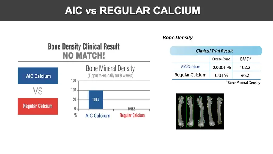
AIC vs REGULAR CALCIUM Bone Density Clinical Result NO MATCH! AIC Calcium VS Regular Calcium Bone Mineral Density (1 ppm taken daily for 9 weeks) Bar chart. 150 100 50 0 % 100.2 0.962 AIC Calcium Regular Calcium Bone Density Clinical Trial Result Dose Conc. BMD* AIC Calcium 0.0001 % 102.2 Regular Calcium 0.01 % 96.2 *Bone Mineral Density Scan image: five femur bones, one outlined in green.
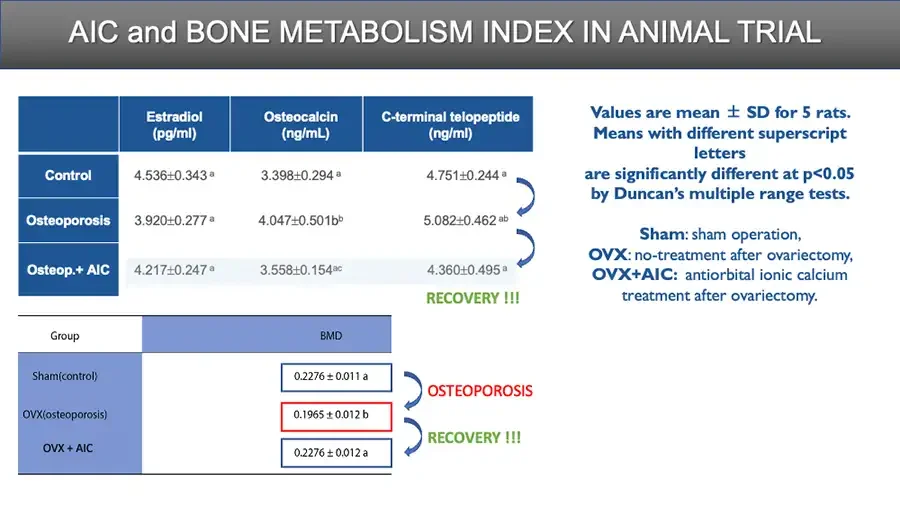
AIC and BONE METABOLISM INDEX IN ANIMAL TRIAL Estradiol (pg/ml) Osteocalcin (ng/mL) C-terminal telopeptide (ng/ml) Control 4.536±0.343ᵃ 3.398±0.294ᵃ 4.751±0.244ᵃ Osteoporosis 3.920±0.277ᵃ 4.047±0.501bᵇ 5.082±0.462ᵃᵇ Osteop.+ AIC 4.217±0.247ᵃ 3.558±0.154ᵃᶜ 4.360±0.495ᵃ Curved arrows linking table rows. RECOVERY !!! Group BMD Sham(control) 0.2276 ± 0.011 a OVX(osteoporosis) 0.1965 ± 0.012 b OVX + AIC 0.2276 ± 0.012 a Curved arrow. OSTEOPOROSIS Curved arrow. RECOVERY !!! Values are mean ± SD for 5 rats. Means with different superscript letters are significantly different at p<0.05 by Duncan’s multiple range tests. Sham: sham operation, OVX: no-treatment after ovariectomy, OVX+AIC: antiorbital ionic calcium treatment after ovariectomy.
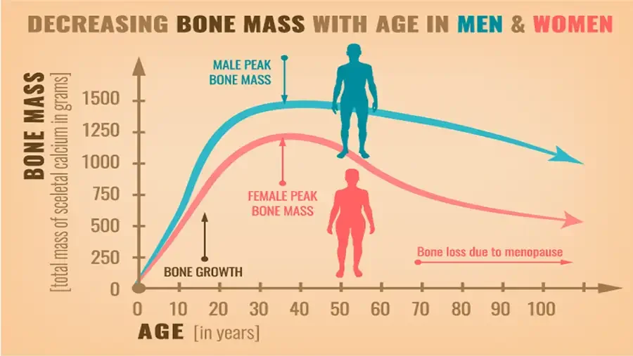
DECREASING BONE MASS WITH AGE IN MEN & WOMEN Line chart: bone mass by age, male and female curves, body silhouettes. BONE MASS total mass of sceletal calcium in grams. 1500 1250 1000 750 500 250 0 MALE PEAK BONE MASS FEMALE PEAK BONE MASS Bone loss due to menopause BONE GROWTH 0 10 20 30 40 50 60 70 80 90 100 AGE in years.
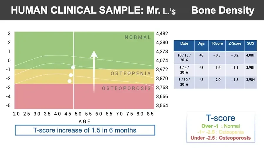
HUMAN CLINICAL SAMPLE: Mr. L.’s Bone Density Chart: T-score by age, colored zones, three dots, upward arrow. 3 2 1 0 -1 -2 -3 -4 -5 NORMAL OSTEOPENIA OSTEOPOROSIS 4,482 4,380 4,278 4,074 3,972 3,870 3,768 3,666 3,564 20 25 30 35 40 45 50 55 60 65 70 75 80 85 AGE T-score increase of 1.5 in 6 months Date Age T-Score Z-Score SOS 10 / 15 / 2016 48 - 0.5 - 0.2 4,081 6 / 4 / 2016 48 - 1.4 - 1.1 3,981 3 / 30 / 2016 48 - 2.0 - 1.8 3,904 T-score Over -1 : Normal -1~ -2.5 : Osteopenia Under -2.5 : Osteoporosis
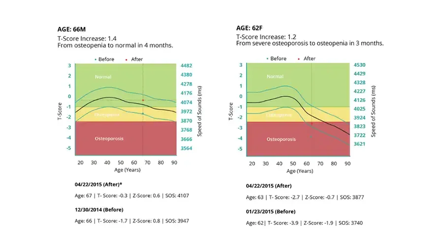
AGE: 66M T-Score Increase: 1.4 From osteopenia to normal in 4 months. Chart: T-score and speed of sound by age, colored zones, before/after dots. Before After T-Score 3 2 1 0 -1 -2 -3 -4 -5 Normal Osteopenia Osteoporosis 4482 4380 4278 4176 4074 3972 3870 3768 3666 3564 Speed of Sounds (ms) 20 30 40 50 60 70 80 90 Age (Years) 04/22/2015 (After)* Age: 67 | T- Score: -0.3 | Z-Score: 0.6 | SOS: 4107 12/30/2014 (Before) Age: 66 | T- Score: -1.7 | Z-Score: 0.8 | SOS: 3947 AGE: 62F T-Score Increase: 1.2 From severe osteoporosis to osteopenia in 3 months. Chart: T-score and speed of sound by age, colored zones, before/after dots. Before After T-Score 3 2 1 0 -1 -2 -3 -4 -5 Normal Osteopenia Osteoporosis 4530 4429 4328 4227 4126 4025 3924 3823 3722 3621 Speed of Sounds (ms) 20 30 40 50 60 70 80 90 Age (Years) 04/22/2015 (After) Age: 63 | T- Score: -2.7 | Z-Score: -0.7 | SOS: 3877 01/23/2015 (Before) Age: 62| T- Score: -3.9 | Z-Score: -1.9 | SOS: 3740
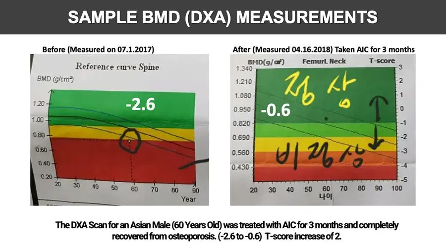
SAMPLE BMD (DXA) MEASUREMENTS Before (Measured on 07.1.2017) Photo: printed BMD reference chart, hand-drawn circle around data point. Reference curve Spine BMD (g/cm²) 1.20 1.00 0.80 0.60 0.40 0.20 -2.6 20 30 40 50 60 70 80 90 Year Cut-off text at bottom edge of photo. After (Measured 04.16.2018) Taken AIC for 3 months Photo: printed BMD chart with handwritten Korean and arrows. BMD(g/㎠) FemurL Neck T-score 1.340 1.210 1.080 0.950 0.820 0.690 0.560 0.430 3 2 1 0 -1 -2 -3 -4 -5 Handwritten, yellow. 정상 -0.6 Handwritten, black. 비정상 20 30 40 50 60 70 80 90 100 나이 The DXA Scan for an Asian Male (60 Years Old) was treated with AIC for 3 months and completely recovered from osteoporosis. (-2.6 to -0.6) T-score increase of 2.
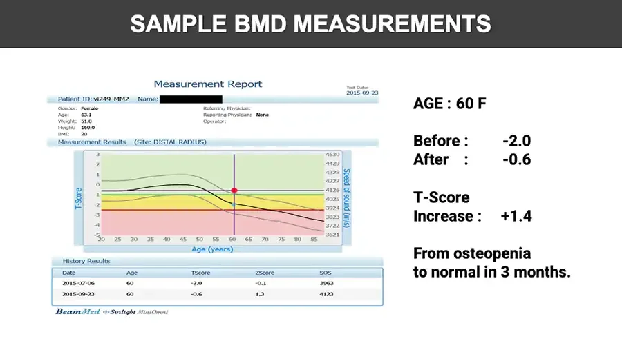
SAMPLE BMD MEASUREMENTS Screenshot of bone density measurement report. Measurement Report Test Date: 2015-09-23 Patient ID: vi249-MM2 Name: Black box covering name. Gender: Female Age: 63.1 Weight: 51.0 Height: 160.0 BMI: 20 Referring Physician: Reporting Physician: None Operator: Measurement Results (Site: DISTAL RADIUS) Chart: T-score and speed of sound by age, colored zones, marked points. T-Score 3 2 1 0 -1 -2 -3 -4 -5 4530 4429 4328 4227 4126 4025 3924 3823 3722 3621 Speed of sound (m/s) 20 25 30 35 40 45 50 55 60 65 70 75 80 85 Age (years) History Results Date Age TScore ZScore SOS 2015-07-06 60 -2.0 -0.1 3963 2015-09-23 60 -0.6 1.3 4123 BeamMed Sunlight MiniOmni AGE : 60 F Before : -2.0 After : -0.6 T-Score Increase : +1.4 From osteopenia to normal in 3 months.
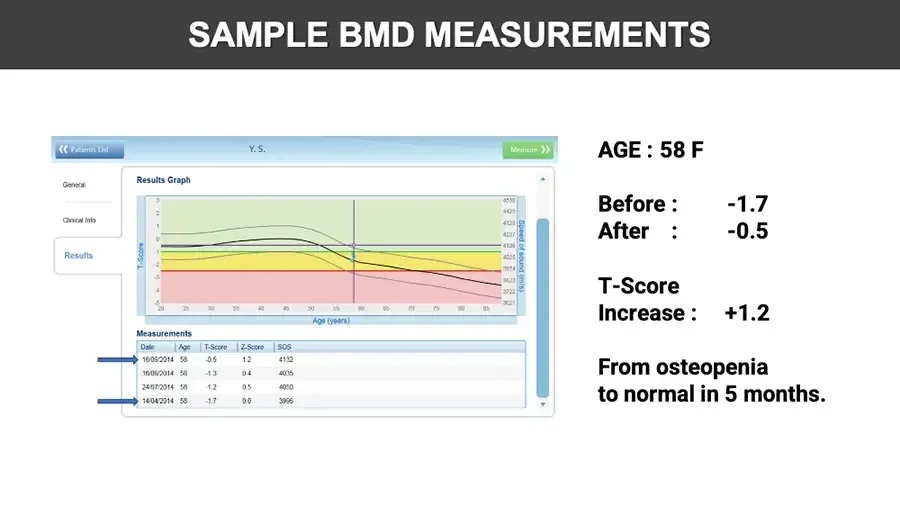
SAMPLE BMD MEASUREMENTS Screenshot of bone density software results screen. Patients List Y. S. Measure General Clinical Info Results Results Graph Chart: T-score and speed of sound by age, colored zones. T-Score 3 2 1 0 -1 -2 -3 -4 -5 4530 4429 4328 4227 4126 4025 3924 3823 3722 3621 Speed of sound (m/s) 20 25 30 35 40 45 50 55 60 65 70 75 80 85 Age (years) Measurements Blue arrows pointing to first and last rows. Date Age T-Score Z-Score SOS 16/09/2014 58 -0.5 1.2 4132 16/09/2014 58 -1.3 0.4 4035 24/07/2014 58 -1.2 0.5 4050 14/04/2014 58 -1.7 0.0 3996 AGE : 58 F Before : -1.7 After : -0.5 T-Score Increase : +1.2 From osteopenia to normal in 5 months.
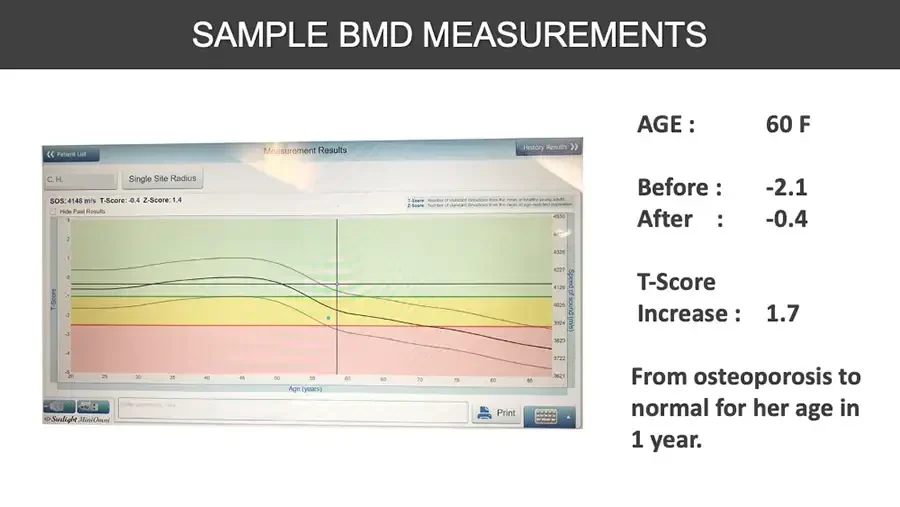
SAMPLE BMD MEASUREMENTS Photo of bone density device screen. Patient List Measurement Results History Results C. H. Single Site Radius SOS: 4148 m/s T-Score: -0.4 Z-Score: 1.4 Hide Past Results T-Score Number of standard deviations from the mean of healthy young adults. Z-Score Number of standard deviations from the mean of age-matched population Chart: T-score and speed of sound by age, colored zones. T-Score 3 2 1 0 -1 -2 -3 -4 -5 4530 4328 4227 4126 4025 3924 3823 3722 3621 Speed of sound (m/s) 20 25 30 35 40 45 50 55 60 65 70 75 80 85 Age (years) Icon buttons. Print Keyboard button. Sunlight MiniOmni AGE : 60 F Before : -2.1 After : -0.4 T-Score Increase : 1.7 From osteoporosis to normal for her age in 1 year.
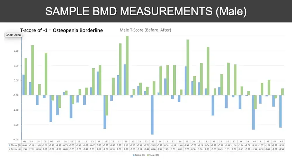
SAMPLE BMD MEASUREMENTS (Male) T-score of -1 = Osteopenia Borderline Chart Area Male T-Score (Before_After) Bar chart: before and after T-scores per subject. 2.00 1.00 0.00 -1.00 -2.00 -3.00 -4.00 01 03 04 05 06 07 08 09 11 12 13 14 15 16 17 19 20 21 22 23 24 25 27 28 29 30 31 32 33 35 36 37 38 39 40 41 43 44 45 TScore (B) 0.40 -0.11 -1.65 -1.19 -2.83 -2.36 -0.79 -2.57 -1.49 -1.66 -0.47 0.60 -3.27 -1.40 0.37 1.10 -1.15 -0.38 -0.72 -3.65 -0.82 0.14 -1.26 -1.46 0.96 -0.05 -0.13 -0.54 -2.37 -0.43 -1.89 -1.14 -1.94 -1.04 -3.33 -1.57 -1.08 -1.77 -3.20 Tscore (A) 1.50 2.39 -0.26 1.87 -1.37 -1.86 -0.69 -1.59 -0.58 -0.49 0.83 1.03 -2.37 0.18 2.51 3.70 -0.60 -0.14 -0.43 -0.06 0.96 1.05 1.03 -0.61 2.77 0.30 1.16 2.26 -0.53 0.43 1.19 1.09 -0.41 -0.71 -1.94 -0.16 0.04 -1.22 -0.54 TScore (B) Tscore (A)
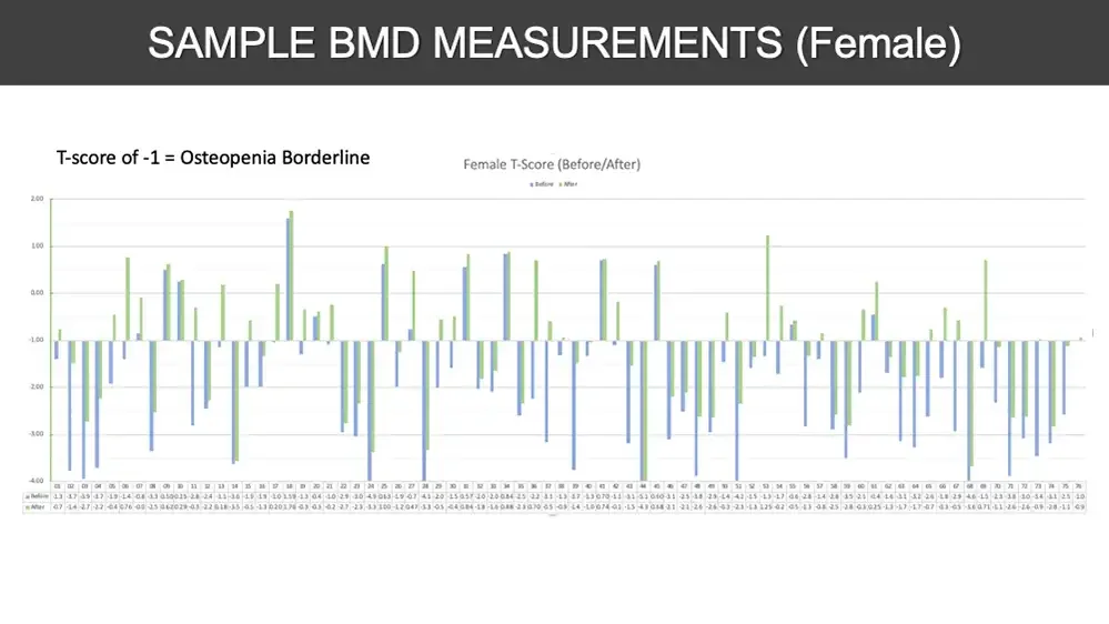
SAMPLE BMD MEASUREMENTS (Female) T-score of -1 = Osteopenia Borderline Female T-Score (Before/After) Before After Bar chart: before and after T-scores per subject. 2.00 1.00 0.00 -1.00 -2.00 -3.00 -4.00 Data table text very small and blurry; values are best readings. 01 02 03 04 05 06 07 08 09 10 11 12 13 14 15 16 17 18 19 20 21 22 23 24 25 26 27 28 29 30 31 32 33 34 35 36 37 38 39 40 41 42 43 44 45 46 47 48 49 50 51 52 53 54 55 56 57 58 59 60 61 62 63 64 65 66 67 68 69 70 71 72 73 74 75 76 Before -1.3 -3.7 -3.9 -3.7 -1.9 -1.4 -0.8 -3.3 0.50 0.25 -2.8 -2.4 -1.1 -3.6 -1.9 -1.9 -1.0 1.59 -1.3 -0.4 -1.0 -2.9 -3.0 -4.9 0.63 -1.9 -0.7 -4.1 -2.0 -1.5 0.57 -2.0 -2.0 0.84 -2.5 -2.2 -3.1 -1.3 -3.7 -1.3 0.70 -1.1 -3.1 -5.1 0.60 -3.1 -2.5 -3.8 -2.9 -1.4 -4.2 -1.5 -1.3 -1.7 -0.6 -2.8 -1.4 -2.8 -3.5 -2.1 -0.4 -1.6 -3.1 -3.2 -2.6 -1.8 -2.9 -4.6 -1.5 -2.3 -3.8 -3.0 -3.4 -3.1 -2.5 -1.0 After -0.7 -1.4 -2.7 -2.2 -0.4 0.76 -0.0 -2.5 0.62 0.29 -0.3 -2.2 0.18 -3.5 -0.5 -1.3 0.20 1.76 -0.3 -0.3 -0.2 -2.7 -2.3 -3.3 1.00 -1.2 0.47 -3.3 -0.5 -0.4 0.84 -1.8 -1.6 0.88 -2.3 0.70 -0.5 -0.9 -1.4 -1.0 0.74 -0.1 -1.5 -4.3 0.68 -2.1 -2.1 -2.6 -2.6 -0.3 -2.3 -1.3 1.25 -0.2 -0.5 -1.3 -0.8 -2.5 -2.8 -0.3 0.25 -1.3 -1.7 -1.7 -0.7 -0.3 -0.5 -3.6 0.71 -1.1 -2.6 -2.6 -0.9 -2.8 -1.1 -0.9
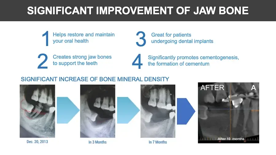
SIGNIFICANT IMPROVEMENT OF JAW BONE 1 Helps restore and maintain your oral health 2 Creates strong jaw bones to support the teeth 3 Great for patients undergoing dental implants 4 Significantly promotes cementogenesis, the formation of cementum SIGNIFICANT INCREASE OF BONE MINERAL DENSITY Dental X-ray with red outline. Dec. 20, 2013 Arrow. Dental X-ray. In 3 Months Arrow. Dental X-ray. In 7 Months Arrow. Dental CT scan. AFTER A Small orange text. After 10 months 10 20
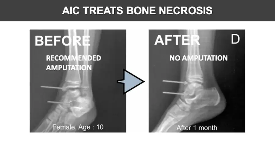
AIC TREATS BONE NECROSIS X-ray: ankle and foot, side view, two arrows. BEFORE RECOMMENDED AMPUTATION Female, Age : 10 Arrow. X-ray: ankle and foot, side view, two arrows. AFTER D NO AMPUTATION After 1 month
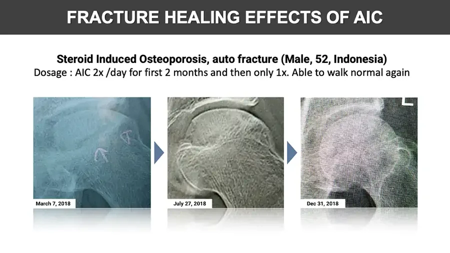
FRACTURE HEALING EFFECTS OF AIC Steroid Induced Osteoporosis, auto fracture (Male, 52, Indonesia) Dosage : AIC 2x /day for first 2 months and then only 1x. Able to walk normal again X-ray: hip joint with hand-drawn arrows. March 7, 2018 Arrow. X-ray: hip joint. July 27, 2018 Arrow. X-ray: hip joint. L Dec 31, 2018

DANGERS OF OSTEOPOROSIS DRUGS
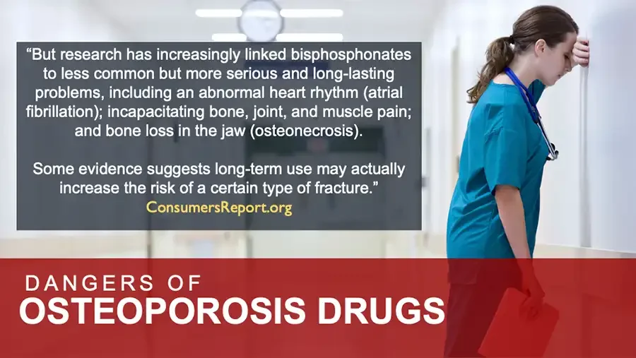
  • Photo: nurse in scrubs leaning head against hospital wall. “But research has increasingly linked bisphosphonates to less common but more serious and long-lasting problems, including an abnormal heart rhythm (atrial fibrillation); incapacitating bone, joint, and muscle pain; and bone loss in the jaw (osteonecrosis). Some evidence suggests long-term use may actually increase the risk of a certain type of fracture.” ConsumersReport.org DANGERS OF OSTEOPOROSIS DRUGS
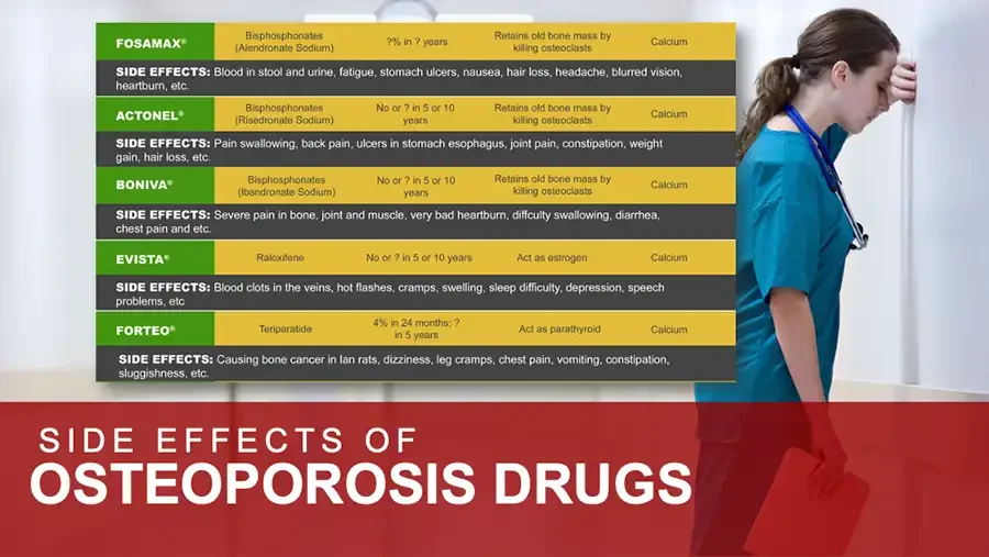
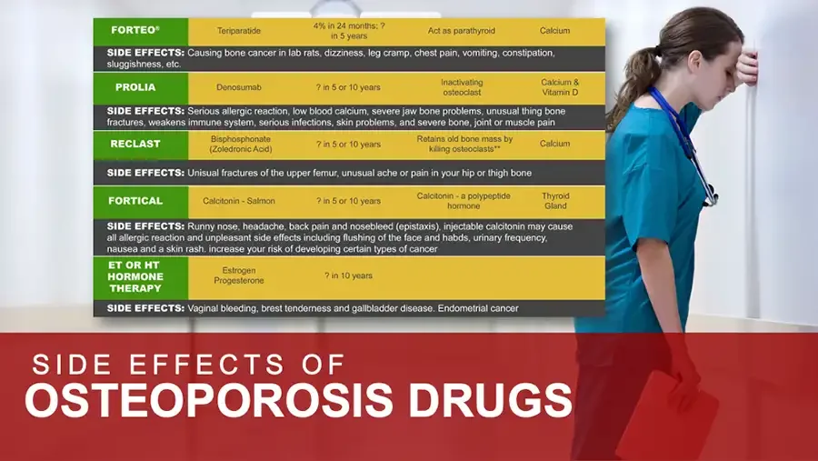
  • Photo: nurse in scrubs leaning head against hospital wall. FOSAMAX® Bisphosphonates (Alendronate Sodium) ?% in ? years Retains old bone mass by killing osteoclasts Calcium SIDE EFFECTS: Blood in stool and urine, fatigue, stomach ulcers, nausea, hair loss, headache, blurred vision, heartburn, etc. ACTONEL® Bisphosphonates (Risedronate Sodium) No or ? in 5 or 10 years Retains old bone mass by killing osteoclasts Calcium SIDE EFFECTS: Pain swallowing, back pain, ulcers in stomach esophagus, joint pain, constipation, weight gain, hair loss, etc. BONIVA® Bisphosphonates (Ibandronate Sodium) No or ? in 5 or 10 years Retains old bone mass by killing osteoclasts Calcium SIDE EFFECTS: Severe pain in bone, joint and muscle, very bad heartburn, diffculty swallowing, diarrhea, chest pain and etc. EVISTA® Raloxifene No or ? in 5 or 10 years Act as estrogen Calcium SIDE EFFECTS: Blood clots in the veins, hot flashes, cramps, swelling, sleep difficulty, depression, speech problems, etc FORTEO® Teriparatide 4% in 24 months; ? in 5 years Act as parathyroid Calcium SIDE EFFECTS: Causing bone cancer in lan rats, dizziness, leg cramps, chest pain, vomiting, constipation, sluggishness, etc. SIDE EFFECTS OF OSTEOPOROSIS DRUGS
  • Photo: nurse in scrubs leaning head against hospital wall. FORTEO® Teriparatide 4% in 24 months; ? in 5 years Act as parathyroid Calcium SIDE EFFECTS: Causing bone cancer in lab rats, dizziness, leg cramp, chest pain, vomiting, constipation, sluggishness, etc. PROLIA Denosumab ? in 5 or 10 years Inactivating osteoclast Calcium & Vitamin D SIDE EFFECTS: Serious allergic reaction, low blood calcium, severe jaw bone problems, unusual thing bone fractures, weakens immune system, serious infections, skin problems, and severe bone, joint or muscle pain RECLAST Bisphosphonate (Zoledronic Acid) ? in 5 or 10 years Retains old bone mass by killing osteoclasts** Calcium SIDE EFFECTS: Unisual fractures of the upper femur, unusual ache or pain in your hip or thigh bone FORTICAL Calcitonin - Salmon ? in 5 or 10 years Calcitonin - a polypeptide hormone Thyroid Gland SIDE EFFECTS: Runny nose, headache, back pain and nosebleed (epistaxis), injectable calcitonin may cause all allergic reaction and unpleasant side effects including flushing of the face and habds, urinary frequency, nausea and a skin rash. increase your risk of developing certain types of cancer ET OR HT HORMONE THERAPY Estrogen Progesterone ? in 10 years SIDE EFFECTS: Vaginal bleeding, brest tenderness and gallbladder disease. Endometrial cancer SIDE EFFECTS OF OSTEOPOROSIS DRUGS
  • Photo: nurse in scrubs leaning head against hospital wall. “The reason why bisphosphonate, estrogen, and parathyroid hormone derivatives cannot increase the bone mineral density is that they lack ionic calcium required for bone cell replication.” INEFFECTIVENESS OF OSTEOPOROSIS DRUGS & CALCIUM SUPPLEMENTS
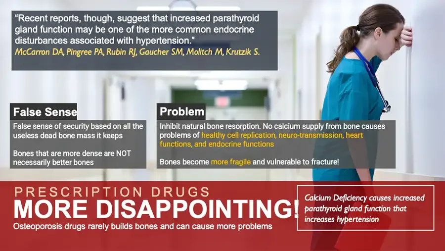
  • Photo: nurse in scrubs leaning head against hospital wall. “Recent reports, though, suggest that increased parathyroid gland function may be one of the more common endocrine disturbances associated with hypertension.” McCarron DA, Pingree PA, Rubin RJ, Gaucher SM, Molitch M, Krutzik S. False Sense False sense of security based on all the useless dead bone mass it keeps Bones that are more dense are NOT necessarily better bones Problem Inhibit natural bone resorption. No calcium supply from bone causes problems of healthy cell replication, neuro-transmission, heart functions, and endocrine functions Bones become more fragile and vulnerable to fracture! PRESCRIPTION DRUGS MORE DISAPPOINTING! Osteoporosis drugs rarely builds bones and can cause more problems Calcium Deficiency causes increased parathyroid gland function that increases hypertension
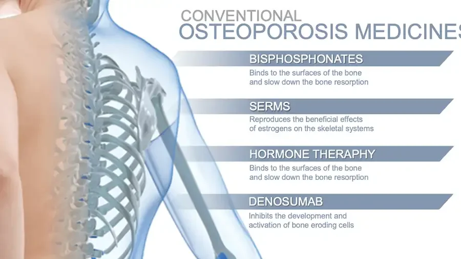
  • Illustration: translucent human shoulder, spine and ribs. CONVENTIONAL OSTEOPOROSIS MEDICINES BISPHOSPHONATES Binds to the surfaces of the bone and slow down the bone resorption SERMS Reproduces the beneficial effects of estrogens on the skeletal systems HORMONE THERAPHY Binds to the surfaces of the bone and slow down the bone resorption DENOSUMAB Inhibits the development and activation of bone eroding cells
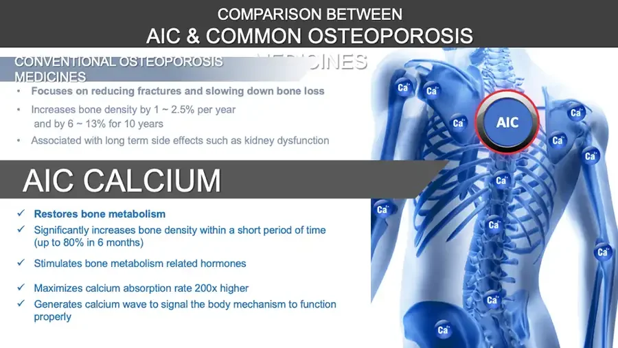
COMPARISON BETWEEN AIC & COMMON OSTEOPOROSIS CONVENTIONAL OSTEOPOROSIS MEDICINES Faint text partly hidden behind banner. MEDICINES Focuses on reducing fractures and slowing down bone loss Increases bone density by 1 ~ 2.5% per year and by 6 ~ 13% for 10 years Associated with long term side effects such as kidney dysfunction AIC CALCIUM Restores bone metabolism Significantly increases bone density within a short period of time (up to 80% in 6 months) Stimulates bone metabolism related hormones Maximizes calcium absorption rate 200x higher Generates calcium wave to signal the body mechanism to function properly Illustration: blue skeleton with round AIC button and Ca²⁺ icons. AIC Ca²⁺ same label on 12 icons.
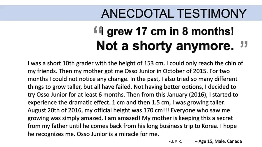
ANECDOTAL TESTIMONY “I grew 17 cm in 8 months! Not a shorty anymore.” I was a short 10th grader with the height of 153 cm. I could only reach the chin of my friends. Then my mother got me Osso Junior in October of 2015. For two months I could not notice any change. In the past, I also tried so many different things to grow taller, but all have failed. Not having better options, I decided to try Osso Junior for at least 6 months. Then from this January (2016), I started to experience the dramatic effect. 1 cm and then 1.5 cm, I was growing taller. August 20th of 2016, my official height was 170 cm!!! Everyone who saw me growing was simply amazed. I am amazed! My mother is keeping this a secret from my father until he comes back from his long business trip to Korea. I hope he recognizes me. Osso Junior is a miracle for me. - J. Y. K. – Age 15, Male, Canada
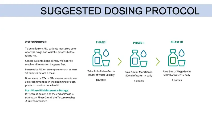
SUGGESTED DOSING PROTOCOL OSTEOPOROSIS: To benefit from AIC, patients must stop osteoporosis drugs and wait 3-6 months before taking AIC. Cancer patient’s bone density will not rise much until remission happens first. Please take AIC on an empty stomach at least 30 minutes before a meal. Bone scans or CTx or NTx measurements are also recommended in the beginning of each phase to monitor bone health. Post-Phase III Maintenance Dosage: If T score is below -1 at the end of Phase 2, staying on Phase 2 until the T score reaches -1 is recommended. PHASE I Icons: bottle, water drop, cup. Take 5ml of MaraGen in 500ml of water 2x daily 8 bottles Arrow. PHASE II Icons: bottle, water drop, cup. Take 5ml of MaraGen in 500ml of water 1x daily 4 bottles Arrow. PHASE III Icons: bottle, water drop, cup. Take 5ml of MegaGen in 500ml of water 1x daily 4 bottles

DEXA vs. REMS

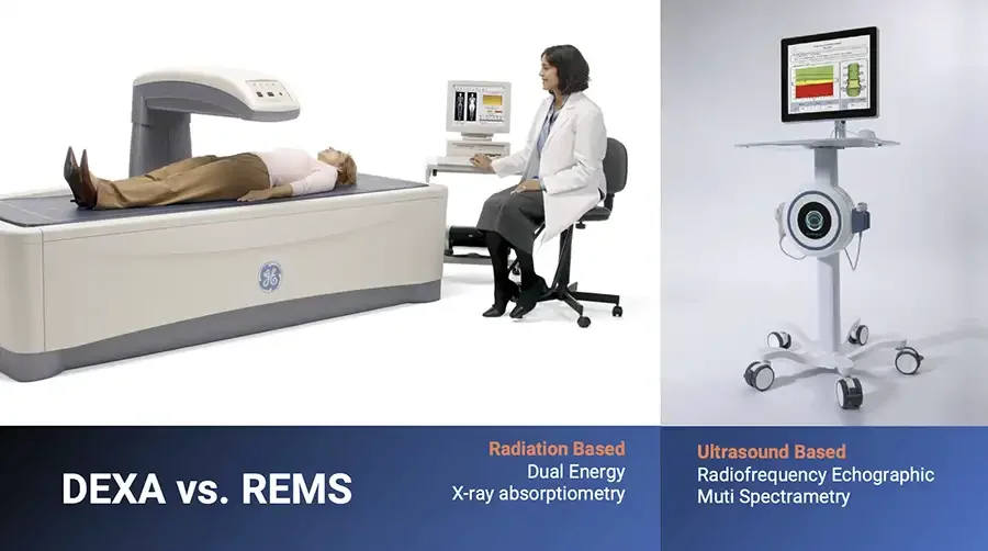
  • Photo: patient lying on DEXA scanner, clinician seated at computer. GE logo on scanner. Photo: ultrasound bone scanner on wheeled stand. DEXA vs. REMS Radiation Based Dual Energy X-ray absorptiometry Ultrasound Based Radiofrequency Echographic Muti Spectrametry
  • DEXA measures bone density, mostly mineral, and is not good measure of bone strength and quality. There are cases where DEXA could get worse but quality and strength get better. Better markers are P1NP which measures osteoblast activity and CTx which measures osteoclast activity. CTx under 600 is target. 400 is better. Estrogen slows down osteoclast. Testasterone and Progesterone has impacts of osteoblasts. https://www.youtube.com/watch?v=BQ0DqR14DUg
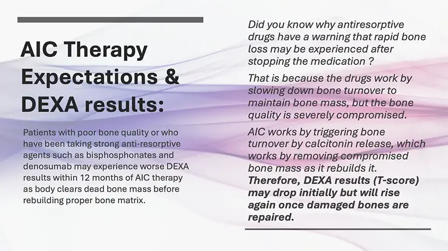
  • AIC Therapy Expectations & DEXA results: Patients with poor bone quality or who have been taking strong anti-resorptive agents such as bisphosphonates and denosumab may experience worse DEXA results within 12 months of AIC therapy as body clears dead bone mass before rebuilding proper bone matrix. Did you know why antiresorptive drugs have a warning that rapid bone loss may be experienced after stopping the medication ? That is because the drugs work by slowing down bone turnover to maintain bone mass, but the bone quality is severely compromised. AIC works by triggering bone turnover by calcitonin release, which works by removing compromised bone mass as it rebuilds it. Therefore, DEXA results (T-score) may drop initially but will rise again once damaged bones are repaired.
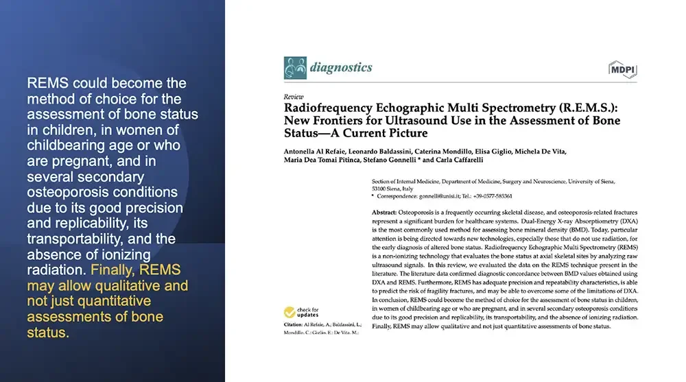
  • REMS could become the method of choice for the assessment of bone status in children, in women of childbearing age or who are pregnant, and in several secondary osteoporosis conditions due to its good precision and replicability, its transportability, and the absence of ionizing radiation. Finally, REMS may allow qualitative and not just quantitative assessments of bone status. First page of journal article. Logo. diagnostics MDPI Review Radiofrequency Echographic Multi Spectrometry (R.E.M.S.): New Frontiers for Ultrasound Use in the Assessment of Bone Status—A Current Picture Antonella Al Refaie, Leonardo Baldassini, Caterina Mondillo, Elisa Giglio, Michela De Vita, Maria Dea Tomai Pitinca, Stefano Gonnelli * and Carla Caffarelli Section of Internal Medicine, Department of Medicine, Surgery and Neuroscience, University of Siena, 53100 Siena, Italy Correspondence: gonnelli@unisi.it; Tel.: +39-0577-585361 check for updates Citation: Al Refaie, A.; Baldassini, L.; Mondillo, C.; Giglio, E.; De Vita, M.; Abstract: Osteoporosis is a frequently occurring skeletal disease, and osteoporosis-related fractures represent a significant burden for healthcare systems. Dual-Energy X-ray Absorptiometry (DXA) is the most commonly used method for assessing bone mineral density (BMD). Today, particular attention is being directed towards new technologies, especially those that do not use radiation, for the early diagnosis of altered bone status. Radiofrequency Echographic Multi Spectrometry (REMS) is a non-ionizing technology that evaluates the bone status at axial skeletal sites by analyzing raw ultrasound signals. In this review, we evaluated the data on the REMS technique present in the literature. The literature data confirmed diagnostic concordance between BMD values obtained using DXA and REMS. Furthermore, REMS has adequate precision and repeatability characteristics, is able to predict the risk of fragility fractures, and may be able to overcome some of the limitations of DXA. In conclusion, REMS could become the method of choice for the assessment of bone status in children, in women of childbearing age or who are pregnant, and in several secondary osteoporosis conditions due to its good precision and replicability, its transportability, and the absence of ionizing radiation. Finally, REMS may allow qualitative and not just quantitative assessments of bone status.
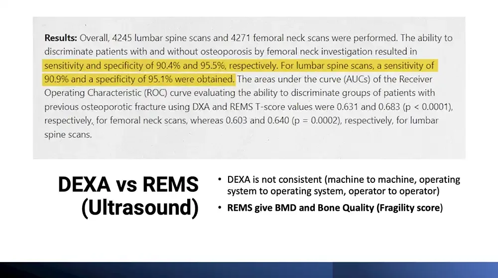
  • Results: Overall, 4245 lumbar spine scans and 4271 femoral neck scans were performed. The ability to discriminate patients with and without osteoporosis by femoral neck investigation resulted in sensitivity and specificity of 90.4% and 95.5%, respectively. For lumbar spine scans, a sensitivity of 90.9% and a specificity of 95.1% were obtained. The areas under the curve (AUCs) of the Receiver Operating Characteristic (ROC) curve evaluating the ability to discriminate groups of patients with previous osteoporotic fracture using DXA and REMS T-score values were 0.631 and 0.683 (p < 0.0001), respectively, for femoral neck scans, whereas 0.603 and 0.640 (p = 0.0002), respectively, for lumbar spine scans. Yellow highlight from "sensitivity and specificity" to "were obtained.". DEXA vs REMS (Ultrasound) DEXA is not consistent (machine to machine, operating system to operating system, operator to operator) REMS give BMD and Bone Quality (Fragility score)
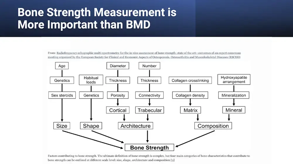
  • Bone Strength Measurement is More Important than BMD From: Radiofrequency echographic multi-spectrometry for the in-vivo assessment of bone strength: state of the art—outcomes of an expert consensus meeting organized by the European Society for Clinical and Economic Aspects of Osteoporosis, Osteoarthritis and Musculoskeletal Diseases (ESCEO) Flowchart: boxes joined by lines and arrows, listed column by column. Age Genetics Sex steroids Size Habitual loads Genetics Shape Diameter Thickness Porosity Cortical Number Thickness Connectivity Trabecular Architecture Collagen crosslinking Collagen density Matrix Hydroxyapatite arrangement Mineralization Mineral Composition Bone Strength Factors contributing to bone strength. The ultimate definition of bone strength is complex, but four main categories of bone characteristics that contribute to bone strength can be outlined at different scale level: size, shape, architecture and composition 3.
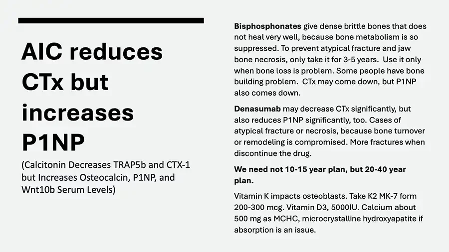
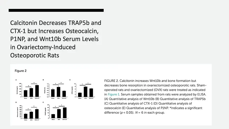
  • AIC reduces CTx but increases P1NP (Calcitonin Decreases TRAP5b and CTX-1 but Increases Osteocalcin, P1NP, and Wnt10b Serum Levels) Bisphosphonates give dense brittle bones that does not heal very well, because bone metabolism is so suppressed. To prevent atypical fracture and jaw bone necrosis, only take it for 3-5 years. Use it only when bone loss is problem. Some people have bone building problem. CTx may come down, but P1NP also comes down. Denasumab may decrease CTx significantly, but also reduces P1NP significantly, too. Cases of atypical fracture or necrosis, because bone turnover or remodeling is compromised. More fractures when discontinue the drug. We need not 10-15 year plan, but 20-40 year plan. Vitamin K impacts osteoblasts. Take K2 MK-7 form 200-300 mcg. Vitamin D3, 5000IU. Calcium about 500 mg as MCHC, microcrystalline hydroxyapatite if absorption is an issue.
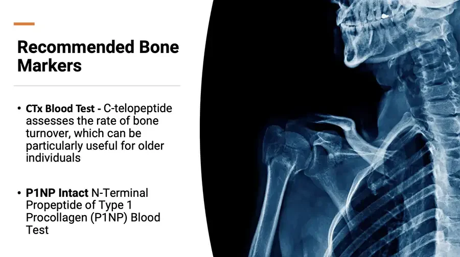
  • Recommended Bone Markers CTx Blood Test - C-telopeptide assesses the rate of bone turnover, which can be particularly useful for older individuals P1NP Intact N-Terminal Propeptide of Type 1 Procollagen (P1NP) Blood Test X-ray: shoulder, neck and upper ribs.
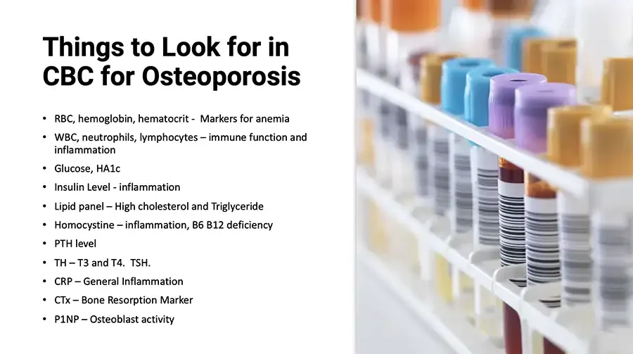
  • Things to Look for in CBC for Osteoporosis RBC, hemoglobin, hematocrit - Markers for anemia WBC, neutrophils, lymphocytes – immune function and inflammation Glucose, HA1c Insulin Level - inflammation Lipid panel – High cholesterol and Triglyceride Homocystine – inflammation, B6 B12 deficiency PTH level TH – T3 and T4. TSH. CRP – General Inflammation CTx – Bone Resorption Marker P1NP – Osteoblast activity Photo: blood sample tubes with colored caps in a rack.

WHAT IS CALCITONIN?
  • Calcitonin based bone building – The best way! AIC triggers robust endogenous calcitonin release Calcitonin is a hormone, triggered by elevated ionic calcium level, inhibits osteoclasts which also triggers osteoblast cells that rebuilds bones. Exogenous calcitonin treatment for bone health (derived from salmon), although considered the best agent to rebuild osteoporotic bones, failed to be the first line of treatment due to difficult in administration and side effects. Despite not being a strong anabolic treatment for bones, is still used in certain medical situations for its anti-resorptive and other properties. Calcitonin can also provide some pain relief in acute bone fractures. It works by reducing bone resorption and stabilizing bone tissue, which can help alleviate pain in the short term. It is effective in treating hypercalcemia, a condition characterized by elevated levels of calcium in the blood. Calcitonin can help lower blood calcium levels by inhibiting the release of calcium from the bone.
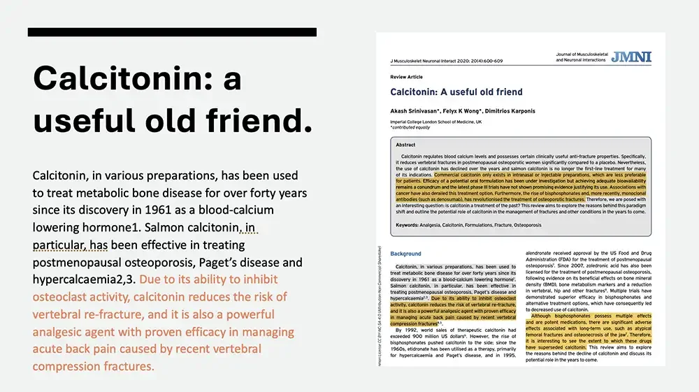
  • Calcitonin: a useful old friend. Calcitonin, in various preparations, has been used to treat metabolic bone disease for over forty years since its discovery in 1961 as a blood-calcium lowering hormone1. Salmon calcitonin, in particular, has been effective in treating postmenopausal osteoporosis, Paget’s disease and hypercalcaemia2,3. Due to its ability to inhibit osteoclast activity, calcitonin reduces the risk of vertebral re-fracture, and it is also a powerful analgesic agent with proven efficacy in managing acute back pain caused by recent vertebral compression fractures. First page of journal article, with yellow highlights. J Musculoskelet Neuronal Interact 2020; 20(4):600-609 Journal of Musculoskeletal and Neuronal Interactions JMNI Review Article Calcitonin: A useful old friend Akash Srinivasan*, Felyx K Wong*, Dimitrios Karponis Imperial College London School of Medicine, UK *contributed equally Abstract Calcitonin regulates blood calcium levels and possesses certain clinically useful anti-fracture properties. Specifically, it reduces vertebral fractures in postmenopausal osteoporotic women significantly compared to a placebo. Nevertheless, the use of calcitonin has declined over the years and salmon calcitonin is no longer the first-line treatment for many of its indications. Commercial calcitonin only exists in intranasal or injectable preparations, which are less preferable for patients. Efficacy of a potential oral formulation has been under investigation but achieving adequate bioavailability remains a conundrum and the latest phase III trials have not shown promising evidence justifying its use. Associations with cancer have also derailed this treatment option. Furthermore, the rise of bisphosphonates and, more recently, monoclonal antibodies (such as denosumab), has revolutionised the treatment of osteoporotic fractures. Therefore, we are posed with an interesting question: is calcitonin a treatment of the past? This review aims to explore the reasons behind this paradigm shift and outline the potential role of calcitonin in the management of fractures and other conditions in the years to come. Yellow highlight from "Commercial calcitonin" to "osteoporotic fractures.". Keywords: Analgesia, Calcitonin, Formulations, Fracture, Osteoporosis Vertical text along left edge, start cut off. mon License CC BY-NC-SA 4.0 (Attribution Non Commercial ShareAlike) Background Calcitonin, in various preparations, has been used to treat metabolic bone disease for over forty years since its discovery in 1961 as a blood-calcium lowering hormone¹. Salmon calcitonin, in particular, has been effective in treating postmenopausal osteoporosis, Paget’s disease and hypercalcaemia²˒³. Due to its ability to inhibit osteoclast activity, calcitonin reduces the risk of vertebral re-fracture, and it is also a powerful analgesic agent with proven efficacy in managing acute back pain caused by recent vertebral compression fractures⁴˒⁵. By 1992, world sales of therapeutic calcitonin had exceeded 900 million US dollars⁶. However, the rise of bisphosphonates pushed calcitonin to the side; since the 1960s, etidronate has been utilised as a therapy, primarily for hypercalcaemia and Paget’s disease, and in 1995, alendronate received approval by the US Food and Drug Administration (FDA) for the treatment of postmenopausal osteoporosis⁷. Since 2007, zoledronic acid has also been licensed for the treatment of postmenopausal osteoporosis, following evidence on its beneficial effects on bone mineral density (BMD), bone metabolism markers and a reduction in vertebral, hip and other fractures⁸. Multiple trials have demonstrated superior efficacy in bisphosphonates and alternative treatment options, which have consequently led to decreased use of calcitonin. Although bisphosphonates possess multiple effects and are potent medications, there are significant adverse effects associated with long-term use, such as atypical femoral fractures and osteonecrosis of the jaw⁷ . Therefore, it is interesting to see the extent to which these drugs have superseded calcitonin. This review aims to explore the reasons behind the decline of calcitonin and discuss its potential role in the years to come. Yellow highlights from "Due to its ability" to "compression fractures" and from "Although bisphosphonates" to "superseded calcitonin.". Superscript reference numbers are tiny; readings uncertain.
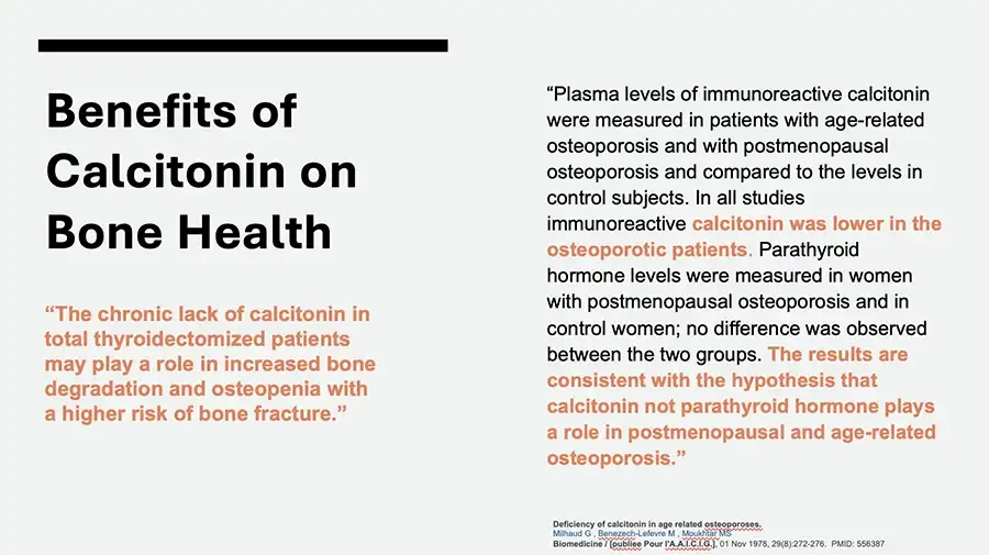
  • Benefits of Calcitonin on Bone Health “The chronic lack of calcitonin in total thyroidectomized patients may play a role in increased bone degradation and osteopenia with a higher risk of bone fracture.” “Plasma levels of immunoreactive calcitonin were measured in patients with age-related osteoporosis and with postmenopausal osteoporosis and compared to the levels in control subjects. In all studies immunoreactive calcitonin was lower in the osteoporotic patients. Parathyroid hormone levels were measured in women with postmenopausal osteoporosis and in control women; no difference was observed between the two groups. The results are consistent with the hypothesis that calcitonin not parathyroid hormone plays a role in postmenopausal and age-related osteoporosis.” Deficiency of calcitonin in age related osteoporoses. Milhaud G , Benezech-Lefevre M , Moukhtar MS Biomedicine / publiee Pour l'A.A.I.C.I.G.. 01 Nov 1978, 29(8):272-276. PMID: 556387
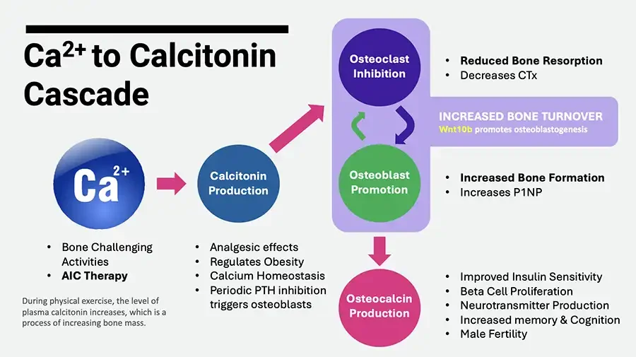
  • Ca²⁺ to Calcitonin Cascade Diagram: Ca²⁺ sphere and colored circles linked by arrows. Ca²⁺ Bone Challenging Activities AIC Therapy During physical exercise, the level of plasma calcitonin increases, which is a process of increasing bone mass. Arrow. Calcitonin Production Analgesic effects Regulates Obesity Calcium Homeostasis Periodic PTH inhibition triggers osteoblasts Arrow. Osteoclast Inhibition Reduced Bone Resorption Decreases CTx Circular arrows between the two circles. INCREASED BONE TURNOVER Wnt10b promotes osteoblastogenesis Osteoblast Promotion Increased Bone Formation Increases P1NP Arrow. Osteocalcin Production Improved Insulin Sensitivity Beta Cell Proliferation Neurotransmitter Production Increased memory & Cognition Male Fertility
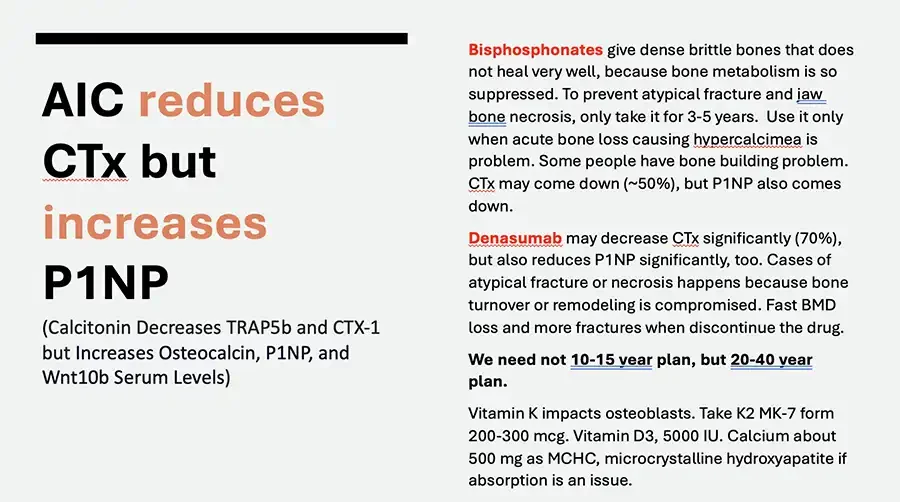
  • AIC reduces CTx but increases P1NP (Calcitonin Decreases TRAP5b and CTX-1 but Increases Osteocalcin, P1NP, and Wnt10b Serum Levels) Bisphosphonates give dense brittle bones that does not heal very well, because bone metabolism is so suppressed. To prevent atypical fracture and jaw bone necrosis, only take it for 3-5 years. Use it only when acute bone loss causing hypercalcimea is problem. Some people have bone building problem. CTx may come down (~50%), but P1NP also comes down. Denasumab may decrease CTx significantly (70%), but also reduces P1NP significantly, too. Cases of atypical fracture or necrosis happens because bone turnover or remodeling is compromised. Fast BMD loss and more fractures when discontinue the drug. We need not 10-15 year plan, but 20-40 year plan. Vitamin K impacts osteoblasts. Take K2 MK-7 form 200-300 mcg. Vitamin D3, 5000 IU. Calcium about 500 mg as MCHC, microcrystalline hydroxyapatite if absorption is an issue. Spell-check underlines visible under some words.
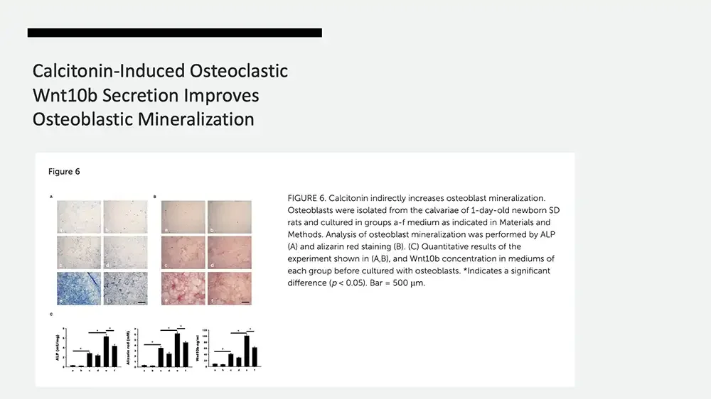
  • Calcitonin-Induced Osteoclastic Wnt10b Secretion Improves Osteoblastic Mineralization Figure 6 A Micrographs: six stained cell culture panels. a b c d e f B Micrographs: six red-stained cell culture panels. a b c d e f C Three bar charts with significance markers. ALP (mU/mg) 8 6 4 2 0 a b c d e f Alizarin red (mM) 7 6 5 4 3 2 1 0 a b c d e f Wnt10b ng/ml 120 100 80 60 40 20 0 a b c d e f on each chart. FIGURE 6. Calcitonin indirectly increases osteoblast mineralization. Osteoblasts were isolated from the calvariae of 1-day-old newborn SD rats and cultured in groups a-f medium as indicated in Materials and Methods. Analysis of osteoblast mineralization was performed by ALP (A) and alizarin red staining (B). (C) Quantitative results of the experiment shown in (A,B), and Wnt10b concentration in mediums of each group before cultured with osteoblasts. *Indicates a significant difference (p < 0.05). Bar = 500 μm.
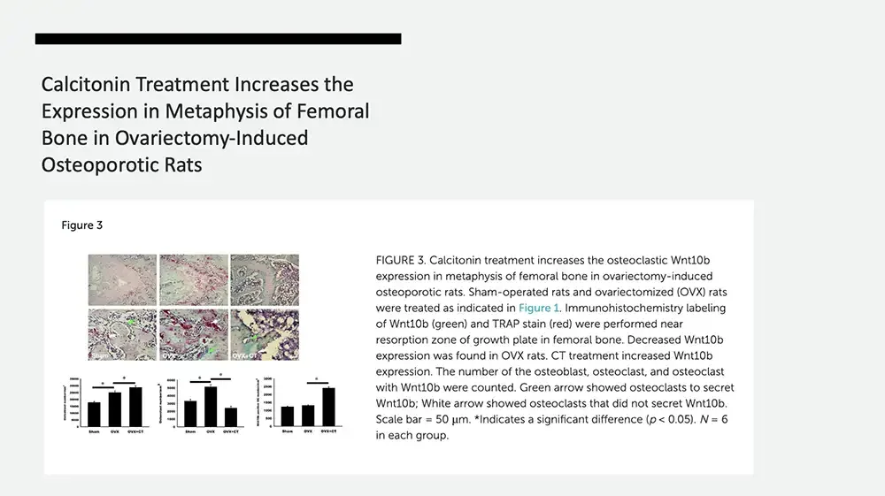
  • Calcitonin Treatment Increases the Expression in Metaphysis of Femoral Bone in Ovariectomy-Induced Osteoporotic Rats Figure 3 Micrographs: six stained bone tissue panels, green and white arrows. Sham OVX OVX+CT Three bar charts with significance markers. Vertical axis label. 35000 30000 25000 20000 15000 10000 5000 0 Sham OVX OVX+CT Vertical axis label. 6000 5000 4000 3000 2000 1000 0 Sham OVX OVX+CT Vertical axis label. 3000 2500 2000 1500 1000 500 0 Sham OVX OVX+CT above bars. FIGURE 3. Calcitonin treatment increases the osteoclastic Wnt10b expression in metaphysis of femoral bone in ovariectomy-induced osteoporotic rats. Sham-operated rats and ovariectomized (OVX) rats were treated as indicated in Figure 1. Immunohistochemistry labeling of Wnt10b (green) and TRAP stain (red) were performed near resorption zone of growth plate in femoral bone. Decreased Wnt10b expression was found in OVX rats. CT treatment increased Wnt10b expression. The number of the osteoblast, osteoclast, and osteoclast with Wnt10b were counted. Green arrow showed osteoclasts to secret Wnt10b; White arrow showed osteoclasts that did not secret Wnt10b. Scale bar = 50 μm. *Indicates a significant difference (p < 0.05). N = 6 in each group.
  • Calcitonin Decreases TRAP5b and CTX-1 but Increases Osteocalcin, P1NP, and Wnt10b Serum Levels in Ovariectomy-Induced Osteoporotic Rats Figure 2 Five bar charts, panels A to E. A Wnt10b (pg/ml) 500 400 300 200 100 0 Sham OVX OVX+CT B TRAP5b (U/l) 1.2 1.0 0.8 0.6 0.4 0.2 0.0 Sham OVX OVX+CT C CTX-1 (ng/ml) 10 8 6 4 2 0 Sham OVX OVX+CT D Osteocalcin (ng/ml) 700 600 500 400 300 200 100 0 Sham OVX OVX+CT E P1NP (ng/ml) 25 20 15 10 5 0 Sham OVX OVX+CT FIGURE 2. Calcitonin increases Wnt10b and bone formation but decreases bone resorption in ovariectomized osteoporotic rats. Sham-operated rats and ovariectomized (OVX) rats were treated as indicated in Figure 1. Serum samples obtained from rats were analyzed by ELISA. (A) Quantitative analysis of Wnt10b (B) Quantitative analysis of TRAP5b (C) Quantitative analysis of CTX-1 (D) Quantitative analysis of osteocalcin (E) Quantitative analysis of P1NP. *Indicates a significant difference (p < 0.05). N = 6 in each group.
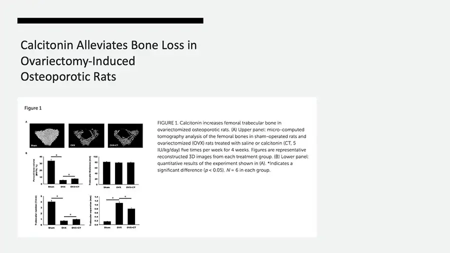
  • Calcitonin Alleviates Bone Loss in Ovariectomy-Induced Osteoporotic Rats Figure 1 A Three micro-CT images of femoral trabecular bone. Sham OVX OVX+CT B Four bar charts. Percent bone volume (BV/TV, %) 40 30 20 10 0 Sham OVX OVX+CT Trabecular thickness (µm) 100 80 60 40 20 0 Sham OVX OVX+CT Trabecular number (1/mm) 5 4 3 2 1 0 Sham OVX OVX+CT Trabecular separation (mm) 1.4 1.2 1.0 0.8 0.6 0.4 0.2 0.0 Sham OVX OVX+CT FIGURE 1. Calcitonin increases femoral trabecular bone in ovariectomized osteoporotic rats. (A) Upper panel: micro-computed tomography analysis of the femoral bones in sham-operated rats and ovariectomized (OVX) rats treated with saline or calcitonin (CT, 5 IU/kg/day) five times per week for 4 weeks. Figures are representative reconstructed 3D images from each treatment group. (B) Lower panel: quantitative results of the experiment shown in (A). *Indicates a significant difference (p < 0.05). N = 6 in each group.
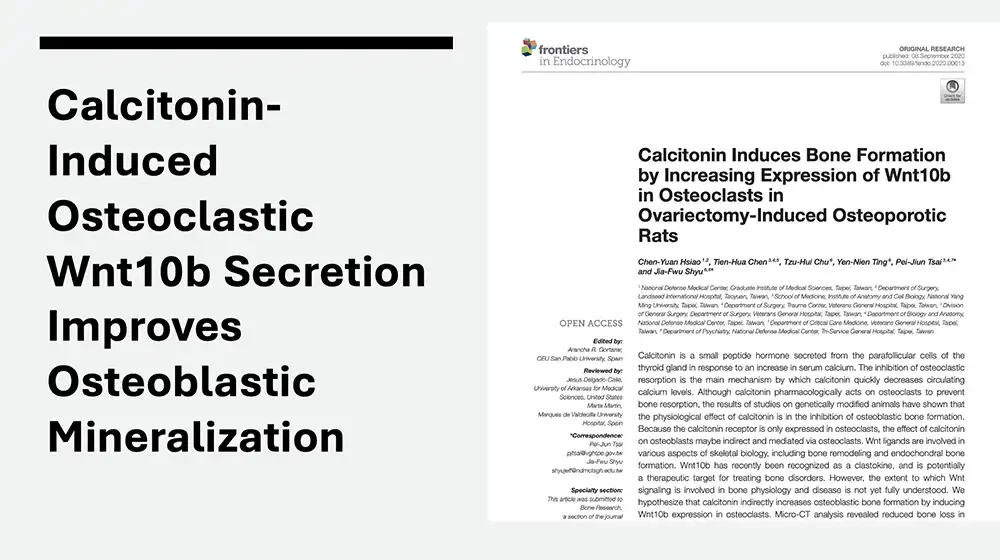
  • Calcitonin-Induced Osteoclastic Wnt10b Secretion Improves Osteoblastic Mineralization Screenshot of journal article first page. frontiers in Endocrinology ORIGINAL RESEARCH published: 08 September 2020 doi: 10.3389/fendo.2020.00613 Badge icon. Check for updates Calcitonin Induces Bone Formation by Increasing Expression of Wnt10b in Osteoclasts in Ovariectomy-Induced Osteoporotic Rats Chen-Yuan Hsiao¹,², Tien-Hua Chen³,⁴,⁵, Tzu-Hui Chu⁶, Yen-Nien Ting⁶, Pei-Jiun Tsai³,⁴,⁷* and Jia-Fwu Shyu⁶,⁸* ¹ National Defense Medical Center, Graduate Institute of Medical Sciences, Taipei, Taiwan, ² Department of Surgery, Landseed International Hospital, Taoyuan, Taiwan, ³ School of Medicine, Institute of Anatomy and Cell Biology, National Yang Ming University, Taipei, Taiwan, ⁴ Department of Surgery, Trauma Center, Veterans General Hospital, Taipei, Taiwan, ⁵ Division of General Surgery, Department of Surgery, Veterans General Hospital, Taipei, Taiwan, ⁶ Department of Biology and Anatomy, National Defense Medical Center, Taipei, Taiwan, ⁷ Department of Critical Care Medicine, Veterans General Hospital, Taipei, Taiwan, ⁸ Department of Psychiatry, National Defense Medical Center, Tri-Service General Hospital, Taipei, Taiwan OPEN ACCESS Edited by: Arancha R. Gortazar , CEU San Pablo University, Spain Reviewed by: Jesus Delgado-Calle, University of Arkansas for Medical Sciences, United States Marta Martin , Marques de Valdecilla University Hospital, Spain *Correspondence: Pei-Jun Tsai pjtsai@vghtpe.gov.tw Jia-Fwu Shyu shyujeff@ndmctsgh.edu.tw Specialty section: This article was submitted to Bone Research, a section of the journal Calcitonin is a small peptide hormone secreted from the parafollicular cells of the thyroid gland in response to an increase in serum calcium. The inhibition of osteoclastic resorption is the main mechanism by which calcitonin quickly decreases circulating calcium levels. Although calcitonin pharmacologically acts on osteoclasts to prevent bone resorption, the results of studies on genetically modified animals have shown that the physiological effect of calcitonin is in the inhibition of osteoblastic bone formation. Because the calcitonin receptor is only expressed in osteoclasts, the effect of calcitonin on osteoblasts maybe indirect and mediated via osteoclasts. Wnt ligands are involved in various aspects of skeletal biology, including bone remodeling and endochondral bone formation. Wnt10b has recently been recognized as a clastokine, and is potentially a therapeutic target for treating bone disorders. However, the extent to which Wnt signaling is involved in bone physiology and disease is not yet fully understood. We hypothesize that calcitonin indirectly increases osteoblastic bone formation by inducing Wnt10b expression in osteoclasts. Micro-CT analysis revealed reduced bone loss in
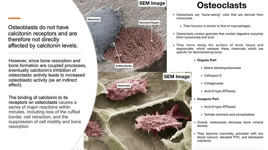
  • Osteoblasts do not have calcitonin receptors and are therefore not directly affected by calcitonin levels. However, since bone resorption and bone formation are coupled processes, eventually calcitonin's inhibition of osteoclastic activity leads to increased osteoblastic activity (as an indirect effect). The binding of calcitonin to its receptors on osteoclasts causes a series of major reactions within minutes, including loss of the ruffled border, cell retraction, and the suppression of cell motility and bone resorption SEM image: osteoclast beside a resorbed bone region. SEM Image Osteoclast Resorbed Region Ruffled Border SEM image: osteoclasts on bone surface. SEM Image Osteoclast Faint diagonal watermark text on lower SEM image. Osteoclasts Osteoclasts are “bone-eating” cells that are derived from monocytes. Their function is similar to that of macrophages. Osteoclasts contain granules that contain digestive enzymes (from lysosomes) and acid. They move along the surface of bone tissue and degranulate, which releases these chemicals which are specific for demineralizing bone. Organic Part Matrix Metalloproteinases Cathepsin K Collagenases Acid (V-type ATPases) Inorganic Part Acid (V-type ATPases) Tartrate-resistant acid phosphatase Overall, osteoclasts decrease bone mineral density. They become maximally activated with low blood calcium, elevated PTH, and decreased calcitonin.

AIC THERAPY In DENTAL ISSUES
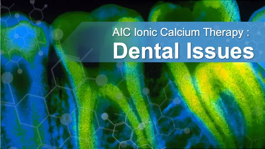
  • Fluorescent micrograph of teeth with molecular structure overlay. AIC Ionic Calcium Therapy : Dental Issues
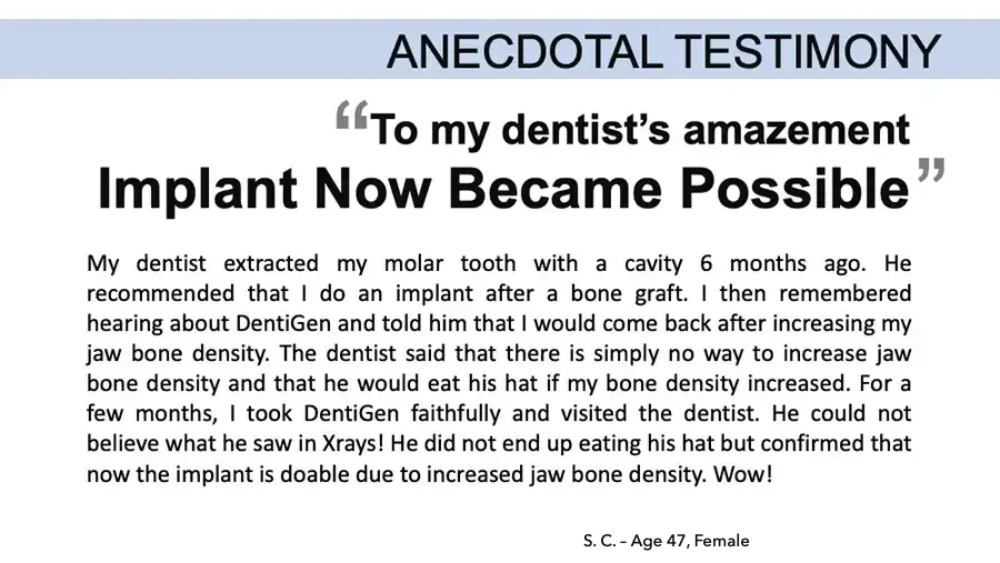
  • ANECDOTAL TESTIMONY “To my dentist’s amazement Implant Now Became Possible” My dentist extracted my molar tooth with a cavity 6 months ago. He recommended that I do an implant after a bone graft. I then remembered hearing about DentiGen and told him that I would come back after increasing my jaw bone density. The dentist said that there is simply no way to increase jaw bone density and that he would eat his hat if my bone density increased. For a few months, I took DentiGen faithfully and visited the dentist. He could not believe what he saw in Xrays! He did not end up eating his hat but confirmed that now the implant is doable due to increased jaw bone density. Wow! S. C. – Age 47, Female
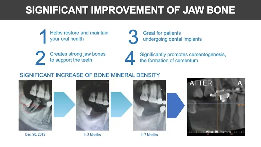
  • SIGNIFICANT IMPROVEMENT OF JAW BONE 1 Helps restore and maintain your oral health 2 Creates strong jaw bones to support the teeth 3 Great for patients undergoing dental implants 4 Significantly promotes cementogenesis, the formation of cementum SIGNIFICANT INCREASE OF BONE MINERAL DENSITY Dental X-ray with red outline drawn on jaw. Dec. 20, 2013 Arrow. Dental X-ray. In 3 Months Arrow. Dental X-ray. In 7 Months Arrow. Dental CT cross-section. AFTER A After 10 months 10 20
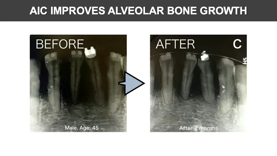
  • AIC IMPROVES ALVEOLAR BONE GROWTH Dental X-ray of lower front teeth. BEFORE Male, Age: 45 Arrow. Dental X-ray of lower front teeth. AFTER C Partial rotated text at right edge of X-ray. H5 After 2 months
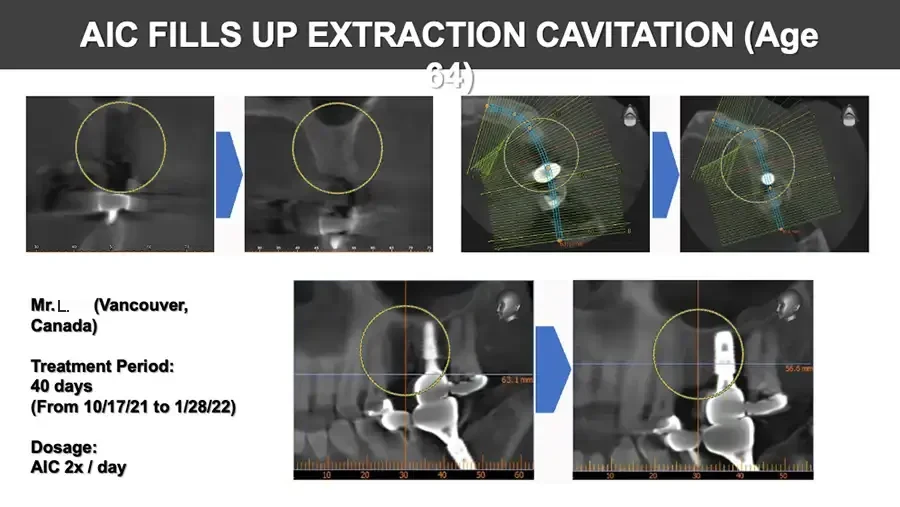
  • AIC FILLS UP EXTRACTION CAVITATION (Age 64) Dental CT section with yellow circle over extraction site. 30 40 50 60 70 Arrow. Dental CT section with yellow circle over extraction site. 35 40 45 55 60 65 70 Axial dental CT view with section lines and yellow circle. Small 3D head orientation icon. Arrow. Axial dental CT view with section lines and yellow circle. Small 3D head orientation icon. Mr. L. (Vancouver, Canada) Treatment Period: 40 days (From 10/17/21 to 1/28/22) Dosage: AIC 2x / day Panoramic CT section with implant and yellow circle. Small 3D head orientation icon. 63.1 mm 10 20 30 40 50 60 Arrow. Panoramic CT section with implant and yellow circle. Small 3D head orientation icon. 56.6 mm 10 20 30 40 50
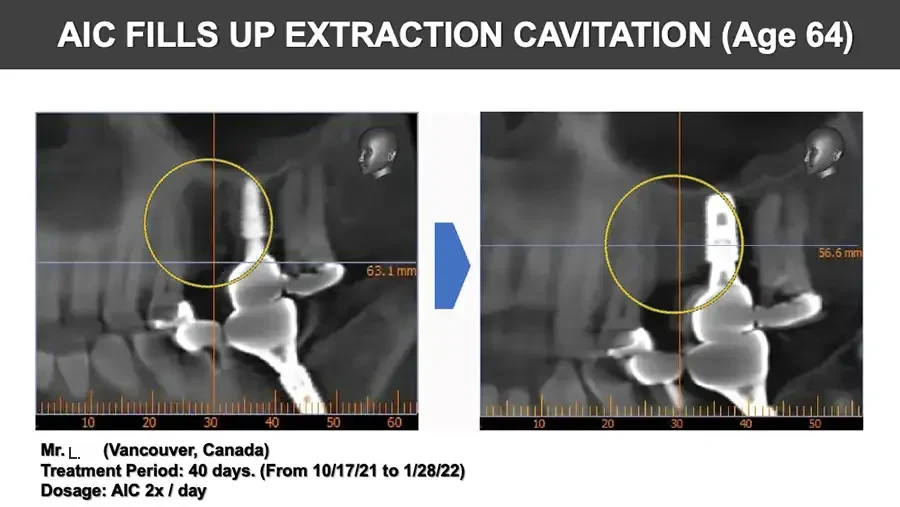
  • AIC FILLS UP EXTRACTION CAVITATION (Age 64) Panoramic CT section with implant and yellow circle. Small 3D head orientation icon. 63.1 mm 10 20 30 40 50 60 Arrow. Panoramic CT section with implant and yellow circle. Small 3D head orientation icon. 56.6 mm 10 20 30 40 50 Mr. L. (Vancouver, Canada) Treatment Period: 40 days. (From 10/17/21 to 1/28/22) Dosage: AIC 2x / day
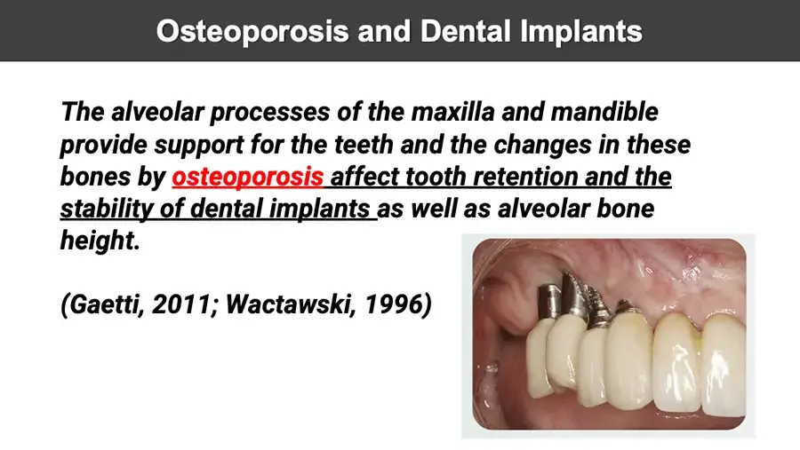
  • Osteoporosis and Dental Implants The alveolar processes of the maxilla and mandible provide support for the teeth and the changes in these bones by osteoporosis affect tooth retention and the stability of dental implants as well as alveolar bone height. (Gaetti, 2011; Wactawski, 1996) Photo: dental implants and crowns in upper jaw.
  • Dental Implants for Type 2 Diabetes During dental implantation, jaw bone problems were observed for T2DM patients Hyperglycaemia increases the risk of T2DM comorbidities and reduces bone turnover; also, impairs bone mineralization and density. The inflammatory response around dental implants was greater in T2DM patients. Hyperglycaemia inhibits osteoblast differentiation and alters parathyroid hormone-mediated regulation of phosphorus and calcium metabolism. AIC is effective in regulating hyperglyceamia and T2DM and therefore strengthen osteoblast performance and healthy calcium metabolism.
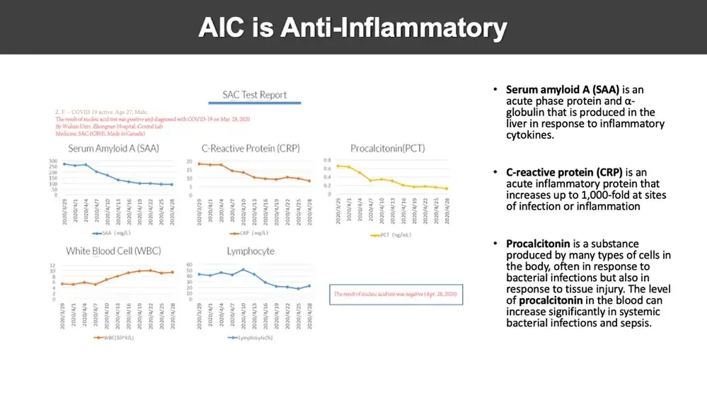
  • AIC is Anti-Inflammatory SAC Test Report Z. F. - COVID 19 active. Age 27, Male. The result of nucleic acid test was positive and diagnosed with COVID-19 on Mar. 28, 2020 By Wuhan Univ. Zhongnan Hospital. Central Lab Medicine: SAC (CBHI , Made in Canada) Five line charts. Serum Amyloid A (SAA) 300 250 200 150 100 50 0 2020/3/29 2020/4/1 2020/4/4 2020/4/7 2020/4/10 2020/4/13 2020/4/16 2020/4/19 2020/4/22 2020/4/25 2020/4/28 SAA (mg/L) C-Reactive Protein (CRP) 20 15 10 5 0 2020/3/29 2020/4/1 2020/4/4 2020/4/7 2020/4/10 2020/4/13 2020/4/16 2020/4/19 2020/4/22 2020/4/25 2020/4/28 CRP (mg/L) Procalcitonin(PCT) 0.8 0.6 0.4 0.2 0 2020/3/29 2020/4/1 2020/4/4 2020/4/7 2020/4/10 2020/4/13 2020/4/16 2020/4/19 2020/4/22 2020/4/25 2020/4/28 PCT (ng/mL) White Blood Cell (WBC) 12 10 8 6 4 2 0 2020/3/29 2020/4/1 2020/4/4 2020/4/7 2020/4/10 2020/4/13 2020/4/16 2020/4/19 2020/4/22 2020/4/25 2020/4/28 WBC(10^9/L) Lymphocyte 60 50 40 30 20 10 0 2020/3/29 2020/4/1 2020/4/4 2020/4/7 2020/4/10 2020/4/13 2020/4/16 2020/4/19 2020/4/22 2020/4/25 2020/4/28 Lymphocyte(%) The result of nucleic acid test was negative (Apr. 28, 2020) Serum amyloid A (SAA) is an acute phase protein and α-globulin that is produced in the liver in response to inflammatory cytokines. C-reactive protein (CRP) is an acute inflammatory protein that increases up to 1,000-fold at sites of infection or inflammation Procalcitonin is a substance produced by many types of cells in the body, often in response to bacterial infections but also in response to tissue injury. The level of procalcitonin in the blood can increase significantly in systemic bacterial infections and sepsis.
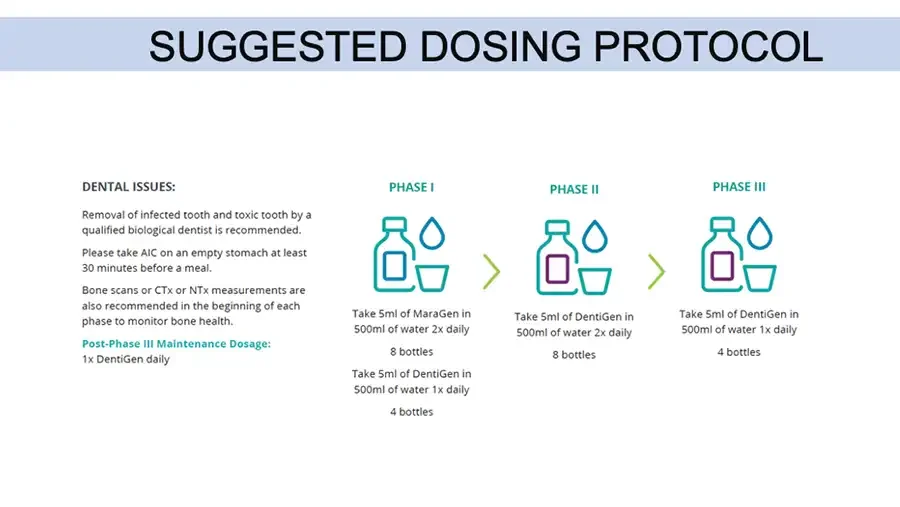
SUGGESTED DOSING PROTOCOL DENTAL ISSUES: Removal of infected tooth and toxic tooth by a qualified biological dentist is recommended. Please take AIC on an empty stomach at least 30 minutes before a meal. Bone scans or CTx or NTx measurements are also recommended in the beginning of each phase to monitor bone health. Post-Phase III Maintenance Dosage: 1x DentiGen daily PHASE I Icon: bottle, water drop and cup. Take 5ml of MaraGen in 500ml of water 2x daily 8 bottles Take 5ml of DentiGen in 500ml of water 1x daily 4 bottles Arrow. PHASE II Icon: bottle, water drop and cup. Take 5ml of DentiGen in 500ml of water 2x daily 8 bottles Arrow. PHASE III Icon: bottle, water drop and cup. Take 5ml of DentiGen in 500ml of water 1x daily 4 bottles

AIC THERAPY IN STEM CELL
  • 3D illustration of stem cells. AIC IONIC CALCIUM THERAPY Title text cut off at right edge. STEM CELL
  • STEM CELL ACTIVITY DEPENDS ON CALCIUM ION SIGNALLING STEM CELLS AND CALCIUM SIGNALING (NIH-PA) Adv Exp Med Biol . 2012 ; 740: 891–916. doi:10.1007/978-94-007-2888-2_40. 3D illustration of stem cells.
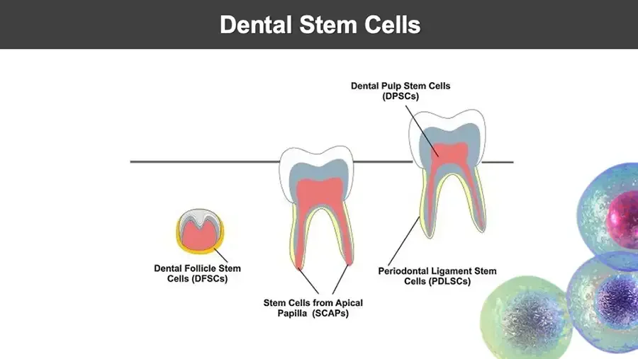
  • Dental Stem Cells Diagram: developing tooth and two erupted teeth with labeled stem cell sites. Dental Pulp Stem Cells (DPSCs) Dental Follicle Stem Cells (DFSCs) Periodontal Ligament Stem Cells (PDLSCs) Stem Cells from Apical Papilla (SCAPs) 3D illustration of stem cells.
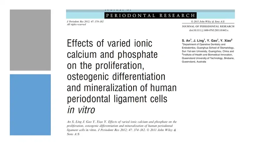
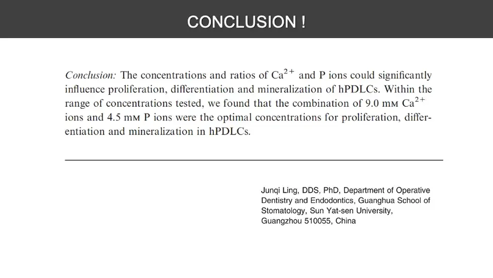
  • Decorative gray and blue blocks. Screenshot of journal article first page. Journal of PERIODONTAL RESEARCH J Periodont Res 2012; 47: 374–382 All rights reserved © 2011 John Wiley & Sons A/S JOURNAL OF PERIODONTAL RESEARCH doi:10.1111/j.1600-0765.2011.01443.x Effects of varied ionic calcium and phosphate on the proliferation, osteogenic differentiation and mineralization of human periodontal ligament cells in vitro An S, Ling J, Gao Y, Xiao Y. Effects of varied ionic calcium and phosphate on the proliferation, osteogenic differentiation and mineralization of human periodontal ligament cells in vitro. J Periodont Res 2012; 47: 374–382. © 2011 John Wiley & Sons A/S S. An¹, J. Ling¹, Y. Gao¹, Y. Xiao² ¹Department of Operative Dentistry and Endodontics, Guanghua School of Stomatology, Sun Yat-sen University, Guangzhou, China and ²Institute of Health and Biomedical Innovation, Queensland University of Technology, Brisbane, Queensland, Australia
  • CONCLUSION ! Junqi Ling, DDS, PhD, Department of Operative Dentistry and Endodontics, Guanghua School of Stomatology, Sun Yat-sen University, Guangzhou 510055, China
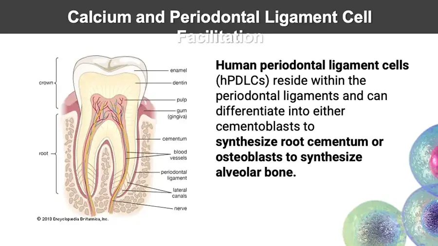
  • Calcium and Periodontal Ligament Cell Facilitation Labeled cross-section diagram of a tooth. crown root enamel dentin pulp gum (gingiva) cementum blood vessels periodontal ligament lateral canals nerve © 2013 Encyclopædia Britannica, Inc. Human periodontal ligament cells (hPDLCs) reside within the periodontal ligaments and can differentiate into either cementoblasts to synthesize root cementum or osteoblasts to synthesize alveolar bone. 3D illustration of stem cells.
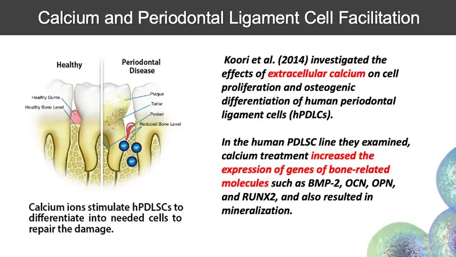
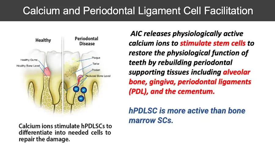
  • Calcium and Periodontal Ligament Cell Facilitation Diagram: healthy teeth versus periodontal disease. Healthy Periodontal Disease Healthy Gums Healthy Bone Level Plaque Tartar Pocket Reduced Bone Level Three blue ion circles. Ca²⁺ Ca²⁺ Ca²⁺ Calcium ions stimulate hPDLSCs to differentiate into needed cells to repair the damage. Koori et al. (2014) investigated the effects of extracellular calcium on cell proliferation and osteogenic differentiation of human periodontal ligament cells (hPDLCs). In the human PDLSC line they examined, calcium treatment increased the expression of genes of bone-related molecules such as BMP-2, OCN, OPN, and RUNX2, and also resulted in mineralization. 3D illustration of stem cells.
  • Calcium and Periodontal Ligament Cell Facilitation Diagram: healthy teeth versus periodontal disease. Healthy Periodontal Disease Healthy Gums Healthy Bone Level Plaque Tartar Pocket Reduced Bone Level Three blue ion circles. Ca²⁺ Ca²⁺ Ca²⁺ Calcium ions stimulate hPDLSCs to differentiate into needed cells to repair the damage. AIC releases physiologically active calcium ions to stimulate stem cells to restore the physiological function of teeth by rebuilding periodontal supporting tissues including alveolar bone, gingiva, periodontal ligaments (PDL), and the cementum. hPDLSC is more active than bone marrow SCs.
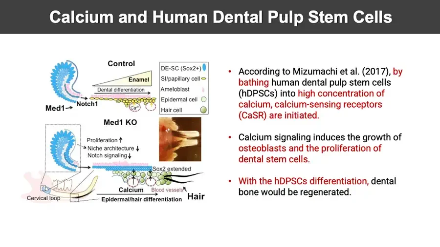
  • Calcium and Human Dental Pulp Stem Cells Diagram: dental stem cell niche, control versus Med1 knockout. Control Enamel Dental differentiation Notch1 Med1 Ca²⁺ DE-SC (Sox2+) SI/papillary cell Ameloblast Epidermal cell Hair cell Med1 KO Photo inset. Proliferation ↑ Niche architecture ↓ Notch signaling ↓ Sox2 extended Calcium Blood vessels Hair Cervical loop Epidermal/hair differentiation According to Mizumachi et al. (2017), by bathing human dental pulp stem cells (hDPSCs) into high concentration of calcium, calcium-sensing receptors (CaSR) are initiated. Calcium signaling induces the growth of osteoblasts and the proliferation of dental stem cells. With the hDPSCs differentiation, dental bone would be regenerated.
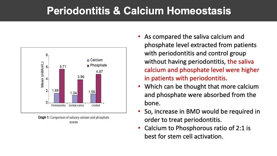
  • Periodontitis & Calcium Homeostasis Bar chart. Mean (mMol/L) 8 6 4 2 0 Calcium Phosphate 1.69 5.71 Periodontitis 1.34 3.96 Dental caries 1.55 4.87 Control Graph 1: Comparison of salivary calcium and phosphate scores As compared the saliva calcium and phosphate level extracted from patients with periodontitis and control group without having periodontitis, the saliva calcium and phosphate level were higher in patients with periodontitis. Which can be thought that more calcium and phosphate were absorbed from the bone. So, increase in BMD would be required in order to treat periodontitis. Calcium to Phosphorous ratio of 2:1 is best for stem cell activation.

AIC THERAPY IN ARTHRITIS
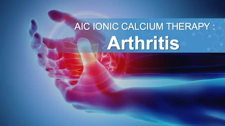
  • Illustration: hand with glowing red inflamed joints. AIC IONIC CALCIUM THERAPY : Arthritis
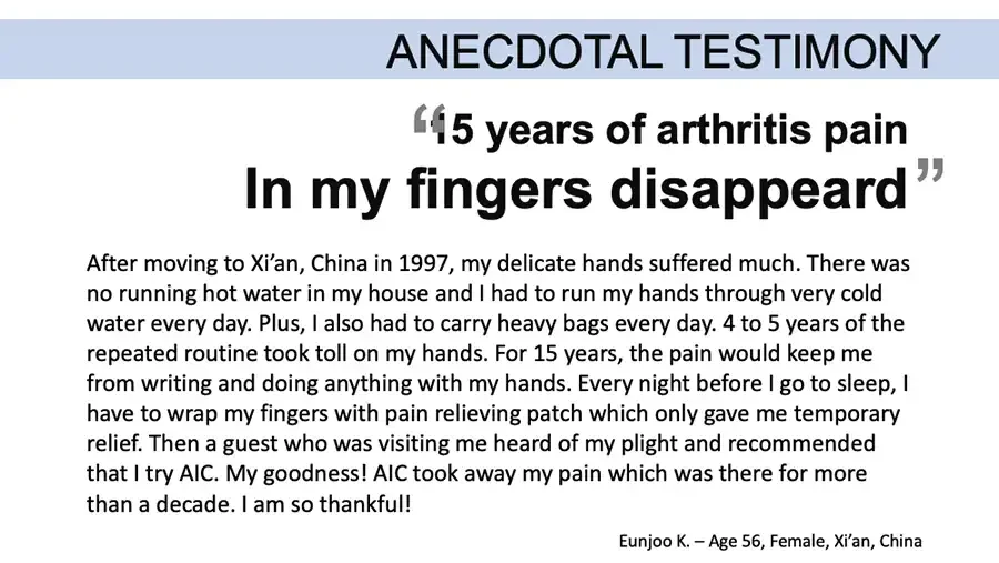
  • ANECDOTAL TESTIMONY “15 years of arthritis pain In my fingers disappeard” After moving to Xi’an, China in 1997, my delicate hands suffered much. There was no running hot water in my house and I had to run my hands through very cold water every day. Plus, I also had to carry heavy bags every day. 4 to 5 years of the repeated routine took toll on my hands. For 15 years, the pain would keep me from writing and doing anything with my hands. Every night before I go to sleep, I have to wrap my fingers with pain relieving patch which only gave me temporary relief. Then a guest who was visiting me heard of my plight and recommended that I try AIC. My goodness! AIC took away my pain which was there for more than a decade. I am so thankful! Eunjoo K. – Age 56, Female, Xi’an, China
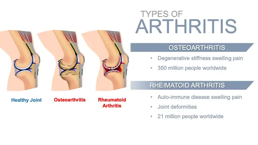
  • TYPES OF ARTHRITIS Three knee joint illustrations. Healthy Joint Osteoarthritis Rheumatoid Arthritis OSTEOARTHRITIS Degenerative stiffness swelling pain 350 million people worldwide RHEIMATOID ARTHRITIS Auto-immune disease swelling pain Joint deformities 21 million people worldwide
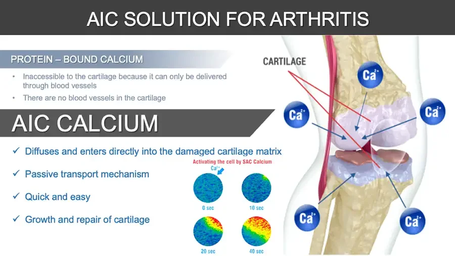
  • AIC SOLUTION FOR ARTHRITIS PROTEIN – BOUND CALCIUM Inaccessible to the cartilage because it can only be delivered through blood vessels There are no blood vessels in the cartilage AIC CALCIUM ✓ Diffuses and enters directly into the damaged cartilage matrix ✓ Passive transport mechanism ✓ Quick and easy ✓ Growth and repair of cartilage Activating the cell by SAC Calcium Four circular cell images changing from blue to red over time. Ca²⁺ 0 sec 10 sec 20 sec 40 sec Knee joint illustration with calcium ions pointing to cartilage. CARTILAGE Ca²⁺ Ca²⁺ Ca²⁺ Ca²⁺ Ca²⁺
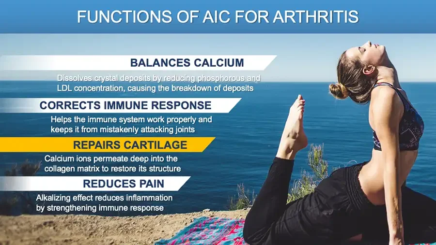
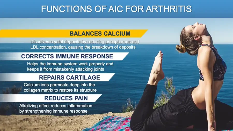
  • FUNCTIONS OF AIC FOR ARTHRITIS Photo: woman doing a yoga backbend by the sea. BALANCES CALCIUM Dissolves crystal deposits by reducing phosphorous and LDL concentration, causing the breakdown of deposits CORRECTS IMMUNE RESPONSE Helps the immune system work properly and keeps it from mistakenly attacking joints REPAIRS CARTILAGE Calcium ions permeate deep into the collagen matrix to restore its structure REDUCES PAIN Alkalizing effect reduces inflammation by strengthening immune response
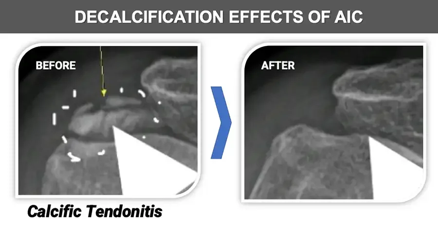
  • DECALCIFICATION EFFECTS OF AIC Shoulder X-ray close-up with dashed circle and arrow. BEFORE Calcific Tendonitis Arrow. Shoulder X-ray close-up. AFTER
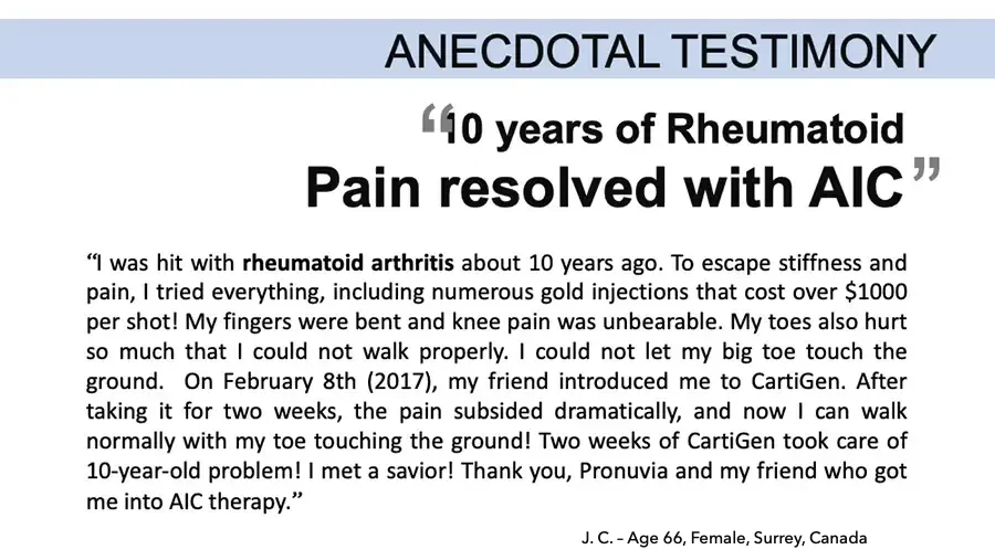
  • ANECDOTAL TESTIMONY “10 years of Rheumatoid Pain resolved with AIC” “I was hit with rheumatoid arthritis about 10 years ago. To escape stiffness and pain, I tried everything, including numerous gold injections that cost over $1000 per shot! My fingers were bent and knee pain was unbearable. My toes also hurt so much that I could not walk properly. I could not let my big toe touch the ground. On February 8th (2017), my friend introduced me to CartiGen. After taking it for two weeks, the pain subsided dramatically, and now I can walk normally with my toe touching the ground! Two weeks of CartiGen took care of 10-year-old problem! I met a savior! Thank you, Pronuvia and my friend who got me into AIC therapy.” J. C. – Age 66, Female, Surrey, Canada
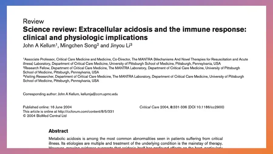
  • Screenshot of journal article first page in a gradient frame. Review Science review: Extracellular acidosis and the immune response: clinical and physiologic implications John A Kellum¹, Mingchen Song² and Jinyou Li³ ¹Associate Professor, Critical Care Medicine and Medicine, Co-Director, The MANTRA (Mechanisms And Novel Therapies for Resuscitation and Acute illness) Laboratory, Department of Critical Care Medicine, University of Pittsburgh School of Medicine, Pittsburgh, Pennsylvania, USA ²Research Fellow, Department of Critical Care Medicine, The MANTRA Laboratory, Department of Critical Care Medicine, University of Pittsburgh School of Medicine, Pittsburgh, Pennsylvania, USA ³Visiting Researcher, Department of Critical Care Medicine, The MANTRA Laboratory, Department of Critical Care Medicine, University of Pittsburgh School of Medicine, Pittsburgh, Pennsylvania, USA Corresponding author: John A Kellum, kellumja@ccm.upmc.edu Published online: 16 June 2004 This article is online at http://ccforum.com/content/8/5/331 © 2004 BioMed Central Ltd Critical Care 2004, 8:331-336 (DOI 10.1186/cc2900) Abstract
  • Large circle on dark background framing a quotation.
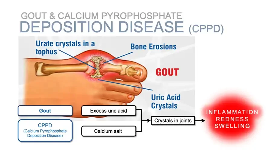
GOUT & CALCIUM PYROPHOSPHATE DEPOSITION DISEASE (CPPD) Illustration: foot with inflamed big toe joint and crystals. Urate crystals in a tophus Bone Erosions GOUT Uric Acid Crystals Flowchart. Gout CPPD (Calcium Pyrophosphate Deposition Disease) Excess uric acid Calcium salt Crystals in joints Red glowing circle. INFLAMMATION REDNESS SWELLING
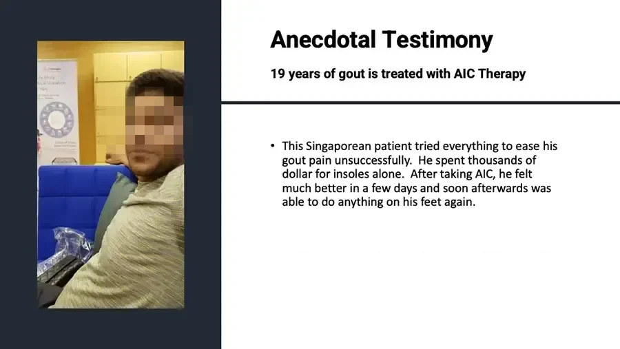
Photo: smiling man seated in an office. Poster in background with small text. Anecdotal Testimony 19 years of gout is treated with AIC Therapy This Singaporean patient tried everything to ease his gout pain unsuccessfully. He spent thousands of dollar for insoles alone. After taking AIC, he felt much better in a few days and soon afterwards was able to do anything on his feet again.
FUNCTIONS OF AIC FOR ARTHRITIS Photo: woman doing a yoga backbend by the sea. BALANCES CALCIUM Dissolves crystal deposits by reducing phosphorous and LDL concentration, causing the breakdown of deposits CORRECTS IMMUNE RESPONSE Helps the immune system work properly and keeps it from mistakenly attacking joints REPAIRS CARTILAGE Calcium ions permeate deep into the collagen matrix to restore its structure REDUCES PAIN Alkalizing effect reduces inflammation by strengthening immune response
ANECDOTAL TESTIMONY “Swelling and pain gone. I can now play golf again!” I was only in my early forties when I was diagnosed with degenerative arthritis in my hands. Ever since, I suffered so much. I could not lift heavy objects and even lifting cooking pots hurt every joints of my fingers. All the prescription drugs did not help much. Then one of my good friend recommended me AIC. I was doubtful in the beginning, but faithfully took AIC as courtesy to my friend. Amazingly, swelling and pain in my fingers decreased noticeably and even experienced increased energy and vitality. Now, I am able to play golf which I had to stop because of arthritis. I recommended AIC to my husband and do not see him fatigued anymore. L. K. – Age 42, Female
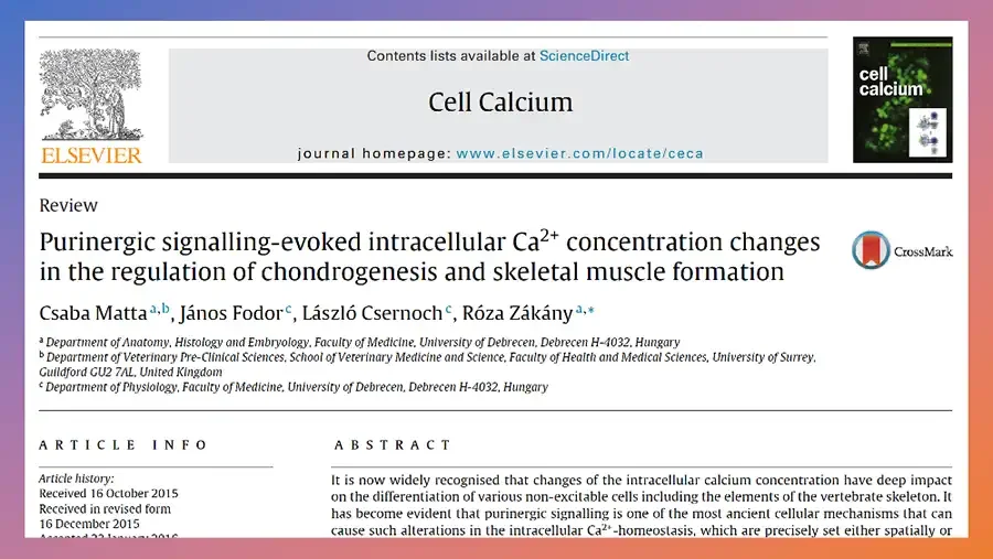
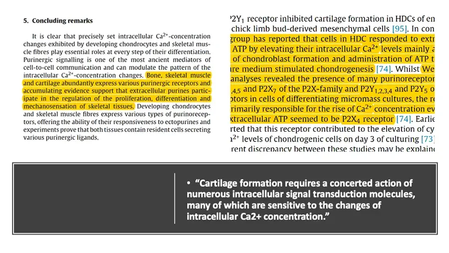
Screenshot of journal article first page in a gradient frame. Elsevier tree logo. ELSEVIER Contents lists available at ScienceDirect Cell Calcium journal homepage: www.elsevier.com/locate/ceca Journal cover thumbnail. cell calcium Review Purinergic signalling-evoked intracellular Ca²⁺ concentration changes in the regulation of chondrogenesis and skeletal muscle formation CrossMark logo. CrossMark Csaba Mattaᵃ,ᵇ, János Fodorᶜ, László Csernochᶜ, Róza Zákányᵃ,* ᵃ Department of Anatomy, Histology and Embryology, Faculty of Medicine, University of Debrecen, Debrecen H-4032, Hungary ᵇ Department of Veterinary Pre-Clinical Sciences, School of Veterinary Medicine and Science, Faculty of Health and Medical Sciences, University of Surrey, Guildford GU2 7AL, United Kingdom ᶜ Department of Physiology, Faculty of Medicine, University of Debrecen, Debrecen H-4032, Hungary ARTICLE INFO Article history: Received 16 October 2015 Received in revised form 16 December 2015 Accepted 22 January 2016 ABSTRACT
Two cropped excerpts of journal article pages, with yellow highlighting. 5. Concluding remarks Dark box with one bullet.
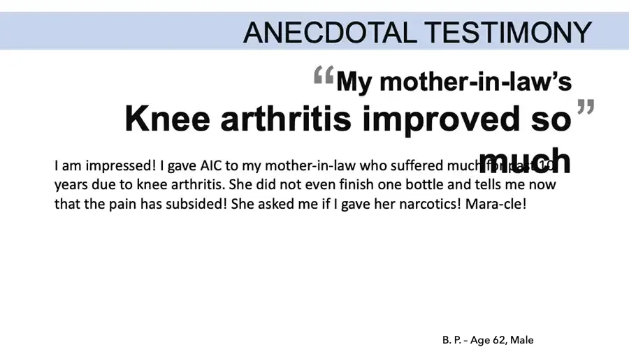
ANECDOTAL TESTIMONY “My mother-in-law’s Knee arthritis improved so” much I am impressed! I gave AIC to my mother-in-law who suffered much for past 10 years due to knee arthritis. She did not even finish one bottle and tells me now that the pain has subsided! She asked me if I gave her narcotics! Mara-cle! B. P. – Age 62, Male
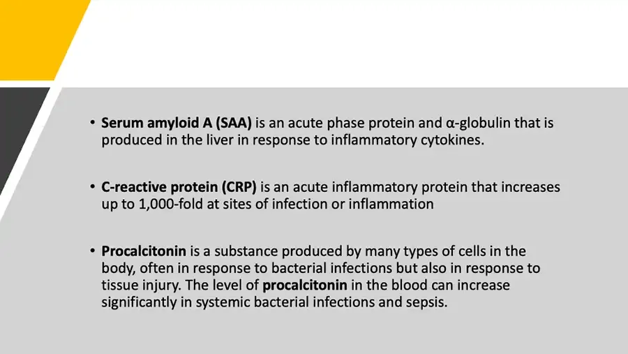
Decorative yellow and dark diagonal shapes. Serum amyloid A (SAA) is an acute phase protein and α-globulin that is produced in the liver in response to inflammatory cytokines. C-reactive protein (CRP) is an acute inflammatory protein that increases up to 1,000-fold at sites of infection or inflammation Procalcitonin is a substance produced by many types of cells in the body, often in response to bacterial infections but also in response to tissue injury. The level of procalcitonin in the blood can increase significantly in systemic bacterial infections and sepsis.
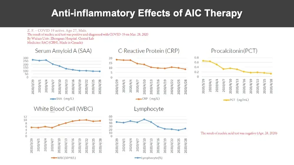
Anti-inflammatory Effects of AIC Therapy Z. F. - COVID 19 active. Age 27, Male. The result of nucleic acid test was positive and diagnosed with COVID-19 on Mar. 28, 2020 By Wuhan Univ. Zhongnan Hospital. Central Lab Medicine: SAC (CBHI , Made in Canada) Five line charts. Serum Amyloid A (SAA) 300 250 200 150 100 50 0 2020/3/29 2020/4/1 2020/4/4 2020/4/7 2020/4/10 2020/4/13 2020/4/16 2020/4/19 2020/4/22 2020/4/25 2020/4/28 SAA (mg/L) C-Reactive Protein (CRP) 20 15 10 5 0 2020/3/29 2020/4/1 2020/4/4 2020/4/7 2020/4/10 2020/4/13 2020/4/16 2020/4/19 2020/4/22 2020/4/25 2020/4/28 CRP (mg/L) Procalcitonin(PCT) 0.8 0.6 0.4 0.2 0 2020/3/29 2020/4/1 2020/4/4 2020/4/7 2020/4/10 2020/4/13 2020/4/16 2020/4/19 2020/4/22 2020/4/25 2020/4/28 PCT (ng/mL) White Blood Cell (WBC) 12 10 8 6 4 2 0 2020/3/29 2020/4/1 2020/4/4 2020/4/7 2020/4/10 2020/4/13 2020/4/16 2020/4/19 2020/4/22 2020/4/25 2020/4/28 WBC(10^9/L) Lymphocyte 60 50 40 30 20 10 0 2020/3/29 2020/4/1 2020/4/4 2020/4/7 2020/4/10 2020/4/13 2020/4/16 2020/4/19 2020/4/22 2020/4/25 2020/4/28 Lymphocyte(%) The result of nucleic acid test was negative (Apr. 28, 2020)
ANECDOTAL TESTIMONY “Swelling and pain of Arthritis improved” significantly I had terrible pain due to knuckle and knee arthritis. Every night, I put heating pads to alleviate the pain, but the continuing pain made the quality of my life so miserable and I came down with depression. Then one of my caring friend recommended Cartigen from Pronuvia. Being desperate after many futile attempts with medicines and supplements, I decided to give it another chance. Remarkably, after taking Cartigen just for ten days, both the pain and swelling were reduced significantly. I was taken aback. My husband who helped me with heating pads every night was surprised at the powerful effect of Cartigen. He is finally free from the task as I am also from suffering! M. K. – Age 53, Female