Notice. The following information is provided for your convenience. ACRI is not directly involved in this research and does not make any medical claims.
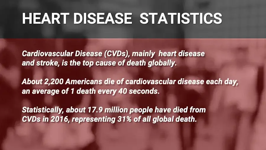
Illustration: glowing heart in blue human torso, ECG trace at right. AIC IONIC CALCIUM THERAPY : Heart Disease
Blurred crowd photo behind red overlay. HEART DISEASE STATISTICS Cardiovascular Disease (CVDs), mainly heart disease and stroke, is the top cause of death globally. About 2,200 Americans die of cardiovascular disease each day, an average of 1 death every 40 seconds. Statistically, about 17.9 million people have died from CVDs in 2016, representing 31% of all global death.
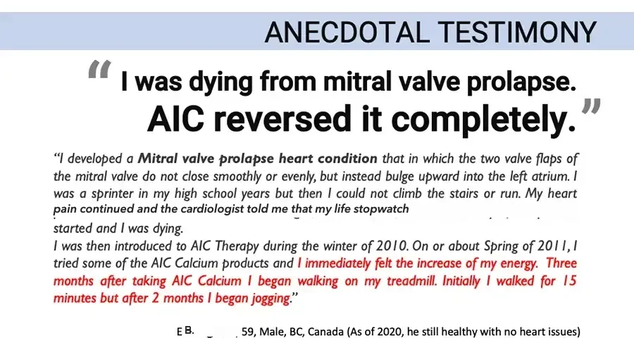
ANECDOTAL TESTIMONY “ I was dying from mitral valve prolapse. AIC reversed it completely. ” “I developed a Mitral valve prolapse heart condition that in which the two valve flaps of the mitral valve do not close smoothly or evenly, but instead bulge upward into the left atrium. I was a sprinter in my high school years but then I could not climb the stairs or run. My heart pain continued and the cardiologist told me that my life stopwatch started and I was dying. I was then introduced to AIC Therapy during the winter of 2010. On or about Spring of 2011, I tried some of the AIC Calcium products and I immediately felt the increase of my energy. Three months after taking AIC Calcium I began walking on my treadmill. Initially I walked for 15 minutes but after 2 months I began jogging.” E. B., 59, Male, BC, Canada (As of 2020, he still healthy with no heart issues)
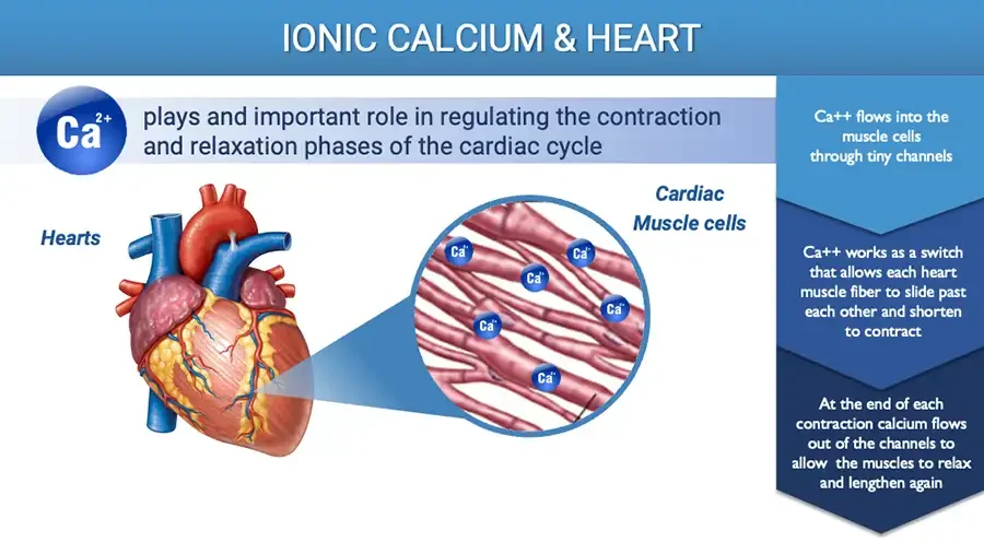
IONIC CALCIUM & HEART Blue sphere with calcium ion symbol. Ca²⁺ plays and important role in regulating the contraction and relaxation phases of the cardiac cycle Hearts Illustration: anatomical heart with magnified view of muscle fibers. Cardiac Muscle cells Ca⁺⁺ repeated on 6 small blue spheres among the fibers. Ca++ flows into the muscle cells through tiny channels Ca++ works as a switch that allows each heart muscle fiber to slide past each other and shorten to contract At the end of each contraction calcium flows out of the channels to allow the muscles to relax and lengthen again
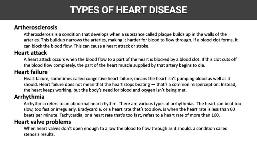
TYPES OF HEART DISEASE Artherosclerosis Atherosclerosis is a condition that develops when a substance called plaque builds up in the walls of the arteries. This buildup narrows the arteries, making it harder for blood to flow through. If a blood clot forms, it can block the blood flow. This can cause a heart attack or stroke. Heart attack A heart attack occurs when the blood flow to a part of the heart is blocked by a blood clot. If this clot cuts off the blood flow completely, the part of the heart muscle supplied by that artery begins to die. Heart failure Heart failure, sometimes called congestive heart failure, means the heart isn’t pumping blood as well as it should. Heart failure does not mean that the heart stops beating — that’s a common misperception. Instead, the heart keeps working, but the body’s need for blood and oxygen isn’t being met. Arrhythmia Arrhythmia refers to an abnormal heart rhythm. There are various types of arrhythmias. The heart can beat too slow, too fast or irregularly. Bradycardia, or a heart rate that’s too slow, is when the heart rate is less than 60 beats per minute. Tachycardia, or a heart rate that’s too fast, refers to a heart rate of more than 100. Heart valve problems When heart valves don’t open enough to allow the blood to flow through as it should, a condition called stenosis results.
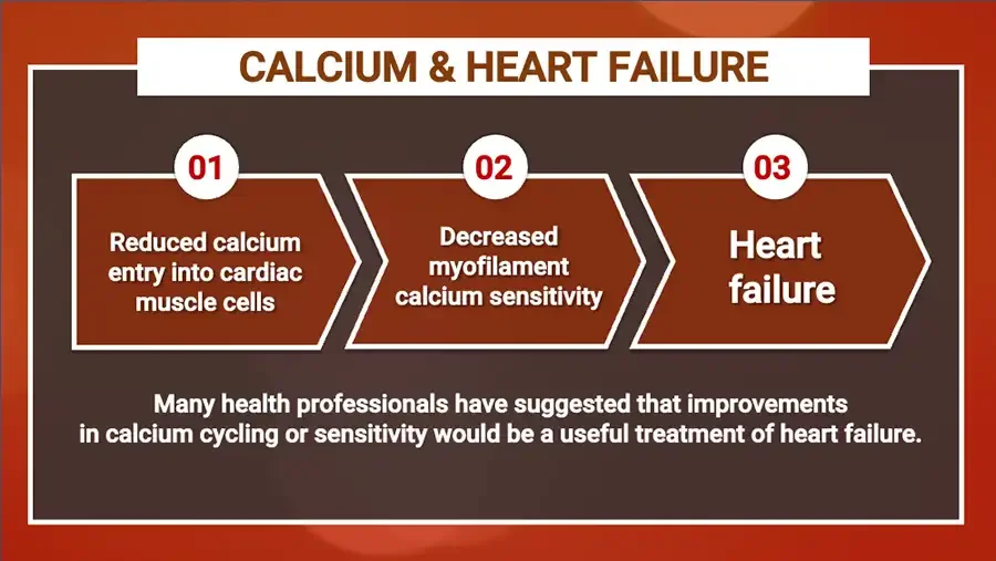
CALCIUM & HEART FAILURE Three numbered arrow steps. 01 Reduced calcium entry into cardiac muscle cells 02 Decreased myofilament calcium sensitivity 03 Heart failure Many health professionals have suggested that improvements in calcium cycling or sensitivity would be a useful treatment of heart failure.
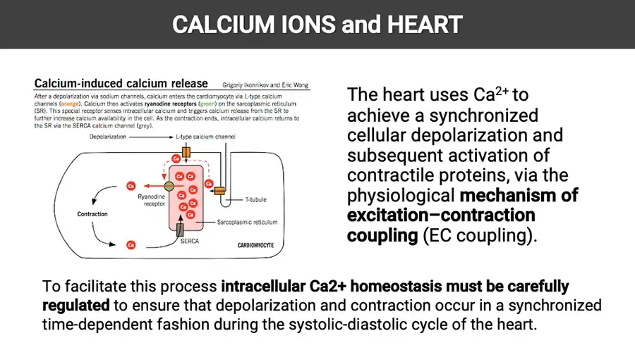
CALCIUM IONS and HEART Calcium-induced calcium release Grigoriy Ikonnikov and Eric Wong After a depolarization via sodium channels, calcium enters the cardiomyocyte via L-type calcium channels (orange). Calcium then activates ryanodine receptors (green) on the sarcoplasmic reticulum (SR). This special receptor senses intracellular calcium and triggers calcium release from the SR to further increase calcium availability in the cell. As the contraction ends, intracellular calcium returns to the SR via the SERCA calcium channel (grey). Diagram: calcium cycling in a cardiomyocyte. Depolarization L-type calcium channel Ryanodine receptor Contraction T-tubule Sarcoplasmic reticulum SERCA CARDIOMYOCYTE Ca repeated on 11 small red circles. The heart uses Ca²⁺ to achieve a synchronized cellular depolarization and subsequent activation of contractile proteins, via the physiological mechanism of excitation–contraction coupling (EC coupling). To facilitate this process intracellular Ca2+ homeostasis must be carefully regulated to ensure that depolarization and contraction occur in a synchronized time-dependent fashion during the systolic-diastolic cycle of the heart.
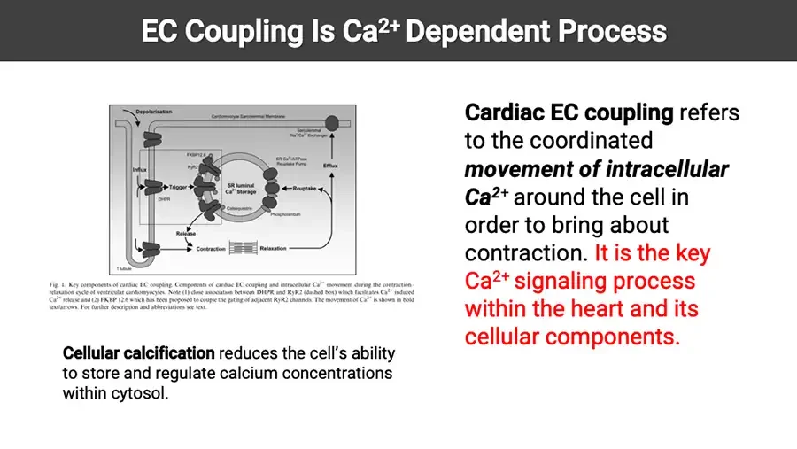
EC Coupling Is Ca²⁺ Dependent Process Journal figure: cardiac EC coupling components in a cardiomyocyte. Depolarisation Cardiomyocyte Sarcolemmal Membrane Sarcolemmal Na⁺/Ca²⁺ Exchanger FKBP12.6 RyR2 SR Ca²⁺/ATPase Reuptake Pump Influx Efflux Trigger SR luminal Ca²⁺ Storage Reuptake DHPR Calsequestrin Phospholamban Release Contraction Relaxation T tubule Fig. 1. Key components of cardiac EC coupling. Cellular calcification reduces the cell’s ability to store and regulate calcium concentrations within cytosol. Cardiac EC coupling refers to the coordinated movement of intracellular Ca²⁺ around the cell in order to bring about contraction. It is the key Ca²⁺ signaling process within the heart and its cellular components.
EC Coupling Is Ca²⁺ Dependent Process The inward Ca²⁺ current (ICa) through DHPR is, on its own, insufficient to bring about the required conformational change in troponin needed for contraction to occur. Additional Ca²⁺ is required and this is obtained from a pool of stored Ca²⁺ within the sarcoplasmic reticulum (SR) of the cell. The initial inward movement of Ca²⁺ acts as an amplification signal for the release of this stored pool of SR.
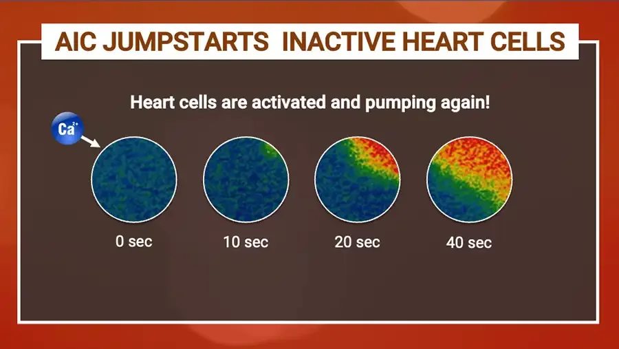
AIC JUMPSTARTS INACTIVE HEART CELLS Heart cells are activated and pumping again! Blue calcium ion sphere with arrow pointing to first image. Ca²⁺ Four circular heat-map images of heart cells activating over time. 0 sec 10 sec 20 sec 40 sec
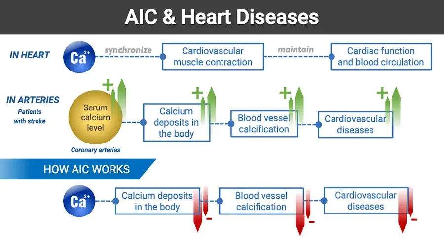
AIC & Heart Diseases Flow diagram with calcium spheres, boxes and up/down arrows. IN HEART Ca²⁺ synchronize Cardiovascular muscle contraction maintain Cardiac function and blood circulation IN ARTERIES Patients with stroke Serum calcium level Coronary arteries + Calcium deposits in the body + Blood vessel calcification + Cardiovascular diseases + HOW AIC WORKS Ca²⁺ Calcium deposits in the body Blood vessel calcification Cardiovascular diseases
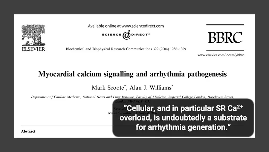
Screenshot of a journal article title block, partly covered by a quote box. Logo: Elsevier tree. ELSEVIER Available online at www.sciencedirect.com SCIENCE DIRECT® Biochemical and Biophysical Research Communications 322 (2004) 1286–1309 BBRC www.elsevier.com/locate/ybbrc Myocardial calcium signalling and arrhythmia pathogenesis Mark Scoote*, Alan J. Williams* Department of Cardiac Medicine, National Heart and Lung Institute, Faculty of Medicine, Imperial College London, Dovehouse Street, London SW3 6LY, UK Received hidden behind quote box. Available online 21 August 2004 Abstract “Cellular, and in particular SR Ca²⁺ overload, is undoubtedly a substrate for arrhythmia generation.”
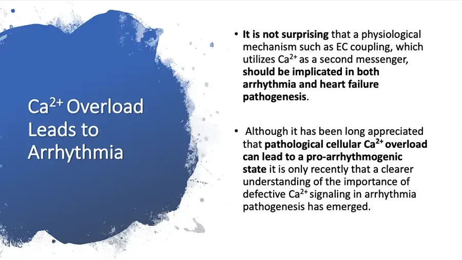
Blue paint splash panel on left. Ca²⁺ Overload Leads to Arrhythmia It is not surprising that a physiological mechanism such as EC coupling, which utilizes Ca²⁺ as a second messenger, should be implicated in both arrhythmia and heart failure pathogenesis. Although it has been long appreciated that pathological cellular Ca²⁺ overload can lead to a pro-arrhythmogenic state it is only recently that a clearer understanding of the importance of defective Ca²⁺ signaling in arrhythmia pathogenesis has emerged.
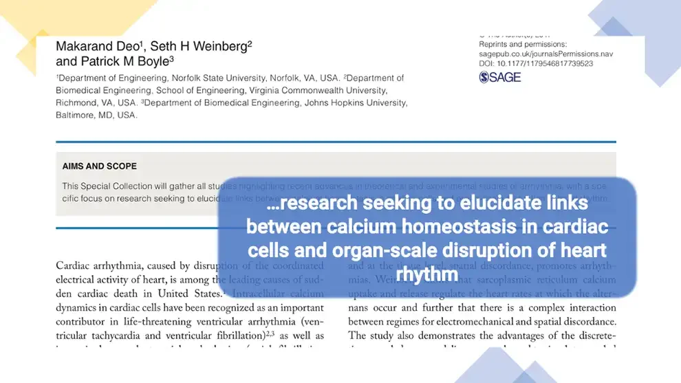
Screenshot of a journal article page, partly covered by a quote box. Makarand Deo¹, Seth H Weinberg² and Patrick M Boyle³ ¹Department of Engineering, Norfolk State University, Norfolk, VA, USA. ²Department of Biomedical Engineering, School of Engineering, Virginia Commonwealth University, Richmond, VA, USA. ³Department of Biomedical Engineering, Johns Hopkins University, Baltimore, MD, USA. © The Author(s) 2017 top half cut off. Reprints and permissions: sagepub.co.uk/journalsPermissions.nav DOI: 10.1177/1179546817739523 Logo: SAGE. SAGE AIMS AND SCOPE …research seeking to elucidate links between calcium homeostasis in cardiac cells and organ-scale disruption of heart rhythm
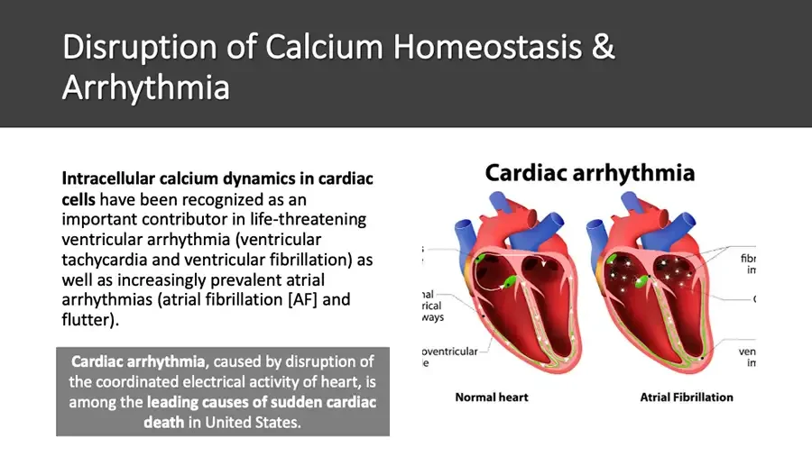
Disruption of Calcium Homeostasis & Arrhythmia Intracellular calcium dynamics in cardiac cells have been recognized as an important contributor in life-threatening ventricular arrhythmia (ventricular tachycardia and ventricular fibrillation) as well as increasingly prevalent atrial arrhythmias (atrial fibrillation AF. and flutter). Cardiac arrhythmia, caused by disruption of the coordinated electrical activity of heart, is among the leading causes of sudden cardiac death in United States. Cardiac arrhythmia Illustration: cross-sections of a normal heart and a fibrillating heart. Label fragments below are cut off at the image edges. nal rical ways oventricular e fibr im ( ven im Normal heart Atrial Fibrillation
Ca²⁺ Channel Blockers Although the widely used Ca²⁺ channel blocking drugs, such as verapamil, have a known anti-arrhythmic action, especially in the context of supraventricular arrhythmias, this effect is primarily related to their blockade of AV nodal conduction rather than a direct molecular modification of defective Ca²⁺ signaling pathways. Ca²⁺ channel blockers come with side effects.
B Blockers The widely publicized beneficial actions of b-blockers appear to result, at least in part, from a reversal of Ca²⁺-mediated signaling defects. This offers an insight into the potential therapeutic benefit that could result from a new era of targeted strategies directed against the Ca²⁺ signaling defects outlined in this article. However, B Blockers come with side-effects.
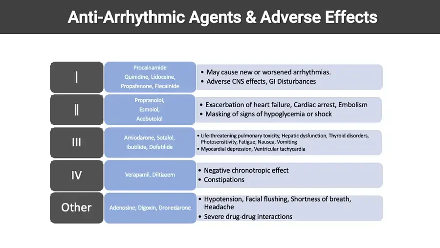
Anti-Arrhythmic Agents & Adverse Effects Table with no header row. I Procainamide Quinidine, Lidocaine, Propafenone, Flecainide • May cause new or worsened arrhythmias. • Adverse CNS effects, GI Disturbances II Propranolol, Esmolol, Acebutolol • Exacerbation of heart failure, Cardiac arrest, Embolism • Masking of signs of hypoglycemia or shock III Amiodarone, Sotalol, Ibutilide, Dofetilide • Life-threatening pulmonary toxicity, Hepatic dysfunction, Thyroid disorders, Photosensitivity, Fatigue, Nausea, Vomiting • Myocardial depression, Ventricular tachycardia IV Verapamil, Diltiazem • Negative chronotropic effect • Constipations Other Adenosine, Digoxin, Dronedarone • Hypotension, Facial flushing, Shortness of breath, Headache • Severe drug-drug interactions
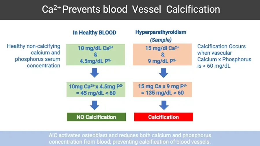
Ca²⁺ Prevents blood Vessel Calcification Healthy non-calcifying calcium and phosphorus serum concentration In Healthy BLOOD 10 mg/dL Ca²⁺ & 4.5mg/dL P³⁻ Down arrow. 10mg Ca²⁺ x 4.5mg P³⁻ = 45 mg/dL < 60 Down arrow. NO Calcification Hyperparathyroidism (Sample) 15 mg/dl Ca²⁺ & 9 mg/dL P³⁻ Down arrow. 15 mg Ca x 9 mg P³⁻ = 135 mg/dL > 60 Down arrow. Calcification Calcification Occurs when vascular Calcium x Phosphorus is > 60 mg/dL AIC activates osteoblast and reduces both calcium and phosphorus concentration from blood, preventing calcification of blood vessels.