Notice. The following information is provided for your convenience. ACRI is not directly involved in this research and does not make any medical claims.

AIC IN INFECTIOUS DISEASES

Ionic Calcium creates adverse environment where pathogens have difficulty proliferating. It also strengthens the immune response against intruders.
Illustration: teal virus particles on dark background. AIC IONIC CALCIUM THERAPY : Infectious Diseases
ANECDOTAL TESTIMONY “Shielded from MERS outbreak” “A high school student with aplastic anemia was going through a rough patch because the only available treatment option for his condition was bone marrow transplant. However, after being introduced to AIC therapy, he fully recovered from aplastic anemia. After graduating from college, he got a job at a hospital as a phlebotomist around 2015 when there was a MERS epidemic in South Korea. His team, who received and diagnosed MERS patients, were all quarantined due to MERS infection, except this young man. It was a mystery to all his colleagues why only he escaped MERs contraction when he was at the riskiest position of drawing the blood of MERS patients as a phlebotomist. The only difference was that he was still taking AIC at a maintenance dosage.” Testimony about a patient by Dr. Paul Lee
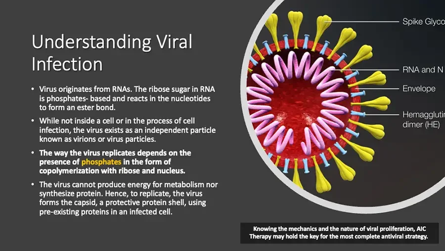
Understanding Viral Infection Virus originates from RNAs. The ribose sugar in RNA is phosphates- based and reacts in the nucleotides to form an ester bond. While not inside a cell or in the process of cell infection, the virus exists as an independent particle known as virions or virus particles. The way the virus replicates depends on the presence of phosphates in the form of copolymerization with ribose and nucleus. The virus cannot produce energy for metabolism nor synthesize protein. Hence, to replicate, the virus forms the capsid, a protective protein shell, using pre-existing proteins in an infected cell. Diagram: cross-section of coronavirus particle with labels. Spike Glyco cut off at image edge. RNA and N cut off at image edge. Envelope Hemagglutin cut off at image edge. dimer (HE) Knowing the mechanics and the nature of viral proliferation, AIC Therapy may hold the key for the most complete antiviral strategy.
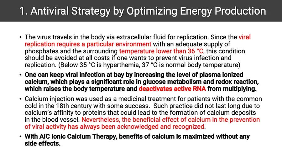
Antiviral Strategy by Optimizing Energy Production The virus travels in the body via extracellular fluid for replication. Since the viral replication requires a particular environment with an adequate supply of phosphates and the surrounding temperature lower than 36 °C, this condition should be avoided at all costs if one wants to prevent virus infection and replication. (Below 35 °C is hyperthemia, 37 °C is normal body temperature) One can keep viral infection at bay by increasing the level of plasma ionized calcium, which plays a significant role in glucose metabolism and redox reaction, which raises the body temperature and deactivates active RNA from multiplying. Calcium injection was used as a medicinal treatment for patients with the common cold in the 18th century with some success. Such practice did not last long due to calcium’s affinity to proteins that could lead to the formation of calcium deposits in the blood vessel. Nevertheless, the beneficial effect of calcium in the prevention of viral activity has always been acknowledged and recognized. With AIC Ionic Calcium Therapy, benefits of calcium is maximized without any side effects.
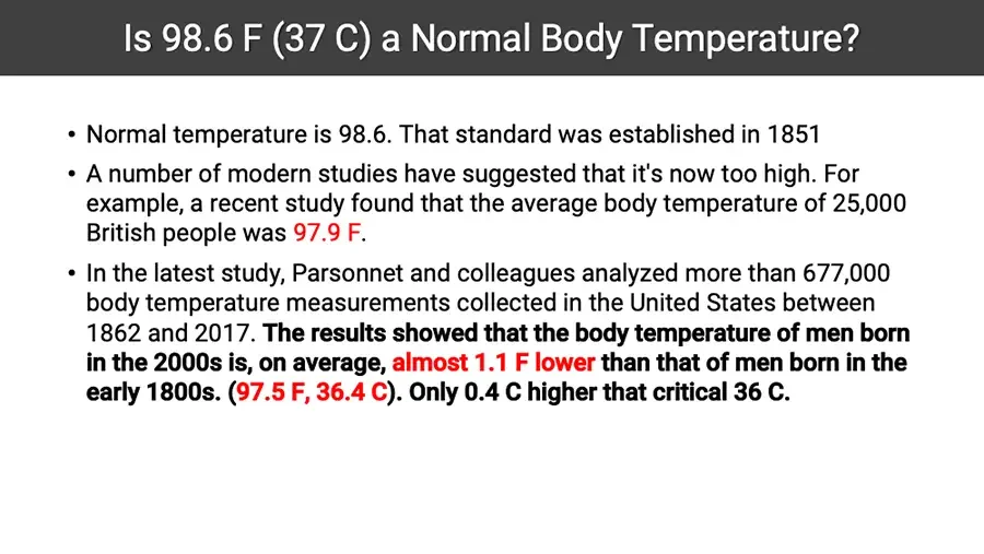
Is 98.6 F (37 C) a Normal Body Temperature? Normal temperature is 98.6. That standard was established in 1851 A number of modern studies have suggested that it's now too high. For example, a recent study found that the average body temperature of 25,000 British people was 97.9 F. In the latest study, Parsonnet and colleagues analyzed more than 677,000 body temperature measurements collected in the United States between 1862 and 2017. The results showed that the body temperature of men born in the 2000s is, on average, almost 1.1 F lower than that of men born in the early 1800s. (97.5 F, 36.4 C). Only 0.4 C higher that critical 36 C.
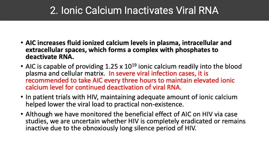
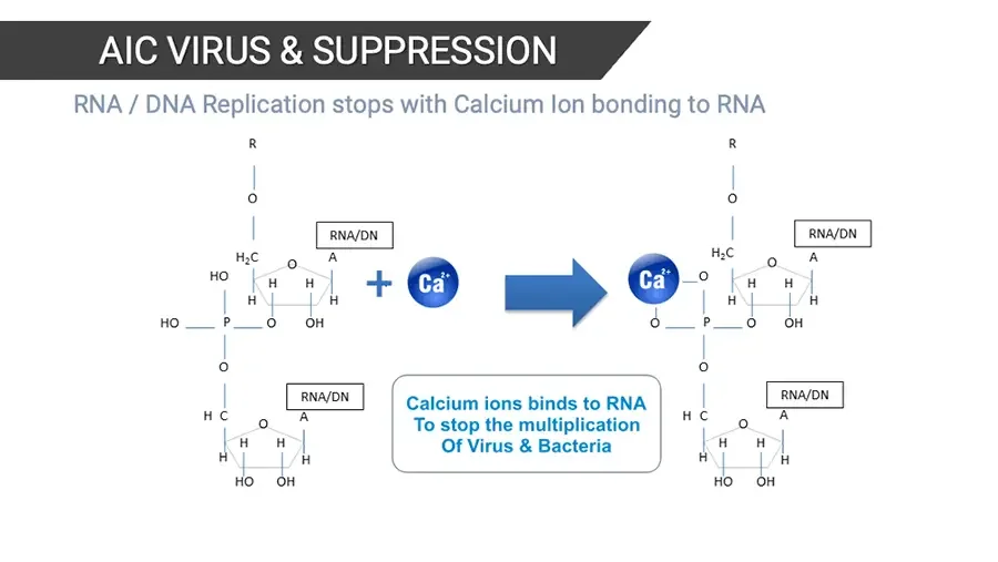
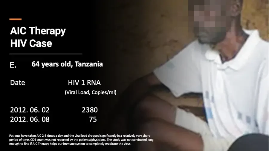
Ionic Calcium Inactivates Viral RNA AIC increases fluid ionized calcium levels in plasma, intracellular and extracellular spaces, which forms a complex with phosphates to deactivate RNA. AIC is capable of providing 1.25 x 10¹⁹ ionic calcium readily into the blood plasma and cellular matrix. In severe viral infection cases, it is recommended to take AIC every three hours to maintain elevated ionic calcium level for continued deactivation of viral RNA. In patient trials with HIV, maintaining adequate amount of ionic calcium helped lower the viral load to practical non-existence. Although we have monitored the beneficial effect of AIC on HIV via case studies, we are uncertain whether HIV is completely eradicated or remains inactive due to the obnoxiously long silence period of HIV.
AIC VIRUS & SUPPRESSION RNA / DNA Replication stops with Calcium Ion bonding to RNA Chemical structure diagram, left: two linked nucleotide units. R O RNA/DN H₂C O A HO H H H H HO P O OH O RNA/DN H C O A H H H H HO OH Ca²⁺ Arrow pointing right. Chemical structure diagram, right: same units with Ca²⁺ bound. R O RNA/DN H₂C O A Ca²⁺ O H H H H O P O OH O RNA/DN H C O A H H H H HO OH Calcium ions binds to RNA To stop the multiplication Of Virus & Bacteria
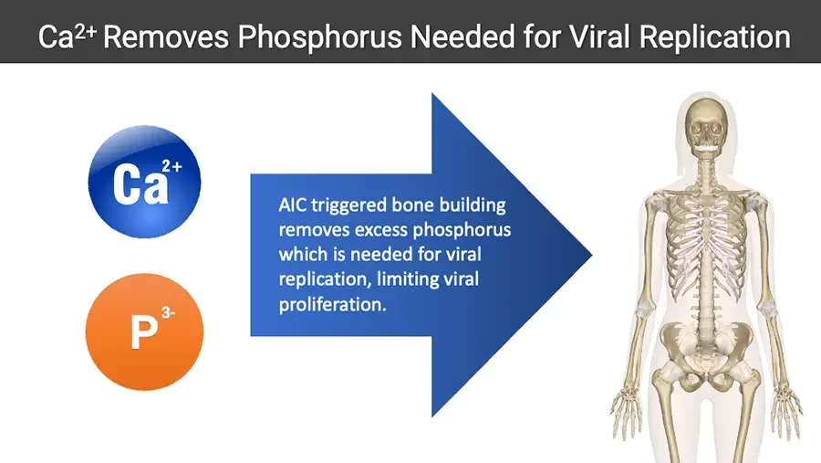
Ca²⁺ Removes Phosphorus Needed for Viral Replication Ca²⁺ P³⁻ Large blue arrow pointing right. AIC triggered bone building removes excess phosphorus which is needed for viral replication, limiting viral proliferation. Illustration: human skeleton within body outline.
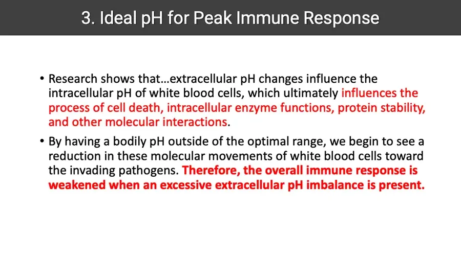
Ideal pH for Peak Immune Response Research shows that…extracellular pH changes influence the intracellular pH of white blood cells, which ultimately influences the process of cell death, intracellular enzyme functions, protein stability, and other molecular interactions. By having a bodily pH outside of the optimal range, we begin to see a reduction in these molecular movements of white blood cells toward the invading pathogens. Therefore, the overall immune response is weakened when an excessive extracellular pH imbalance is present.
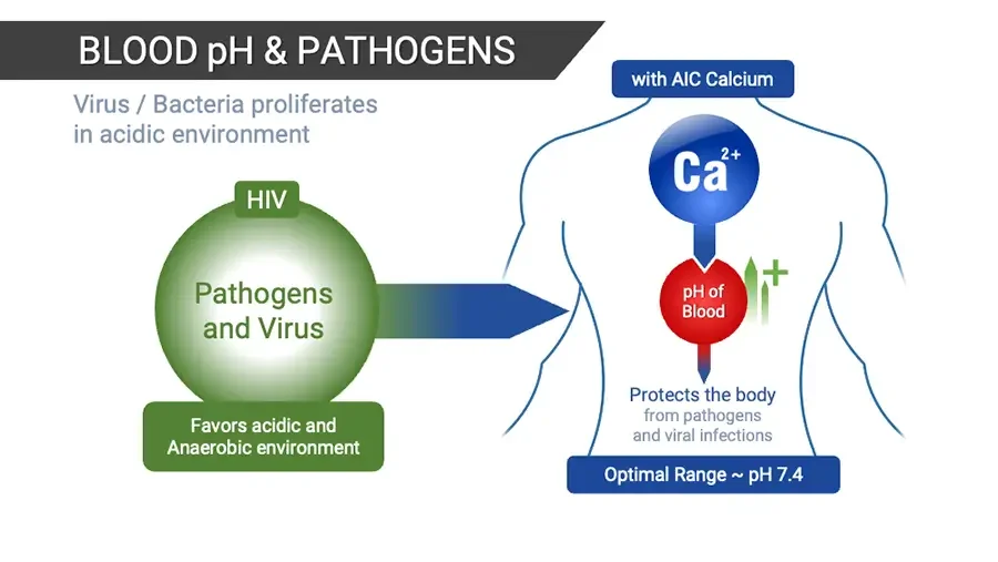
BLOOD pH & PATHOGENS Virus / Bacteria proliferates in acidic environment HIV Pathogens and Virus Favors acidic and Anaerobic environment Arrow pointing right into body outline. with AIC Calcium Ca²⁺ pH of Blood Green upward arrows with plus sign. Protects the body from pathogens and viral infections Optimal Range ~ pH 7.4
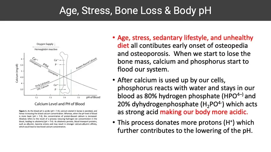
Age, Stress, Bone Loss & Body pH Line chart: calcium level versus blood pH, with bone drawing. Oxygen Supply ↓ Left-pointing arrow. Hemoglobin inactive Calcium (mg/dl) Y-axis. 6 5.5 5 4.5 4 3.5 Ca⁺⁺ Osteoporosis PTH Ca⁺⁺ P⁻⁻⁻ Protein-Bound Calcium Osteopinia TH & Calcitonin SAC Calcium Ca⁺⁺ P⁻⁻⁻ X-axis. 7.2 7.3 7.4 7.5 pH of Blood Calcium Level and PH of Blood Figure 1. As the blood pH is acidic (pH < 7.4), calcium stored in bones is secreted, and hence increasing the blood calcium concentration. Whereas, when the pH level of blood is more basic (pH > 7.4), the concentration of protein-bound calcium is increased. Alkalosis refers to the result of a process reducing hydrogen ion concentration in the blood, leading to alkalemia (pH > 7.4). As alkalemia persists, blood transport proteins, such as albumin, become anions and may result in stronger calcium-albumin affinity, which could lead to low blood calcium concentration. Age, stress, sedantary lifestyle, and unhealthy diet all contibutes early onset of osteopedia and osteoporosis. When we start to lose the bone mass, calcium and phosphorus start to flood our system. After calcium is used up by our cells, phosphorus reacts with water and stays in our blood as 80% hydrogen phosphate (HPO⁴⁻⁾ and 20% dyhydrogenphosphate (H₂PO⁴⁻) which acts as strong acid making our body more acidic. This process donates more protons (H⁺) which further contributes to the lowering of the pH.
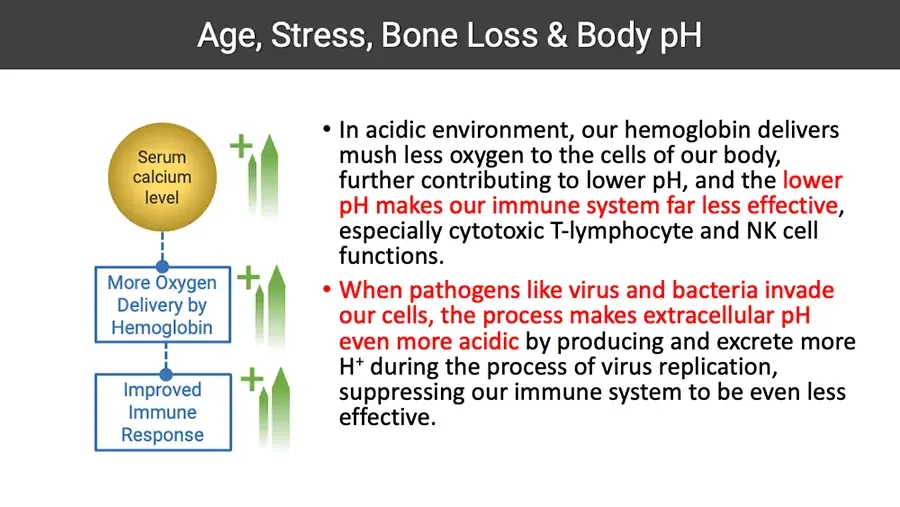
Age, Stress, Bone Loss & Body pH Serum calcium level Green upward arrows with plus sign. More Oxygen Delivery by Hemoglobin Green upward arrows with plus sign. Improved Immune Response Green upward arrows with plus sign. In acidic environment, our hemoglobin delivers mush less oxygen to the cells of our body, further contributing to lower pH, and the lower pH makes our immune system far less effective, especially cytotoxic T-lymphocyte and NK cell functions. When pathogens like virus and bacteria invade our cells, the process makes extracellular pH even more acidic by producing and excrete more H⁺ during the process of virus replication, suppressing our immune system to be even less effective.
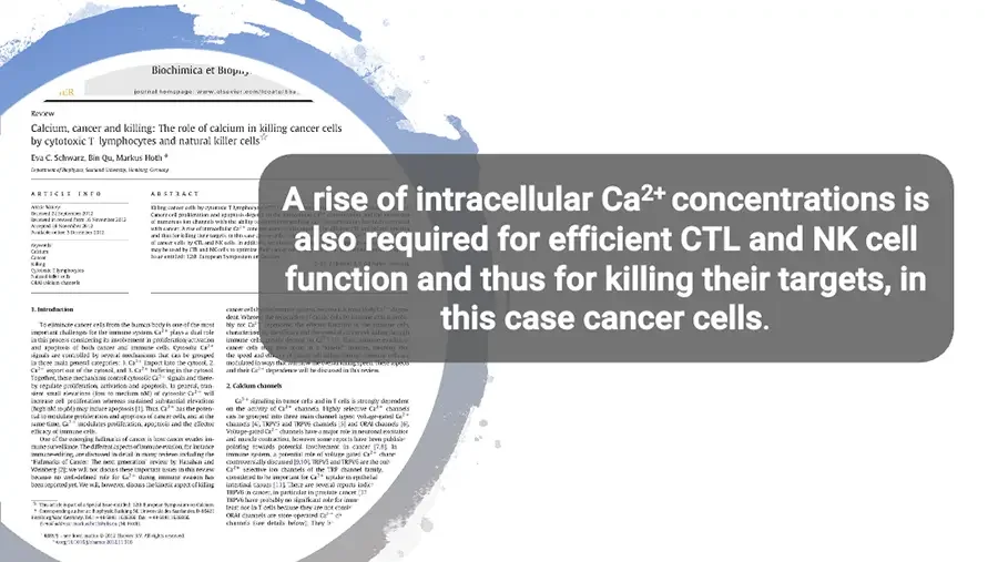
Overlay text box on top of background journal page. A rise of intracellular Ca²⁺ concentrations is also required for efficient CTL and NK cell function and thus for killing their targets, in this case cancer cells. Background: journal article page, partly hidden; small blurred text. Biochimica et Biop cut off by brush stroke. journal homepage: www.elsevier.com/locate/bba Review Calcium, cancer and killing: The role of calcium in killing cancer cells by cytotoxic T lymphocytes and natural killer cells☆ Eva C. Schwarz, Bin Qu, Markus Hoth * Department of Biophysics, Saarland University, Homburg, Germany ARTICLE INFO Article history: Received 21 September 2012 Received in revised form 16 November 2012 Accepted 18 November 2012 Available online 3 December 2012 Keywords: Calcium Cancer Killing Cytotoxic T lymphocytes Natural killer cells ORAI calcium channels ABSTRACT Killing cancer cells by cytotoxic T lymphocytes Cancer cell proliferation and apoptosis depend of numerous ion channels with the ability with cancer. A rise of intracellular Ca²⁺ con and thus for killing their targets, in this case of cancer cells by CTL and NK cells. In addition may be used by CTL and NK cells to optimize Issue entitled: 12th European Symposium on Calcium. Introduction To eliminate cancer cells from the human body is one of the most important challenges for the immune system. Ca²⁺ plays a dual role in this process considering its involvement in proliferation/activation and apoptosis of both cancer and immune cells. Cytosolic Ca²⁺ signals are controlled by several mechanisms that can be grouped in three main general categories: 1. Ca²⁺ import into the cytosol, 2. Ca²⁺ export out of the cytosol, and 3. Ca²⁺ buffering in the cytosol. Together, these mechanisms control cytosolic Ca²⁺ signals and thereby regulate proliferation, activation and apoptosis. In general, transient small elevations (low to medium nM) of cytosolic Ca²⁺ will increase cell proliferation whereas sustained substantial elevations (high nM to µM) may induce apoptosis 1.. Thus, Ca²⁺ has the potential to modulate proliferation and apoptosis of cancer cells, and at the same time, Ca²⁺ modulates proliferation, apoptosis and the effector efficacy of immune cells. One of the emerging hallmarks of cancer is how cancer evades immune surveillance. The different aspects of immune evasion, for instance immunoediting, are discussed in detail in many reviews including the 'Hallmarks of Cancer: The next generation' review by Hanahan and Weinberg 2.; we will not discuss these important issues in this review because no well-defined role for Ca²⁺ during immune evasion has been reported yet. We will, however, discuss the kinetic aspect of killing that the overall killing speed. These aspects dependence will be discussed in this review. Calcium channels Ca²⁺ signaling in tumor cells and in T cells is strongly dependent on the activity of Ca²⁺ channels. Highly selective Ca²⁺ channels can be grouped into three main channel types: Voltage-gated Ca²⁺ channels 4. , TRPV5 and TRPV6 channels 5. and ORAI channels 6. . Voltage-gated Ca²⁺ channels have a major role in neuronal excitation and muscle contractions, however some reports have been published pointing towards potential involvement in cancer 7,8.. In immune system, a potential role of voltage gated Ca²⁺ channels is controversially discussed . TRPV5 and TRPV6 are the only Ca²⁺ selective TRP channel family, considered to be important for Ca²⁺ uptake in epithelial intestinal cells . There are several reports linking TRPV6 in cancer, in particular in prostate cancer TRPV6 have probably no significant role for immune least not in T cells because they are not ORAI channels are store-operated Ca²⁺ channels (see details below). They ☆ This article is part of a Special Issue entitled: 12th European Symposium on Calcium. Corresponding author at: Germany. Tel.: ; fax: E-mail address: (M. Hoth). – see front matter © 2012 Elsevier B.V. All rights reserved.
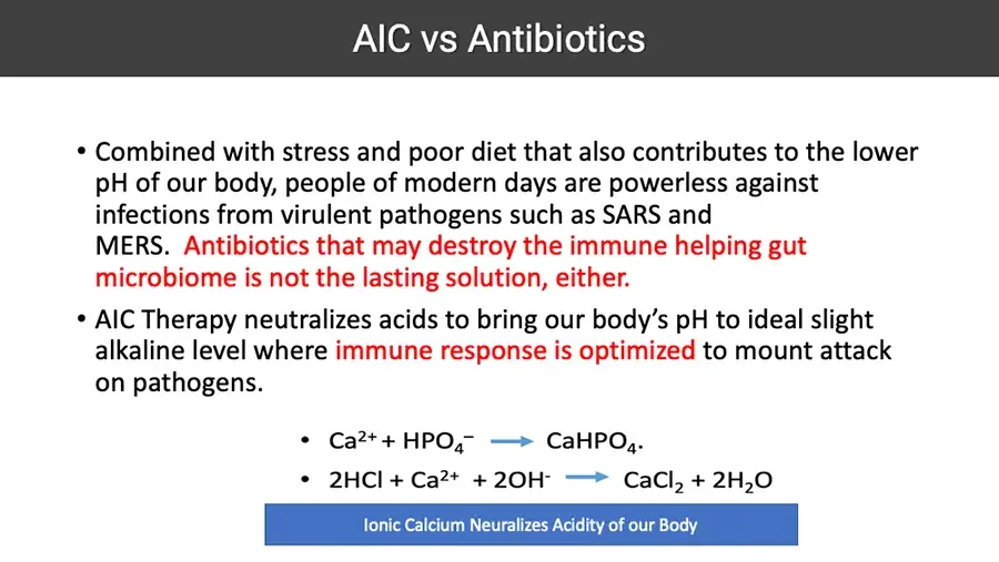
AIC vs Antibiotics Combined with stress and poor diet that also contributes to the lower pH of our body, people of modern days are powerless against infections from virulent pathogens such as SARS and MERS. Antibiotics that may destroy the immune helping gut microbiome is not the lasting solution, either. AIC Therapy neutralizes acids to bring our body’s pH to ideal slight alkaline level where immune response is optimized to mount attack on pathogens. Ca²⁺ + HPO₄⁻ → CaHPO₄. 2HCl + Ca²⁺ + 2OH⁻ → CaCl₂ + 2H₂O Ionic Calcium Neuralizes Acidity of our Body
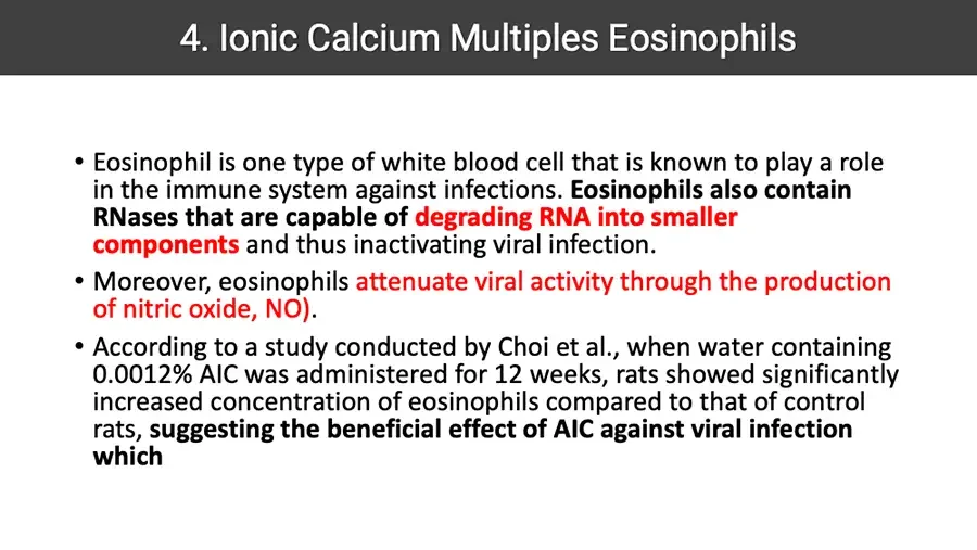
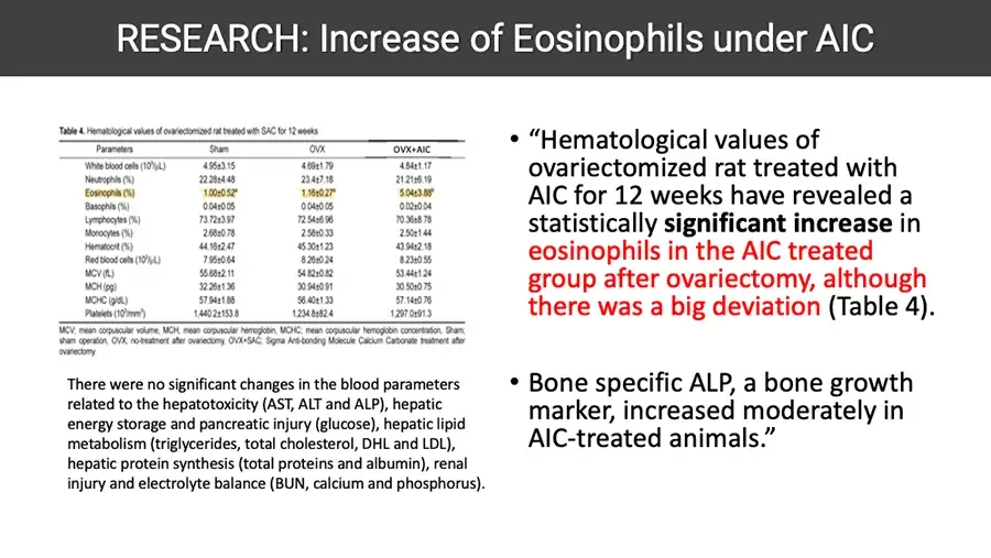
Ionic Calcium Multiples Eosinophils Eosinophil is one type of white blood cell that is known to play a role in the immune system against infections. Eosinophils also contain RNases that are capable of degrading RNA into smaller components and thus inactivating viral infection. Moreover, eosinophils attenuate viral activity through the production of nitric oxide, NO). According to a study conducted by Choi et al., when water containing 0.0012% AIC was administered for 12 weeks, rats showed significantly increased concentration of eosinophils compared to that of control rats, suggesting the beneficial effect of AIC against viral infection which
RESEARCH: Increase of Eosinophils under AIC Table 4. Hematological values of ovariectomized rat treated with SAC for 12 weeks Eosinophils row highlighted in yellow. Parameters Sham OVX OVX+AIC White blood cells (10³/µL) 4.95±3.15 4.69±1.79 4.84±1.17 Neutrophils (%) 22.28±4.48 23.4±7.18 21.21±6.19 Eosinophils (%) 1.00±0.52ᵃ 1.16±0.27ᵃ 5.04±3.88ᵇ Basophils (%) 0.04±0.05 0.04±0.05 0.02±0.04 Lymphocytes (%) 73.72±3.97 72.54±6.96 70.36±8.78 Monocytes (%) 2.68±0.78 2.58±0.33 2.50±1.44 Hematocrit (%) 44.16±2.47 45.30±1.23 43.94±2.18 Red blood cells (10⁶/µL) 7.95±0.64 8.26±0.24 8.23±0.55 MCV (fL) 55.68±2.11 54.82±0.82 53.44±1.24 MCH (pg) 32.26±1.36 30.94±0.91 30.50±0.75 MCHC (g/dL) 57.94±1.88 56.40±1.33 57.14±0.76 Platelets (10³/mm³) 1,440.2±153.8 1,234.8±82.4 1,297.0±91.3 MCV; mean corpuscular volume, MCH; mean corpuscular hemoglobin, MCHC; mean corpuscular hemoglobin concentration, Sham; sham operation, OVX; no-treatment after ovariectomy, OVX+SAC; Sigma Anti-bonding Molecule Calcium Carbonate treatment after ovariectomy. There were no significant changes in the blood parameters related to the hepatotoxicity (AST, ALT and ALP), hepatic energy storage and pancreatic injury (glucose), hepatic lipid metabolism (triglycerides, total cholesterol, DHL and LDL), hepatic protein synthesis (total proteins and albumin), renal injury and electrolyte balance (BUN, calcium and phosphorus). “Hematological values of ovariectomized rat treated with AIC for 12 weeks have revealed a statistically significant increase in eosinophils in the AIC treated group after ovariectomy, although there was a big deviation (Table 4). Bone specific ALP, a bone growth marker, increased moderately in AIC-treated animals.”
RESEARCH: Eosinophils Reduce Viral Infectivity “However, experiments carried out in vitro and in vivo suggest positive roles for eosinophils, as they have been shown to reduce virus infectivity in tissue culture and promote clearance of the human pathogen, respiratory syncytial virus (RSV) in a mouse challenge model.” “When eosinophils were added in increasing concentration to a fixed number of viruses, dose-dependent inhibition of virus infectivity was observed” “Thus, not only is this another clear demonstration of antiviral effects of mouse eosinophils in vivo, but the first suggestion of a role for TLRs – specifically TLR7, in promoting eosinophil-mediated antiviral activity” Journal article screenshot; right edge cut off, lines kept as shown. ] NIH Public Access Author Manuscript Immunol Res. Author manuscript; available in PMC 2009 November 16. Published in final edited form as: Immunol Res. 2009 ; 43(1-3): 128–137. doi:10.1007/s12026-008-8058-5. Eosinophils and their Interactions with Respiratory Pathogens Helene F. Rosenberg¹,³, Kimberly D. Dyer¹, and Joseph B. Domachowsl cut off. ¹ Eosinophil Biology Section, Laboratory of Allergic Diseases, National Institut cut off. Infectious Diseases, National Institutes of Health, Bethesda, Maryland ² Depa cut off. SUNY Upstate Medical University, Syracuse, New York Abstract Eosinophils are implicated in the pathophysiology of respiratory virus infection, cut off. negative roles, such as promoting wheezing and bronchoconstriction in conjuncti cut off. induced exacerbations of reactive airways disease and in association with aberra cut off. responses to antiviral vaccines. However, experiments carried out in vitro and in vi cut off. roles for eosinophils, as they have been shown to reduce virus infectivity in tissue c cut off. clearance of the human pathogen, respiratory syncytial virus (RSV) in a mouse cha cut off. related natural rodent pathogen, pneumonia virus of mice (PVM) is highly virule cut off. not readily cleared by eosinophils in vivo. Interestingly, PVM replicates in eosino cut off. cytokine release. The molecular basis of virus infection in eosinophils and its rela cut off. outcome is currently under study.
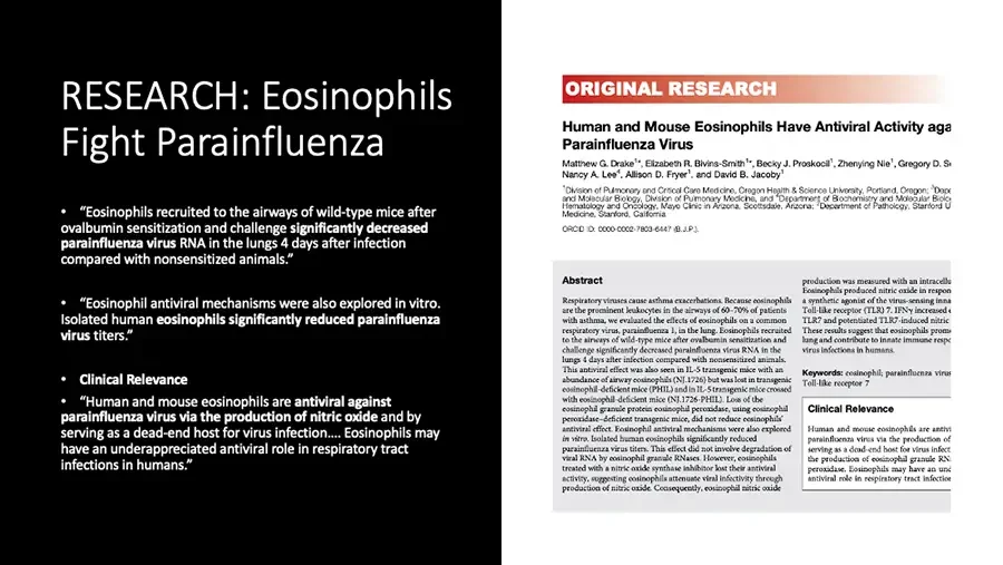
RESEARCH: Eosinophils Fight Parainfluenza “Eosinophils recruited to the airways of wild-type mice after ovalbumin sensitization and challenge significantly decreased parainfluenza virus RNA in the lungs 4 days after infection compared with nonsensitized animals.” “Eosinophil antiviral mechanisms were also explored in vitro. Isolated human eosinophils significantly reduced parainfluenza virus titers.” Clinical Relevance “Human and mouse eosinophils are antiviral against parainfluenza virus via the production of nitric oxide and by serving as a dead-end host for virus infection…. Eosinophils may have an underappreciated antiviral role in respiratory tract infections in humans.” Journal article screenshot; right edge cut off, lines kept as shown. ORIGINAL RESEARCH Human and Mouse Eosinophils Have Antiviral Activity aga cut off. Parainfluenza Virus Matthew G. Drake¹*, Elizabeth R. Bivins-Smith¹*, Becky J. Proskocil¹, Zhenying Nie¹, Gregory D. S cut off. Nancy A. Lee⁴, Allison D. Fryer¹, and David B. Jacoby¹ ¹Division of Pulmonary and Critical Care Medicine, Oregon Health & Science University, Portland, Oregon; ³Depa cut off. and Molecular Biology, Division of Pulmonary Medicine, and ⁴Department of Biochemistry and Molecular Biolo cut off. Hematology and Oncology, Mayo Clinic in Arizona, Scottsdale, Arizona; ⁵Department of Pathology, Stanford U cut off. Medicine, Stanford, California ORCID ID: 0000-0002-7803-6447 (B.J.P.). Abstract Respiratory viruses cause asthma exacerbations. Because eosinophils are the prominent leukocytes in the airways of 60–70% of patients with asthma, we evaluated the effects of eosinophils on a common respiratory virus, parainfluenza 1, in the lung. Eosinophils recruited to the airways of wild-type mice after ovalbumin sensitization and challenge significantly decreased parainfluenza virus RNA in the lungs 4 days after infection compared with nonsensitized animals. This antiviral effect was also seen in IL-5 transgenic mice with an abundance of airway eosinophils (NJ.1726) but was lost in transgenic eosinophil-deficient mice (PHIL) and in IL-5 transgenic mice crossed with eosinophil-deficient mice (NJ.1726-PHIL). Loss of the eosinophil granule protein eosinophil peroxidase, using eosinophil peroxidase–deficient transgenic mice, did not reduce eosinophils’ antiviral effect. Eosinophil antiviral mechanisms were also explored in vitro. Isolated human eosinophils significantly reduced parainfluenza virus titers. This effect did not involve degradation of viral RNA by eosinophil granule RNases. However, eosinophils treated with a nitric oxide synthase inhibitor lost their antiviral activity, suggesting eosinophils attenuate viral infectivity through production of nitric oxide. Consequently, eosinophil nitric oxide production was measured with an intracellu cut off. Eosinophils produced nitric oxide in respon cut off. a synthetic agonist of the virus-sensing inna cut off. Toll-like receptor (TLR) 7. IFNγ increased e cut off. TLR7 and potentiated TLR7-induced nitric cut off. These results suggest that eosinophils prom cut off. lung and contribute to innate immune respo cut off. virus infections in humans. Keywords: eosinophil; parainfluenza virus cut off. Toll-like receptor 7 Clinical Relevance Human and mouse eosinophils are antivi cut off. parainfluenza virus via the production of cut off. serving as a dead-end host for virus infect cut off. the production of eosinophil granule RN cut off. peroxidase. Eosinophils may have an und cut off. antiviral role in respiratory tract infectio cut off.

HIV Case Studies

  • AIC Therapy HIV Case E., 64 years old, Tanzania Date HIV 1 RNA (Viral Load, Copies/ml) 2012. 06. 02 2380 2012. 06. 08 75 Patients have taken AIC 2-3 times a day and the viral load dropped significantly in a relatively very short period of time. CD4 count was not reported by the patients/physicians. The study was not conducted long enough to find if AIC Therapy helps our immune system to completely eradicate the virus. Photo: older man in white shirt, seated.
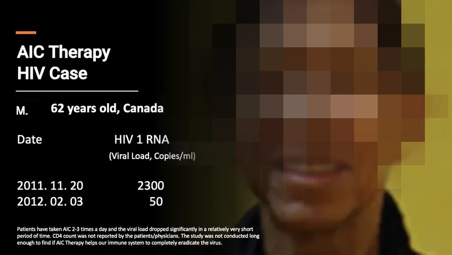
  • AIC Therapy HIV Case M., 62 years old, Canada Date HIV 1 RNA (Viral Load, Copies/ml) 2011. 11. 20 2300 2012. 02. 03 50 Patients have taken AIC 2-3 times a day and the viral load dropped significantly in a relatively very short period of time. CD4 count was not reported by the patients/physicians. The study was not conducted long enough to find if AIC Therapy helps our immune system to completely eradicate the virus. Photo: smiling man, eyes pixelated.
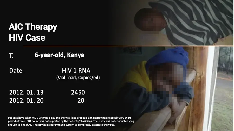
  • AIC Therapy HIV Case T., 6-year-old, Kenya Date HIV 1 RNA (Vial Load, Copies/ml) 2012. 01. 13 2450 2012. 01. 20 20 Patients have taken AIC 2-3 times a day and the viral load dropped significantly in a relatively very short period of time. CD4 count was not reported by the patients/physicians. The study was not conducted long enough to find if AIC Therapy helps our immune system to completely eradicate the virus. Photo: child lying under striped blanket. Photo: child in blue hat leaning on wooden railing.

AIC In Lyme Disease

  • Photo: tick over faint molecular-structure background. AIC Ionic Calcium Therapy Lyme Disease
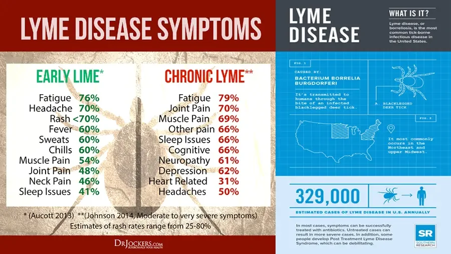
  • LYME DISEASE SYMPTOMS Faint tick drawing in background. EARLY LIME* Fatigue 76% Headache 70% Rash <70% Fever 60% Sweats 60% Chills 60% Muscle Pain 54% Joint Pain 48% Neck Pain 46% Sleep Issues 41% CHRONIC LYME** Fatigue 79% Joint Pain 70% Muscle Pain 69% Other pain 66% Sleep Issues 66% Cognitive 66% Neuropathy 61% Depression 62% Heart Related 31% Headaches 50% (Aucott 2013) **(Johnson 2014, Moderate to very severe symptoms) Estimates of rash rates range from 25-80% DrJockers.com SUPERCHARGE YOUR HEALTH LYME DISEASE WHAT IS IT? Lyme disease, or borreliosis, is the most common tick-borne infectious disease in the United States. FIG. 1 CAUSED BY: BACTERIUM BORRELIA BURGDORFERI It’s transmitted to humans through the bite of an infected blacklegged deer tick. Line drawing: tick. A. BLACKLEGGED DEER TICK FIG. 2 Map: United States with Northeast and upper Midwest hatched. Location pin icon. It most commonly occurs in the Northeast and upper Midwest. 329,000 Icons: tick, arrow, person. ESTIMATED CASES OF LYME DISEASE IN U.S. ANNUALLY In most cases, symptoms can be successfully treated with antibiotics. Untreated cases can result in more severe cases. In addition, some people develop Post Treatment Lyme Disease Syndrome, which can be debilitating. SR SOUTHERN RESEARCH
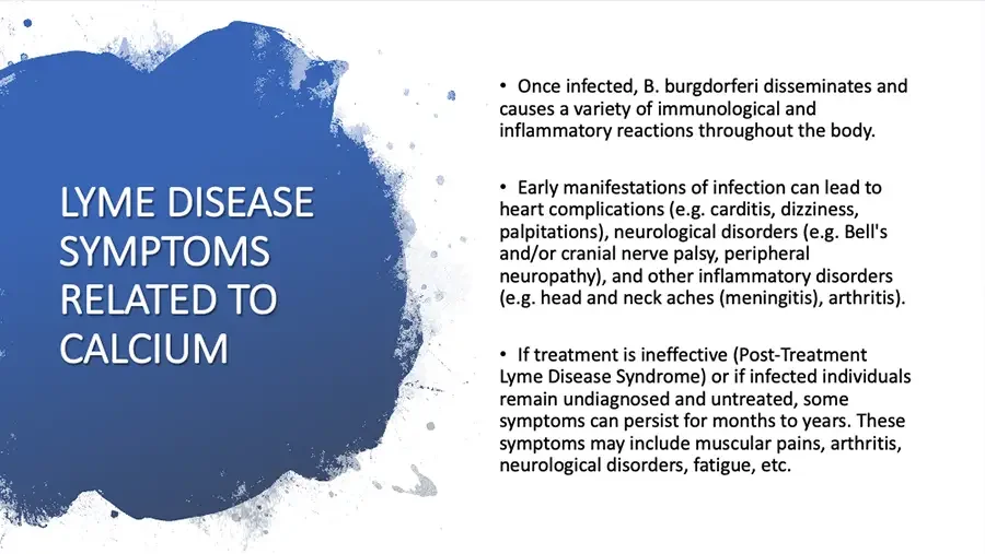
  • Blue paint-splash shape behind heading. LYME DISEASE SYMPTOMS RELATED TO CALCIUM Once infected, B. burgdorferi disseminates and causes a variety of immunological and inflammatory reactions throughout the body. Early manifestations of infection can lead to heart complications (e.g. carditis, dizziness, palpitations), neurological disorders (e.g. Bell's and/or cranial nerve palsy, peripheral neuropathy), and other inflammatory disorders (e.g. head and neck aches (meningitis), arthritis). If treatment is ineffective (Post-Treatment Lyme Disease Syndrome) or if infected individuals remain undiagnosed and untreated, some symptoms can persist for months to years. These symptoms may include muscular pains, arthritis, neurological disorders, fatigue, etc.
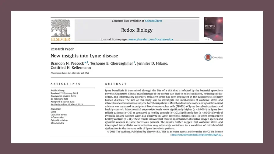
  • Screenshot of journal article first page on a dark mauve frame. Elsevier tree logo. ELSEVIER Contents lists available at ScienceDirect Redox Biology journal homepage: www.elsevier.com/locate/redox Journal cover thumbnail. REDOX BIOLOGY Research Paper New insights into Lyme disease CrossMark logo. CrossMark Brandon N. Peacock*,¹, Teshome B. Gherezghiher¹, Jennifer D. Hilario, Gottfried H. Kellermann Pharmasan Labs, Inc., Osceola, WI, USA ARTICLE INFO Article history: Received 12 February 2015 Received in revised form 26 February 2015 Accepted 4 March 2015 Available online 16 March 2015 Keywords: Lyme Oxidative stress Inflammation Cytosolic calcium Mitochondria ABSTRACT Lyme borreliosis is transmitted through the bite of a tick that is infected by the bacterial spirochete Borrelia burgdorferi. Clinical manifestation of the disease can lead to heart conditions, neurological disorders, and inflammatory disorders. Oxidative stress has been implicated in the pathogenesis of many human diseases. The aim of this study was to investigate the mechanisms of oxidative stress and intracellular communication in Lyme borreliosis patients. Mitochondrial superoxide and cytosolic ionized calcium was measured in peripheral blood mononuclear cells (PBMCs) of Lyme borreliosis patients and healthy controls. Mitochondrial superoxide levels were significantly higher (p < 0.0001) in Lyme borreliosis patients (n=32) as compared to healthy controls (n=30). Significantly low (p < 0.0001) levels of cytosolic ionized calcium were also observed in Lyme borreliosis patients (n=11) when compared to healthy controls (n=11). These results indicate that there is an imbalance of reactive oxygen species and cytosolic calcium in Lyme borreliosis patients. The results further suggest that oxidative stress and interrupted intracellular communication may ultimately contribute to a condition of mitochondrial dysfunction in the immune cells of Lyme borreliosis patients. © 2015 The Authors. Published by Elsevier B.V. This is an open access article under the CC BY license (http://creativecommons.org/licenses/by/4.0/).
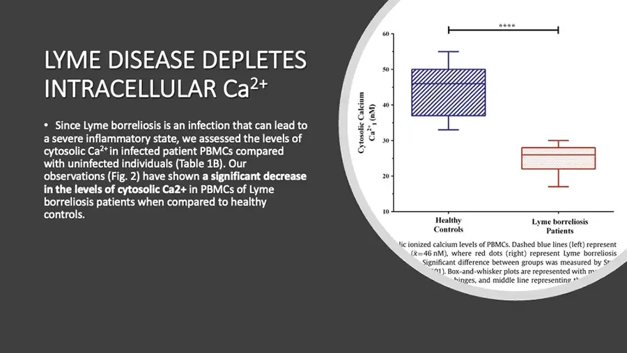
  • LYME DISEASE DEPLETES INTRACELLULAR Ca²⁺ Since Lyme borreliosis is an infection that can lead to a severe inflammatory state, we assessed the levels of cytosolic Ca²⁺ in infected patient PBMCs compared with uninfected individuals (Table 1B). Our observations (Fig. 2) have shown a significant decrease in the levels of cytosolic Ca2+ in PBMCs of Lyme borreliosis patients when compared to healthy controls. Box plot from journal figure, circular crop, edges cut off. Cytosolic Calcium Ca²⁺ᵢ (nM) Y-axis. 60 50 40 30 20 10 Healthy Controls Lyme borreliosis Patients lic ionized calcium levels of PBMCs. Dashed blue lines (left) represent cut off at both ends. (x̄=46 nM), where red dots (right) represent Lyme borreliosis cut off. Significant difference between groups was measured by St cut off at both ends. 01). Box-and-whisker plots are represented with m cut off at both ends. hinges, and middle line representing th cut off at both ends.
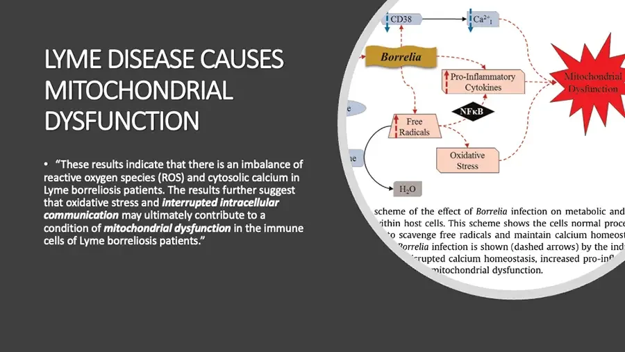
  • LYME DISEASE CAUSES MITOCHONDRIAL DYSFUNCTION “These results indicate that there is an imbalance of reactive oxygen species (ROS) and cytosolic calcium in Lyme borreliosis patients. The results further suggest that oxidative stress and interrupted intracellular communication may ultimately contribute to a condition of mitochondrial dysfunction in the immune cells of Lyme borreliosis patients.” Pathway diagram from journal figure, circular crop, edges cut off. CD38 Ca²⁺ᵢ Borrelia Mitochondrial Dysfunction Pro-Inflammatory Cytokines e label cut off at circle edge. NFκB Free Radicals ne label cut off at circle edge. Oxidative Stress H₂O scheme of the effect of Borrelia infection on metabolic and cut off at both ends. ithin host cells. This scheme shows the cells normal proce cut off at both ends. to scavenge free radicals and maintain calcium homeost cut off at both ends. Borrelia infection is shown (dashed arrows) by the ind cut off at both ends. rupted calcium homeostasis, increased pro-inf cut off at both ends. mitochondrial dysfunction. cut off at start.
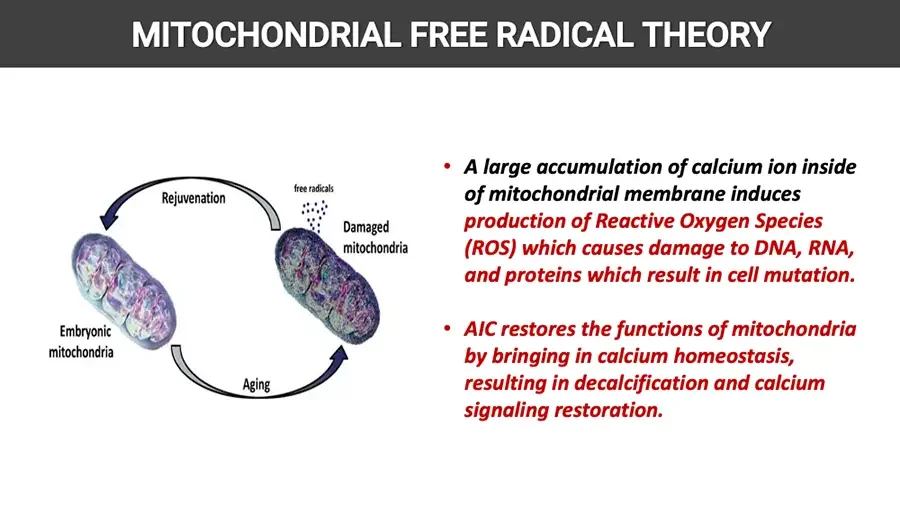
  • MITOCHONDRIAL FREE RADICAL THEORY Cycle diagram: two mitochondria linked by curved arrows. Rejuvenation free radicals Damaged mitochondria Embryonic mitochondria Aging A large accumulation of calcium ion inside of mitochondrial membrane induces production of Reactive Oxygen Species (ROS) which causes damage to DNA, RNA, and proteins which result in cell mutation. AIC restores the functions of mitochondria by bringing in calcium homeostasis, resulting in decalcification and calcium signaling restoration.
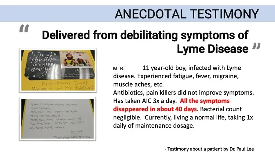
  • ANECDOTAL TESTIMONY “Delivered from debilitating symptoms of Lyme Disease” Photo: Christmas card with photo of two children, handwritten note. MERRY christmas & a HAPPY new year Jesus Christ is the today and forever. small print on card, partly hidden by glare. Happy Birthday, Jesus♡ We are so glad it's Christmas! Photo: handwritten letter on gray paper. M. K.. 11 year-old boy, infected with Lyme disease. Experienced fatigue, fever, migraine, muscle aches, etc. Antibiotics, pain killers did not improve symptoms. Has taken AIC 3x a day. All the symptoms disappeared in about 40 days. Bacterial count negligible. Currently, living a normal life, taking 1x daily of maintenance dosage. Testimony about a patient by Dr. Paul Lee

AIC In COVID Cases

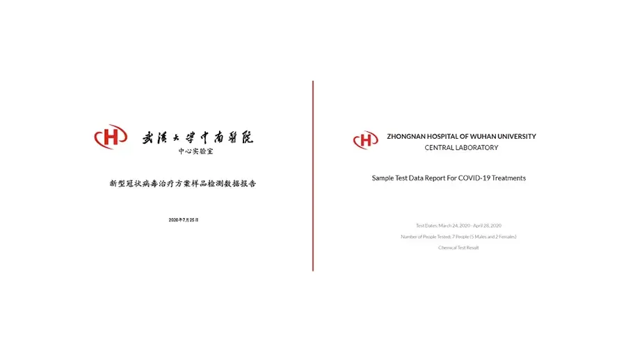
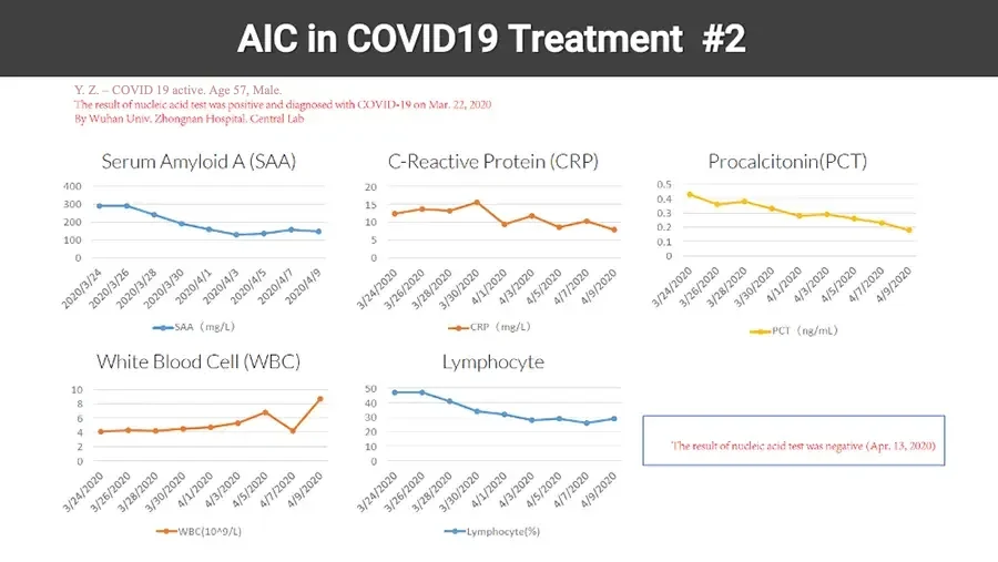
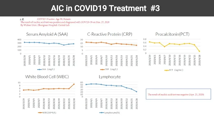
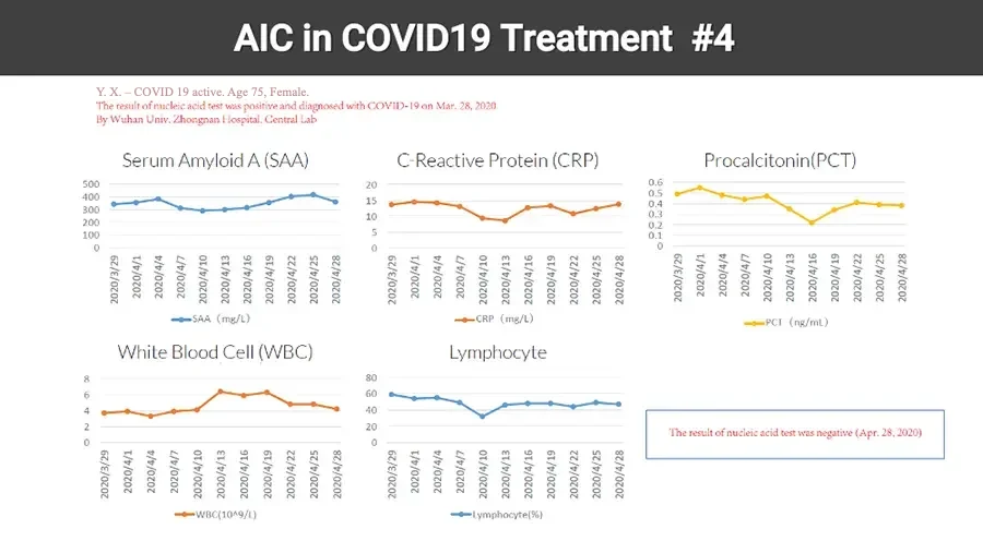
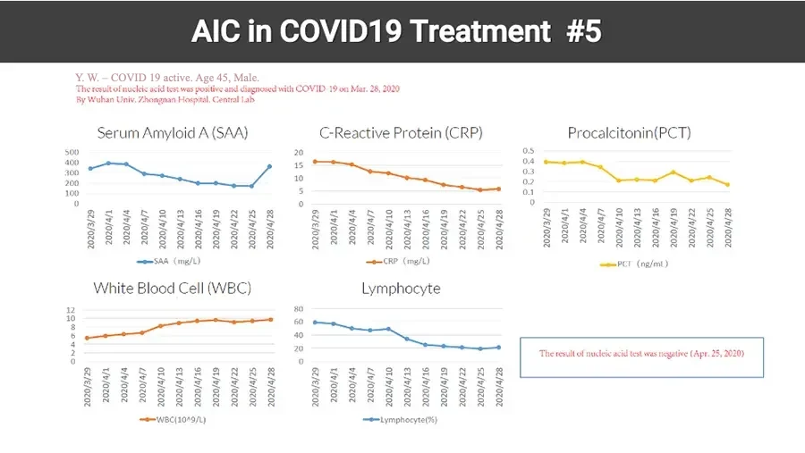
  • Two report cover pages side by side, red divider line. Logo: red H with curved swoosh. 武漢大學中南醫院 calligraphy. 中心实验室 新型冠状病毒治疗方案样品检测数据报告 2020年7月25日 Logo: red H with curved swoosh. ZHONGNAN HOSPITAL OF WUHAN UNIVERSITY CENTRAL LABORATORY Sample Test Data Report For COVID-19 Treatments Test Dates: March 24, 2020 - April 28, 2020 Number of People Tested: 7 People (5 Males and 2 Females) Chemical Test Result
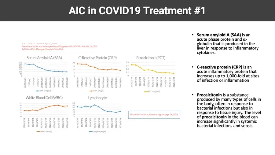
  • AIC in COVID19 Treatment #1 Z. F. - COVID 19 active. Age 27, Male. The result of nucleic acid test was positive and diagnosed with COVID-19 on Mar. 28, 2020 By Wuhan Univ. Zhongnan Hospital, Central Lab Serum Amyloid A (SAA) Line chart, decreasing. Y-axis. 300 250 200 150 100 50 0 X-axis. 2020/3/29 2020/4/1 2020/4/4 2020/4/7 2020/4/10 2020/4/13 2020/4/16 2020/4/19 2020/4/22 2020/4/25 2020/4/28 SAA (mg/L) C-Reactive Protein (CRP) Line chart, decreasing. Y-axis. 20 15 10 5 0 X-axis. 2020/3/29 2020/4/1 2020/4/4 2020/4/7 2020/4/10 2020/4/13 2020/4/16 2020/4/19 2020/4/22 2020/4/25 2020/4/28 CRP (mg/L) Procalcitonin(PCT) Line chart, decreasing. Y-axis. 0.8 0.6 0.4 0.2 0 X-axis. 2020/3/29 2020/4/1 2020/4/4 2020/4/7 2020/4/10 2020/4/13 2020/4/16 2020/4/19 2020/4/22 2020/4/25 2020/4/28 PCT (ng/mL) White Blood Cell (WBC) Line chart, increasing. Y-axis. 12 10 8 6 4 2 0 X-axis. 2020/3/29 2020/4/1 2020/4/4 2020/4/7 2020/4/10 2020/4/13 2020/4/16 2020/4/19 2020/4/22 2020/4/25 2020/4/28 WBC(10^9/L) Lymphocyte Line chart, decreasing. Y-axis. 60 50 40 30 20 10 0 X-axis. 2020/3/29 2020/4/1 2020/4/4 2020/4/7 2020/4/10 2020/4/13 2020/4/16 2020/4/19 2020/4/22 2020/4/25 2020/4/28 Lymphocyte(%) The result of nucleic acid test was negative (Apr. 28, 2020) Serum amyloid A (SAA) is an acute phase protein and α-globulin that is produced in the liver in response to inflammatory cytokines. C-reactive protein (CRP) is an acute inflammatory protein that increases up to 1,000-fold at sites of infection or inflammation Procalcitonin is a substance produced by many types of cells in the body, often in response to bacterial infections but also in response to tissue injury. The level of procalcitonin in the blood can increase significantly in systemic bacterial infections and sepsis.
  • AIC in COVID19 Treatment #2 Y. Z. - COVID 19 active. Age 57, Male. The result of nucleic acid test was positive and diagnosed with COVID-19 on Mar. 22, 2020 By Wuhan Univ. Zhongnan Hospital, Central Lab Serum Amyloid A (SAA) Line chart. Y-axis. 400 300 200 100 0 X-axis. 2020/3/24 2020/3/26 2020/3/28 2020/3/30 2020/4/1 2020/4/3 2020/4/5 2020/4/7 2020/4/9 SAA (mg/L) C-Reactive Protein (CRP) Line chart. Y-axis. 20 15 10 5 0 X-axis. 3/24/2020 3/26/2020 3/28/2020 3/30/2020 4/1/2020 4/3/2020 4/5/2020 4/7/2020 4/9/2020 CRP (mg/L) Procalcitonin(PCT) Line chart. Y-axis. 0.5 0.4 0.3 0.2 0.1 0 X-axis. 3/24/2020 3/26/2020 3/28/2020 3/30/2020 4/1/2020 4/3/2020 4/5/2020 4/7/2020 4/9/2020 PCT (ng/mL) White Blood Cell (WBC) Line chart. Y-axis. 10 8 6 4 2 0 X-axis. 3/24/2020 3/26/2020 3/28/2020 3/30/2020 4/1/2020 4/3/2020 4/5/2020 4/7/2020 4/9/2020 WBC(10^9/L) Lymphocyte Line chart. Y-axis. 50 40 30 20 10 0 X-axis. 3/24/2020 3/26/2020 3/28/2020 3/30/2020 4/1/2020 4/3/2020 4/5/2020 4/7/2020 4/9/2020 Lymphocyte(%) The result of nucleic acid test was negative (Apr. 13, 2020)
  • AIC in COVID19 Treatment #3 Jingfang Zhang - COVID 19 active. Age 58, Female. The result of nucleic acid test was positive and diagnosed with COVID-19 on Mar. 27, 2020 By Wuhan Univ. Zhongnan Hospital, Central Lab Serum Amyloid A (SAA) Line chart. Y-axis. 400 300 200 100 0 X-axis. 2020/3/29 2020/4/1 2020/4/4 2020/4/7 2020/4/10 2020/4/13 2020/4/16 2020/4/19 2020/4/22 2020/4/25 2020/4/28 SAA (mg/L) C-Reactive Protein (CRP) Line chart. Y-axis. 20 15 10 5 0 X-axis. 2020/3/29 2020/4/1 2020/4/4 2020/4/7 2020/4/10 2020/4/13 2020/4/16 2020/4/19 2020/4/22 2020/4/25 2020/4/28 CRP (mg/L) Procalcitonin(PCT) Line chart. Y-axis. 0.8 0.6 0.4 0.2 0 X-axis. 2020/3/29 2020/4/1 2020/4/4 2020/4/7 2020/4/10 2020/4/13 2020/4/16 2020/4/19 2020/4/22 2020/4/25 2020/4/28 PCT (ng/mL) White Blood Cell (WBC) Line chart. Y-axis. 10 8 6 4 2 0 X-axis. 2020/3/29 2020/4/1 2020/4/4 2020/4/7 2020/4/10 2020/4/13 2020/4/16 2020/4/19 2020/4/22 2020/4/25 2020/4/28 WBC(10^9/L) Lymphocyte Line chart. Y-axis. 60 50 40 30 20 10 0 X-axis. 2020/3/29 2020/4/1 2020/4/4 2020/4/7 2020/4/10 2020/4/13 2020/4/16 2020/4/19 2020/4/22 2020/4/25 2020/4/28 Lymphocyte(%) The result of nucleic acid test was negative (Apr. 25, 2020)
  • AIC in COVID19 Treatment #4 Y. X. - COVID 19 active. Age 75, Female. The result of nucleic acid test was positive and diagnosed with COVID-19 on Mar. 28, 2020 By Wuhan Univ. Zhongnan Hospital, Central Lab Serum Amyloid A (SAA) Line chart. Y-axis. 500 400 300 200 100 0 X-axis. 2020/3/29 2020/4/1 2020/4/4 2020/4/7 2020/4/10 2020/4/13 2020/4/16 2020/4/19 2020/4/22 2020/4/25 2020/4/28 SAA (mg/L) C-Reactive Protein (CRP) Line chart. Y-axis. 20 15 10 5 0 X-axis. 2020/3/29 2020/4/1 2020/4/4 2020/4/7 2020/4/10 2020/4/13 2020/4/16 2020/4/19 2020/4/22 2020/4/25 2020/4/28 CRP (mg/L) Procalcitonin(PCT) Line chart. Y-axis. 0.6 0.5 0.4 0.3 0.2 0.1 0 X-axis. 2020/3/29 2020/4/1 2020/4/4 2020/4/7 2020/4/10 2020/4/13 2020/4/16 2020/4/19 2020/4/22 2020/4/25 2020/4/28 PCT (ng/mL) White Blood Cell (WBC) Line chart. Y-axis. 8 6 4 2 0 X-axis. 2020/3/29 2020/4/1 2020/4/4 2020/4/7 2020/4/10 2020/4/13 2020/4/16 2020/4/19 2020/4/22 2020/4/25 2020/4/28 WBC(10^9/L) Lymphocyte Line chart. Y-axis. 80 60 40 20 0 X-axis. 2020/3/29 2020/4/1 2020/4/4 2020/4/7 2020/4/10 2020/4/13 2020/4/16 2020/4/19 2020/4/22 2020/4/25 2020/4/28 Lymphocyte(%) The result of nucleic acid test was negative (Apr. 28, 2020)
  • AIC in COVID19 Treatment #5 Y. W. - COVID 19 active. Age 45, Male. The result of nucleic acid test was positive and diagnosed with COVID-19 on Mar. 28, 2020 By Wuhan Univ. Zhongnan Hospital, Central Lab Serum Amyloid A (SAA) Line chart. Y-axis. 500 400 300 200 100 0 X-axis. 2020/3/29 2020/4/1 2020/4/4 2020/4/7 2020/4/10 2020/4/13 2020/4/16 2020/4/19 2020/4/22 2020/4/25 2020/4/28 SAA (mg/L) C-Reactive Protein (CRP) Line chart. Y-axis. 20 15 10 5 0 X-axis. 2020/3/29 2020/4/1 2020/4/4 2020/4/7 2020/4/10 2020/4/13 2020/4/16 2020/4/19 2020/4/22 2020/4/25 2020/4/28 CRP (mg/L) Procalcitonin(PCT) Line chart. Y-axis. 0.5 0.4 0.3 0.2 0.1 0 X-axis. 2020/3/29 2020/4/1 2020/4/4 2020/4/7 2020/4/10 2020/4/13 2020/4/16 2020/4/19 2020/4/22 2020/4/25 2020/4/28 PCT (ng/ml) White Blood Cell (WBC) Line chart. Y-axis. 12 10 8 6 4 2 0 X-axis. 2020/3/29 2020/4/1 2020/4/4 2020/4/7 2020/4/10 2020/4/13 2020/4/16 2020/4/19 2020/4/22 2020/4/25 2020/4/28 WBC(10^9/L) Lymphocyte Line chart. Y-axis. 80 60 40 20 0 X-axis. 2020/3/29 2020/4/1 2020/4/4 2020/4/7 2020/4/10 2020/4/13 2020/4/16 2020/4/19 2020/4/22 2020/4/25 2020/4/28 Lymphocyte(%) The result of nucleic acid test was negative (Apr. 25, 2020)
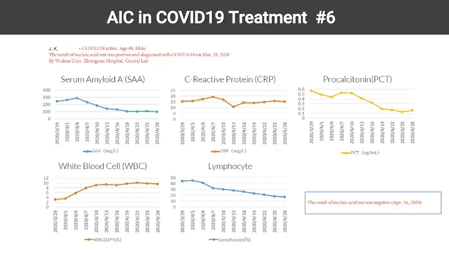
  • AIC in COVID19 Treatment #6 J. K. - COVID 19 active. Age 49, Male. The result of nucleic acid test was positive and diagnosed with COVID-19 on Mar. 28, 2020 By Wuhan Univ. Zhongnan Hospital, Central Lab Serum Amyloid A (SAA) Line chart. Y-axis. 400 300 200 100 0 X-axis. 2020/3/29 2020/4/1 2020/4/4 2020/4/7 2020/4/10 2020/4/13 2020/4/16 2020/4/19 2020/4/22 2020/4/25 2020/4/28 SAA (mg/L) C-Reactive Protein (CRP) Line chart. Y-axis. 25 20 15 10 5 0 X-axis. 2020/3/29 2020/4/1 2020/4/4 2020/4/7 2020/4/10 2020/4/13 2020/4/16 2020/4/19 2020/4/22 2020/4/25 2020/4/28 CRP (mg/L) Procalcitonin(PCT) Line chart. Y-axis. 0.6 0.5 0.4 0.3 0.2 0.1 0 X-axis. 2020/3/29 2020/4/1 2020/4/4 2020/4/7 2020/4/10 2020/4/13 2020/4/16 2020/4/19 2020/4/22 2020/4/25 2020/4/28 PCT (ng/mL) White Blood Cell (WBC) Line chart. Y-axis. 12 10 8 6 4 2 0 X-axis. 2020/3/29 2020/4/1 2020/4/4 2020/4/7 2020/4/10 2020/4/13 2020/4/16 2020/4/19 2020/4/22 2020/4/25 2020/4/28 WBC(10^9/L) Lymphocyte Line chart. Y-axis. 50 40 30 20 10 0 X-axis. 2020/3/29 2020/4/1 2020/4/4 2020/4/7 2020/4/10 2020/4/13 2020/4/16 2020/4/19 2020/4/22 2020/4/25 2020/4/28 Lymphocyte(%) The result of nucleic acid test was negative (Apr. 16, 2020)
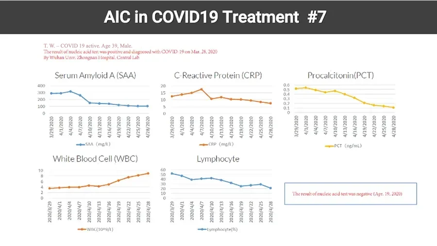
  • AIC in COVID19 Treatment #7 T. W. - COVID 19 active. Age 39, Male. The result of nucleic acid test was positive and diagnosed with COVID-19 on Mar. 28, 2020 By Wuhan Univ. Zhongnan Hospital, Central Lab Serum Amyloid A (SAA) Line chart. Y-axis. 400 300 200 100 0 X-axis. 3/29/2020 4/1/2020 4/4/2020 4/7/2020 4/10/2020 4/13/2020 4/16/2020 4/19/2020 4/22/2020 4/25/2020 4/28/2020 SAA (mg/L) C-Reactive Protein (CRP) Line chart. Y-axis. 20 15 10 5 0 X-axis. 3/29/2020 4/1/2020 4/4/2020 4/7/2020 4/10/2020 4/13/2020 4/16/2020 4/19/2020 4/22/2020 4/25/2020 4/28/2020 CRP (mg/L) Procalcitonin(PCT) Line chart. Y-axis. 0.6 0.5 0.4 0.3 0.2 0.1 0 X-axis. 3/29/2020 4/1/2020 4/4/2020 4/7/2020 4/10/2020 4/13/2020 4/16/2020 4/19/2020 4/22/2020 4/25/2020 4/28/2020 PCT (ng/mL) White Blood Cell (WBC) Line chart. Y-axis. 10 8 6 4 2 0 X-axis. 2020/3/29 2020/4/1 2020/4/4 2020/4/7 2020/4/10 2020/4/13 2020/4/16 2020/4/19 2020/4/22 2020/4/25 2020/4/28 WBC(10^9/L) Lymphocyte Line chart. Y-axis. 60 50 40 30 20 10 0 X-axis. 2020/3/29 2020/4/1 2020/4/4 2020/4/7 2020/4/10 2020/4/13 2020/4/16 2020/4/19 2020/4/22 2020/4/25 2020/4/28 Lymphocyte(%) The result of nucleic acid test was negative (Apr. 19, 2020)

AIC IN AUTOIMMUNE DISEASES

Modulating correct calcium signals in immune cells prevent the onset of the disease

AIC in Autoimmunity
Illustration: neuron and virus-like cells on magenta background. AIC IONIC CALCIUM THERAPY : Autoimmunity
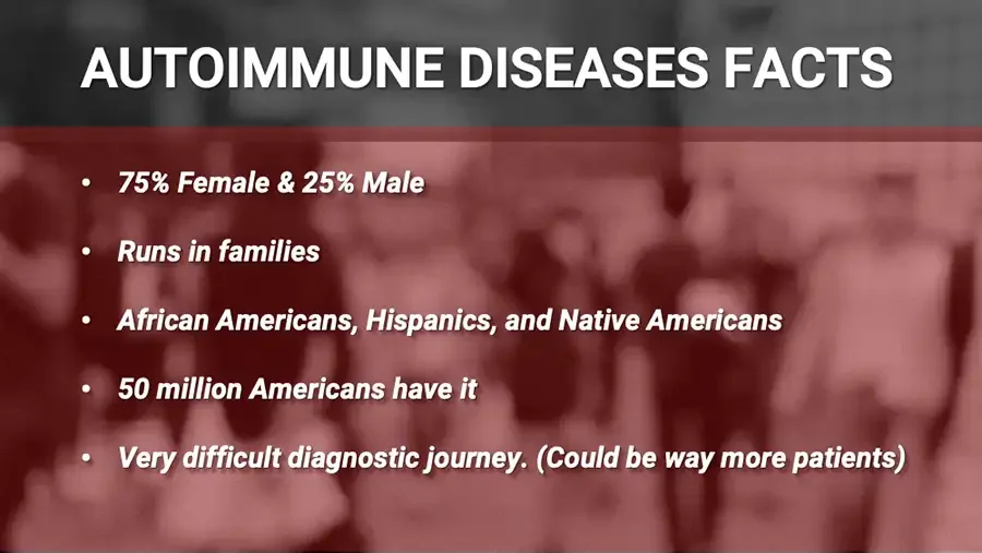
Background: blurred photo of a crowd of people. AUTOIMMUNE DISEASES FACTS 75% Female & 25% Male Runs in families African Americans, Hispanics, and Native Americans 50 million Americans have it Very difficult diagnostic journey. (Could be way more patients)
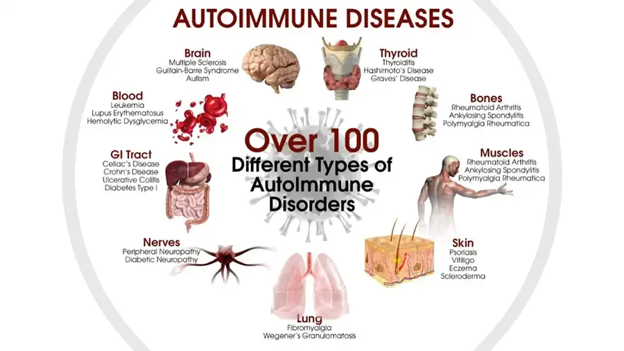
Infographic: organ illustrations arranged in a circle around a virus. AUTOIMMUNE DISEASES Brain Multiple Sclerosis Guillain-Barre Syndrome Autism Blood Leukemia Lupus Erythematosus Hemolytic Dysglycemia GI Tract Celiac’s Disease Crohn’s Disease Ulcerative Colitis Diabetes Type I Nerves Peripheral Neuropathy Diabetic Neuropathy Over 100 Different Types of AutoImmune Disorders Lung Fibromyalgia Wegener’s Granulomatosis Thyroid Thyroiditis Hashimoto’s Disease Graves’ Disease Bones Rheumatoid Arthritis Ankylosing Spondylitis Polymyalgia Rheumatica Muscles Rheumatoid Arthritis Ankylosing Spondylitis Polymyalgia Rheumatica Skin Psoriasis Vitiligo Eczema Scleroderma
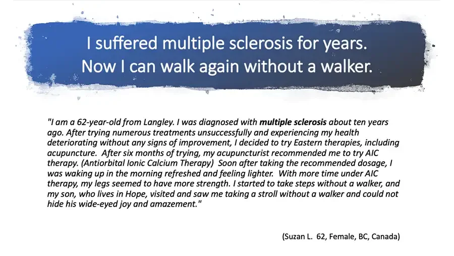
I suffered multiple sclerosis for years. Now I can walk again without a walker. "I am a 62-year-old from Langley. I was diagnosed with multiple sclerosis about ten years ago. After trying numerous treatments unsuccessfully and experiencing my health deteriorating without any signs of improvement, I decided to try Eastern therapies, including acupuncture. After six months of trying, my acupuncturist recommended me to try AIC therapy. (Antiorbital Ionic Calcium Therapy) Soon after taking the recommended dosage, I was waking up in the morning refreshed and feeling lighter. With more time under AIC therapy, my legs seemed to have more strength. I started to take steps without a walker, and my son, who lives in Hope, visited and saw me taking a stroll without a walker and could not hide his wide-eyed joy and amazement." (Suzan L. 62, Female, BC, Canada)
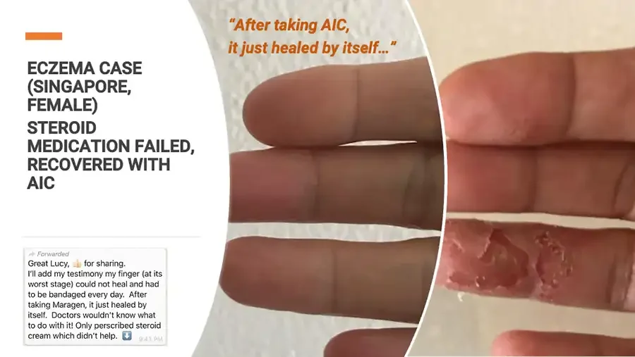
ECZEMA CASE (SINGAPORE, FEMALE) STEROID MEDICATION FAILED, RECOVERED WITH AIC Screenshot of a forwarded chat message. Forwarded Great Lucy, 👍 for sharing. I’ll add my testimony my finger (at its worst stage) could not heal and had to be bandaged every day. After taking Maragen, it just healed by itself. Doctors wouldn’t know what to do with it! Only perscribed steroid cream which didn’t help. ⬇️ 9:41 PM “After taking AIC, it just healed by itself…” Photo: fingers with healed skin. Photo: finger with severely peeling, cracked skin.
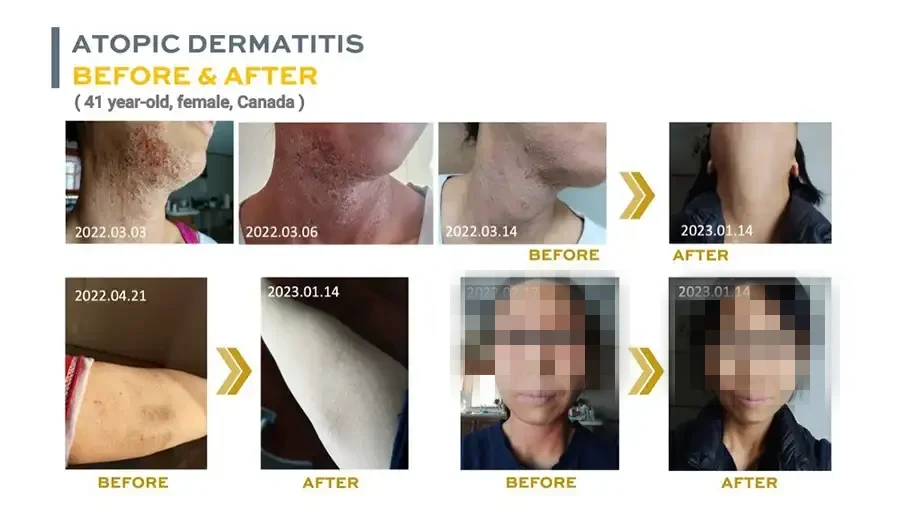
ATOPIC DERMATITIS BEFORE & AFTER ( 41 year-old, female, Canada ) Photo: neck with rash. 2022.03.03 Photo: neck with rash. 2022.03.06 Photo: neck with rash. 2022.03.14 BEFORE Arrow. Photo: neck with clear skin. 2023.01.14 AFTER Photo: arm with dark rash patches. 2022.04.21 BEFORE Arrow. Photo: arm with clear skin. 2023.01.14 AFTER Photo: woman's face with redness, eyes covered by a bar. 2022.02.17 BEFORE Arrow. Photo: woman's face with clearer skin, eyes covered by a bar. 2023.01.14 AFTER
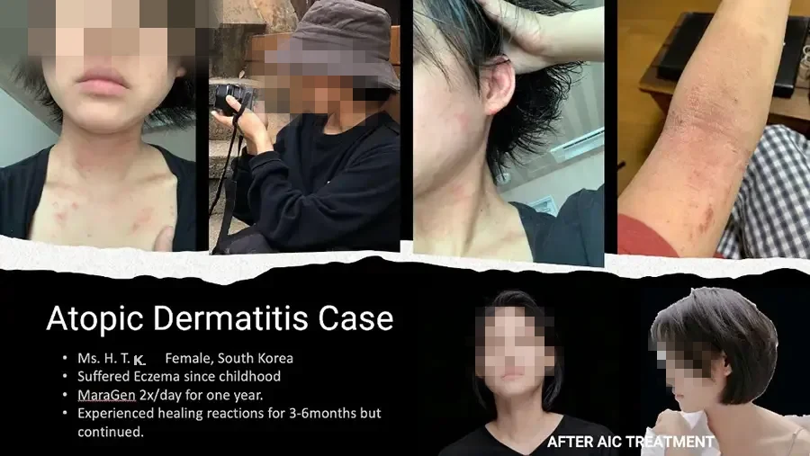
Photo: young woman with red rash on neck and chest. Photo: person in bucket hat holding a camera. Photo: neck and ear with red rash. Photo: forearm with red rash. Atopic Dermatitis Case Ms. H. T. K., Female, South Korea Suffered Eczema since childhood MaraGen 2x/day for one year. Experienced healing reactions for 3-6months but continued. Photo: woman with clear skin, front view. Photo: woman with short hair, side view. AFTER AIC TREATMENT
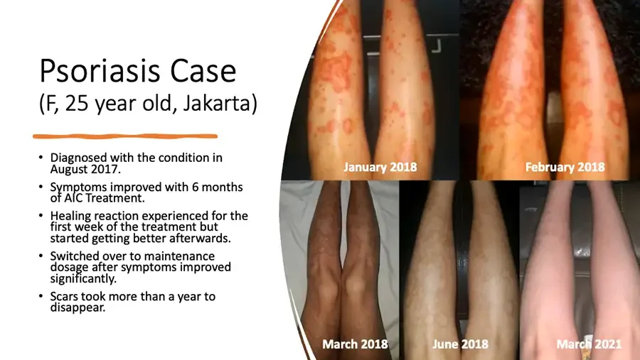
Psoriasis Case (F, 25 year old, Jakarta) Diagnosed with the condition in August 2017. Symptoms improved with 6 months of AIC Treatment. Healing reaction experienced for the first week of the treatment but started getting better afterwards. Switched over to maintenance dosage after symptoms improved significantly. Scars took more than a year to disappear. Photo: legs with red psoriasis patches. January 2018 Photo: legs with red psoriasis patches. February 2018 Photo: legs with darkened skin. March 2018 Photo: legs with pale patches. June 2018 Photo: legs with clear skin. March 2021
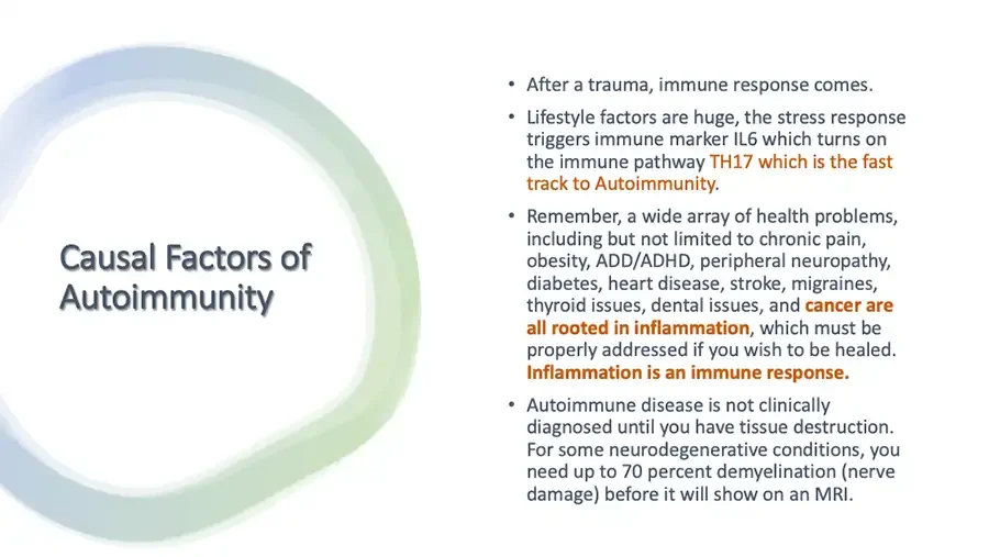
Causal Factors of Autoimmunity After a trauma, immune response comes. Lifestyle factors are huge, the stress response triggers immune marker IL6 which turns on the immune pathway TH17 which is the fast track to Autoimmunity. Remember, a wide array of health problems, including but not limited to chronic pain, obesity, ADD/ADHD, peripheral neuropathy, diabetes, heart disease, stroke, migraines, thyroid issues, dental issues, and cancer are all rooted in inflammation, which must be properly addressed if you wish to be healed. Inflammation is an immune response. Autoimmune disease is not clinically diagnosed until you have tissue destruction. For some neurodegenerative conditions, you need up to 70 percent demyelination (nerve damage) before it will show on an MRI.
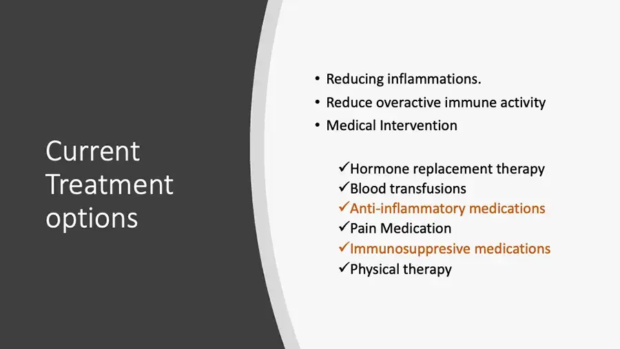
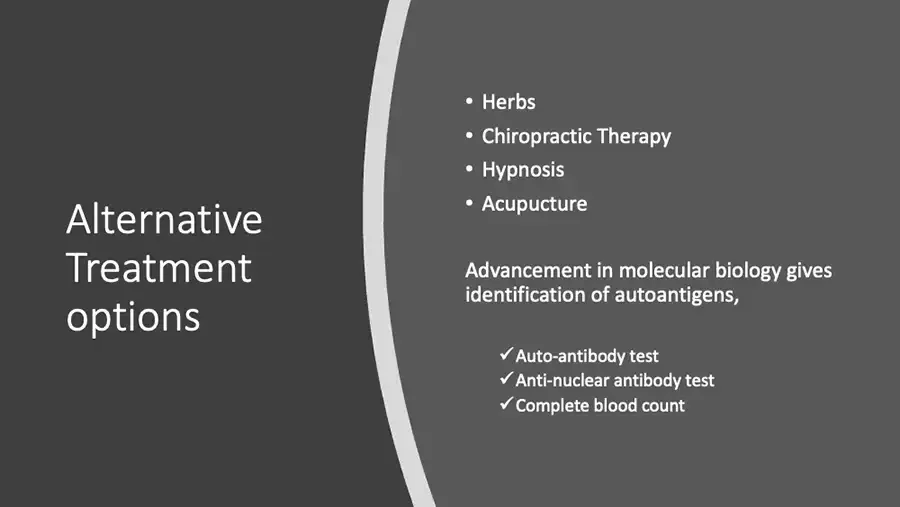
Current Treatment options Reducing inflammations. Reduce overactive immune activity Medical Intervention ✓ Hormone replacement therapy ✓ Blood transfusions ✓ Anti-inflammatory medications ✓ Pain Medication ✓ Immunosuppresive medications ✓ Physical therapy
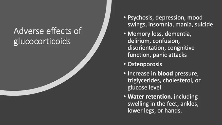
Adverse effects of glucocorticoids Psychosis, depression, mood swings, insomnia, mania, suicide Memory loss, dementia, delirium, confusion, disorientation, congnitive function, panic attacks Osteoporosis Increase in blood pressure, triglycerides, cholesterol, or glucose level Water retention, including swelling in the feet, ankles, lower legs, or hands.
Alternative Treatment options Herbs Chiropractic Therapy Hypnosis Acupucture Advancement in molecular biology gives identification of autoantigens, ✓ Auto-antibody test ✓ Anti-nuclear antibody test ✓ Complete blood count
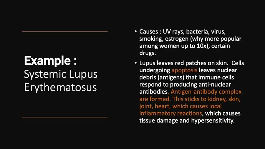
Example : Systemic Lupus Erythematosus Causes : UV rays, bacteria, virus, smoking, estrogen (why more popular among women up to 10x), certain drugs. Lupus leaves red patches on skin. Cells undergoing apoptosis leaves nuclear debris (antigens) that immune cells respond to producing anti-nuclear antibodies. Antigen-antibody complex are formed. This sticks to kidney, skin, joint, heart, which causes local inflammatory reactions, which causes tissue damage and hypersensitivity.
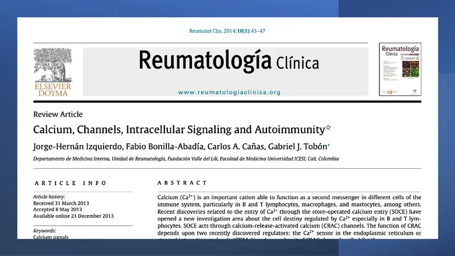
Screenshot of a journal article first page on blue background. Reumatol Clin. 2014;10(1):43–47 Elsevier tree logo. ELSEVIER DOYMA Reumatología Clínica www.reumatologiaclinica.org Journal cover thumbnail. Reumatología Clínica Review Article Calcium, Channels, Intracellular Signaling and Autoimmunity☆ Jorge-Hernán Izquierdo, Fabio Bonilla-Abadía, Carlos A. Cañas, Gabriel J. Tobón* Departamento de Medicina Interna, Unidad de Reumatología, Fundación Valle del Lili, Facultad de Medicina Universidad ICESI, Cali, Colombia ARTICLE INFO Article history: Received 31 March 2013 Accepted 8 May 2013 Available online 21 December 2013 Keywords: Calcium signals ABSTRACT
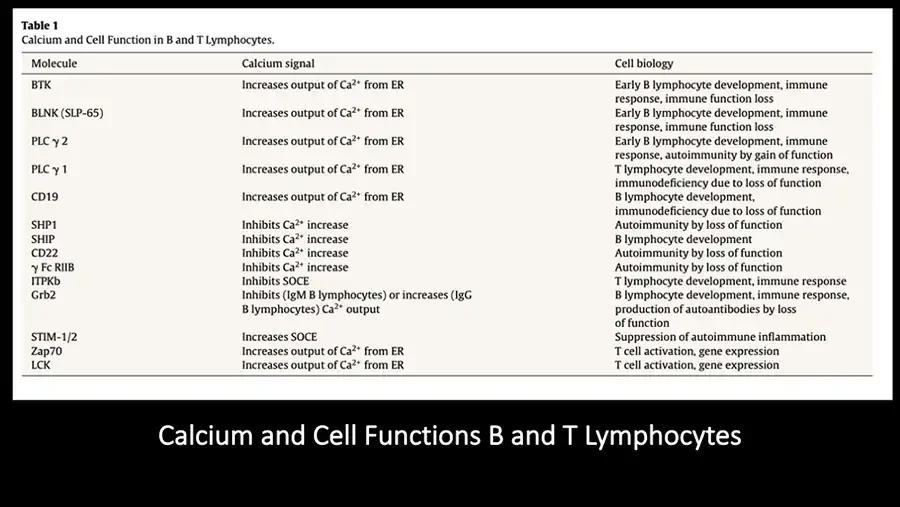
Table 1 Calcium and Cell Function in B and T Lymphocytes. Molecule Calcium signal Cell biology BTK Increases output of Ca²⁺ from ER Early B lymphocyte development, immune response, immune function loss BLNK (SLP-65) Increases output of Ca²⁺ from ER Early B lymphocyte development, immune response, immune function loss PLC γ 2 Increases output of Ca²⁺ from ER Early B lymphocyte development, immune response, autoimmunity by gain of function PLC γ 1 Increases output of Ca²⁺ from ER T lymphocyte development, immune response, immunodeficiency due to loss of function CD19 Increases output of Ca²⁺ from ER B lymphocyte development, immunodeficiency due to loss of function SHP1 Inhibits Ca²⁺ increase Autoimmunity by loss of function SHIP Inhibits Ca²⁺ increase B lymphocyte development CD22 Inhibits Ca²⁺ increase Autoimmunity by loss of function γ Fc RIIB Inhibits Ca²⁺ increase Autoimmunity by loss of function ITPKb Inhibits SOCE T lymphocyte development, immune response Grb2 Inhibits (IgM B lymphocytes) or increases (IgG B lymphocytes) Ca²⁺ output B lymphocyte development, immune response, production of autoantibodies by loss of function STIM-1/2 Increases SOCE Suppression of autoimmune inflammation Zap70 Increases output of Ca²⁺ from ER T cell activation, gene expression LCK Increases output of Ca²⁺ from ER T cell activation, gene expression Calcium and Cell Functions B and T Lymphocytes
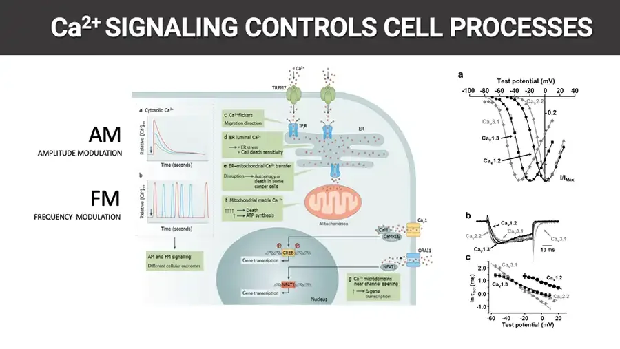
Ca²⁺ SIGNALING CONTROLS CELL PROCESSES AM AMPLITUDE MODULATION FM FREQUENCY MODULATION Diagram: calcium signaling pathways inside a cell. a Cytosolic Ca²⁺ Relative Ca²⁺.cyt Time (seconds) b Relative Ca²⁺.cyt Time (seconds) AM and FM signalling Different cellular outcomes Ca²⁺ TRPM7 c Ca²⁺ flickers Migration direction IP₃R ER d ER luminal Ca²⁺ → • ER stress • Cell death sensitivity e ER–mitochondrial Ca²⁺ transfer Disruption → Autophagy or death in some cancer cells f Mitochondrial matrix Ca²⁺ ↑↑↑↑ → Death ↑ → ATP synthesis Mitochondrion CaM CaMKIIγ CaV1 ORAI1 NFAT1 P P CREB Gene transcription NFAT1 Gene transcription Nucleus g Ca²⁺ microdomains near channel opening ↑ → Δ gene transcription Line graphs: calcium channel current measurements. a Test potential (mV) -100 -80 -60 -40 -20 0 20 40 CaV2.2 0.2 CaV3.1 CaV1.3 CaV1.2 I/IMax b CaV1.2 CaV2.2 CaV3.1 CaV1.3 CaV3.1 10 ms c CaV3.1 2.0 1.0 0.0 -1.0 ln τact (ms) CaV1.2 CaV1.3 CaV2.2 -60 -40 -20 0 20 Test potential (mV)
Intracellular Ca²⁺ signaling is a component of signaling cascades that are generated in different cells after exposure of its receptors to a specific stimulus. In the case of T and B lymphocytes, these signals give rise to multiple changes in intracellular DNA expression, with the consequent production of various inflammatory markers. Research in this area also has established the importance of Ca²⁺ pathways in the development of autoimmune diseases, both due to overproduction as well as inactivity. As progress is made in the study of all these concepts, it becomes important to consider intervention on these pathways and receptors for therapeutic purposes. DNA icon in a circle on blue side band.
Signaling through intracellular Ca²⁺ has been implicated in the pathogenesis of autoimmune and congenital immunodeficiencies. Diseases such as systemic lupus erythematosus (SLE), rheumatoid arthritis (RA), multiple sclerosis (MS) and type 1 diabetes mellitus are related in their pathophysiology by the presence of autoreactive T and B cells that contribute in the generation of the inflammatory process. DNA icon in a circle on blue side band.
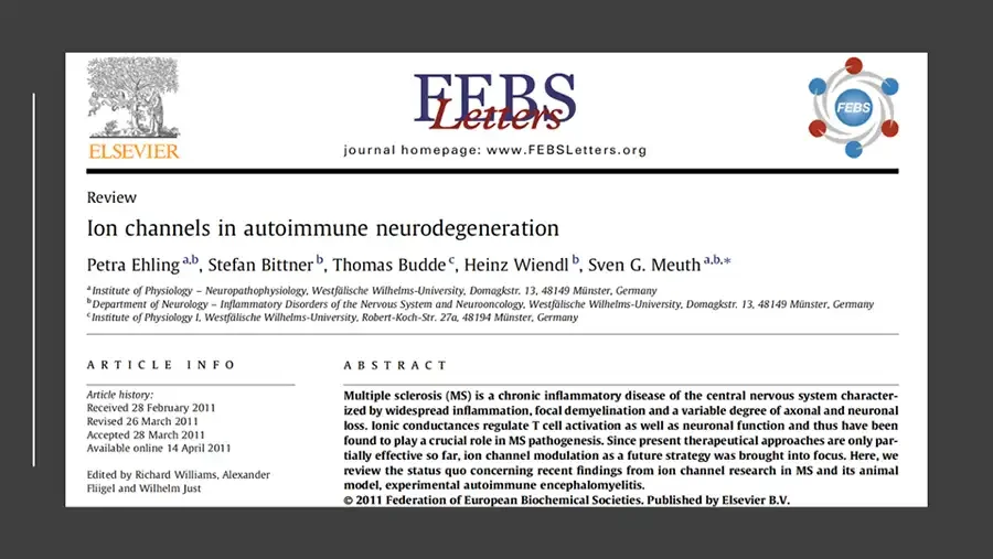
Screenshot of a journal article first page on dark background. Elsevier tree logo. ELSEVIER FEBS Letters journal homepage: www.FEBSLetters.org FEBS logo. FEBS Review Ion channels in autoimmune neurodegeneration Petra Ehling a,b, Stefan Bittner b, Thomas Budde c, Heinz Wiendl b, Sven G. Meuth a,b,* a Institute of Physiology – Neuropathophysiology, Westfälische Wilhelms-University, Domagkstr. 13, 48149 Münster, Germany b Department of Neurology – Inflammatory Disorders of the Nervous System and Neurooncology, Westfälische Wilhelms-University, Domagkstr. 13, 48149 Münster, Germany c Institute of Physiology I, Westfälische Wilhelms-University, Robert-Koch-Str. 27a, 48194 Münster, Germany ARTICLE INFO Article history: Received 28 February 2011 Revised 26 March 2011 Accepted 28 March 2011 Available online 14 April 2011 Edited by Richard Williams, Alexander Flügel and Wilhelm Just ABSTRACT © 2011 Federation of European Biochemical Societies. Published by Elsevier B.V.
Currently, MS is considered to be due to axonal and neuronal degeneration. In this context, it can be assumed that one leading mechanism is the reorganization and altered gene expression of different ion channels which occurs in demyelinated axons. Therefore, MS may be defined as a member of the group of channelopathies, i.e., disorders in which abnormal ion channel function leads to the appearance of clinical signs and symptoms. These diseases can be further subdivided in genetic, autoimmune and transcriptional/translational channelopathies. Icon: head silhouette with brain.
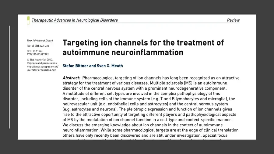
Screenshot of a journal article first page on dark background. Journal logo icon. Therapeutic Advances in Neurological Disorders Review Ther Adv Neurol Disord (2013) 6(5) 322–336 DOI: 10.1177/1756285613487782 © The Author(s), 2013. Reprints and permissions: http://www.sagepub.co.uk/journalsPermissions.nav Targeting ion channels for the treatment of autoimmune neuroinflammation Stefan Bittner and Sven G. Meuth Abstract:
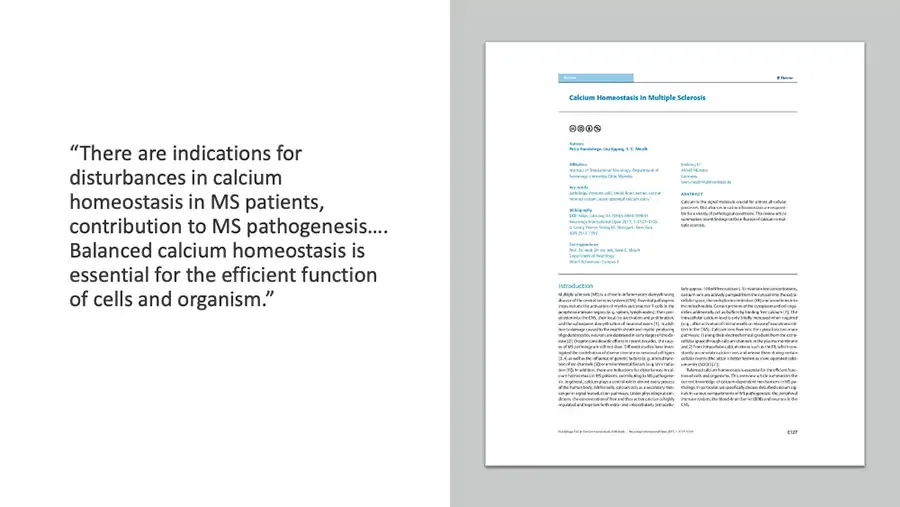
Thumbnail of a journal article first page. Calcium Homeostasis in Multiple Sclerosis Creative Commons license icons. Introduction
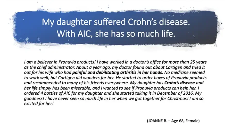
My daughter suffered Crohn’s disease. With AIC, she has so much life. I am a believer in Pronuvia products! I have worked in a doctor’s office for more than 25 years as the chief administrator. About a year ago, my doctor found out about Cartigen and tried it out for his wife who had painful and debilitating arthritis in her hands. No medicine seemed to work well, but Cartigen did wonders for her. He started to order boxes of Pronuvia products and recommended to many of his friends everywhere. My daughter has Crohn’s disease and her life simply has been miserable, and I wanted to see if Pronuvia products can help her. I ordered 4 bottles of AIC for my daughter and she started taking it in December of 2016. My goodness! I have never seen so much life in her when we got together for Christmas! I am so excited for her! (JOANNE B. – Age 68, Female)
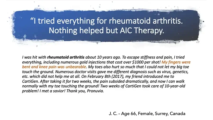
“I tried everything for rheumatoid arthritis. Nothing helped but AIC Therapy. I was hit with rheumatoid arthritis about 10 years ago. To escape stiffness and pain, I tried everything, including numerous gold injections that cost over $1000 per shot! My fingers were bent and knee pain was unbearable. My toes also hurt so much that I could not let my big toe touch the ground. Numerous doctor visits gave me different diagnosis such as virus, genetics, etc. which did not help me at all. On February 8th (2017), my friend introduced me to CartiGen. After taking it for two weeks, the pain subsided dramatically, and now I can walk normally with my toe touching the ground! Two weeks of CartiGen took care of 10-year-old problem! I met a savior! Thank you, Pronuvia. J. C. – Age 66, Female, Surrey, Canada
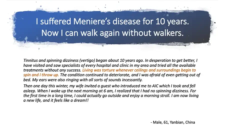
I suffered Meniere’s disease for 10 years. Now I can walk again without walkers. Tinnitus and spinning dizziness (vertigo) began about 10 years ago. In desperation to get better, I have visited and saw specialists of every hospital and clinic in my area and tried all the available treatments without any success. Living was torture whenever ceilings and surroundings begin to spin and I throw up. The condition continued to deteriorate, and I was afraid of even getting out of bed. My ears were also ringing with all sorts of sounds incessantly. Then one day this winter, my wife invited a guest who introduced me to AIC which I took and fell asleep. When I woke up the next morning at 6 am, I realized that I had no spinning dizziness. For the first time in a long time, I could actually go outside and enjoy a morning stroll. I am now living a new life, and it feels like a dream!! Male, 61, Yanbian, China
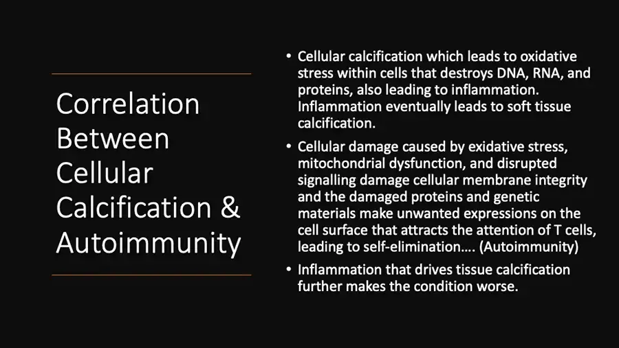
Correlation Between Cellular Calcification & Autoimmunity Cellular calcification which leads to oxidative stress within cells that destroys DNA, RNA, and proteins, also leading to inflammation. Inflammation eventually leads to soft tissue calcification. Cellular damage caused by exidative stress, mitochondrial dysfunction, and disrupted signalling damage cellular membrane integrity and the damaged proteins and genetic materials make unwanted expressions on the cell surface that attracts the attention of T cells, leading to self-elimination…. (Autoimmunity) Inflammation that drives tissue calcification further makes the condition worse.
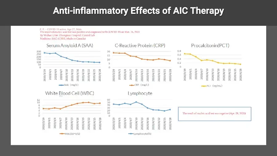
Anti-inflammatory Effects of AIC Therapy Z. F. - COVID 19 active. Age 27, Male. The result of nucleic acid test was positive and diagnosed with COVID-19 on Mar. 26 , 2020 By Wuhan Univ. Zhongnan Hospital, Central Lab Medicine: SAC (C3HI , Made in Canada) Line chart. Serum Amyloid A (SAA) 300 250 200 150 100 50 0 2020/3/29 2020/4/1 2020/4/4 2020/4/7 2020/4/10 2020/4/13 2020/4/16 2020/4/19 2020/4/22 2020/4/25 2020/4/28 SAA (mg/L) Line chart. C-Reactive Protein (CRP) 20 15 10 5 0 2020/3/29 2020/4/1 2020/4/4 2020/4/7 2020/4/10 2020/4/13 2020/4/16 2020/4/19 2020/4/22 2020/4/25 2020/4/28 CRP (mg/L) Line chart. Procalcitonin(PCT) 0.8 0.6 0.4 0.2 0 2020/3/29 2020/4/1 2020/4/4 2020/4/7 2020/4/10 2020/4/13 2020/4/16 2020/4/19 2020/4/22 2020/4/25 2020/4/28 PCT (ng/mL) Line chart. White Blood Cell (WBC) 12 10 8 6 4 2 0 2020/3/29 2020/4/1 2020/4/4 2020/4/7 2020/4/10 2020/4/13 2020/4/16 2020/4/19 2020/4/22 2020/4/25 2020/4/28 WBC(10^9/L) Line chart. Lymphocyte 60 50 40 30 20 10 0 2020/3/29 2020/4/1 2020/4/4 2020/4/7 2020/4/10 2020/4/13 2020/4/16 2020/4/19 2020/4/22 2020/4/25 2020/4/28 Lymphocyte(%) The result of nucleic acid test was negative (Apr. 28, 2020)
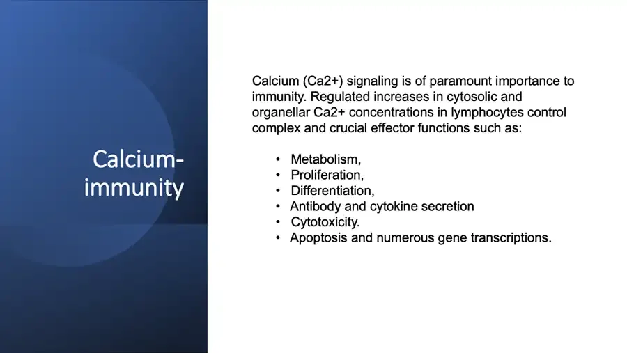
Calcium-immunity Calcium (Ca2+) signaling is of paramount importance to immunity. Regulated increases in cytosolic and organellar Ca2+ concentrations in lymphocytes control complex and crucial effector functions such as: Metabolism, Proliferation, Differentiation, Antibody and cytokine secretion Cytotoxicity. Apoptosis and numerous gene transcriptions.
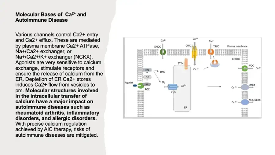
Molecular Bases of Ca²⁺ and Autoimmune Disease Various channels control Ca2+ entry and Ca2+ efflux. These are mediated by plasma membrane Ca2+ ATPase, Na+/Ca2+ exchanger, or Na+/Ca2+/K+ exchanger (NCKX). Agonists are very sensitive to calcium exchange, stimulate receptors and ensure the release of calcium from the ER. Depletion of ER Ca2+ stores induces Ca2+ flow from vesicles to pm. Molecular structures involved in the intracellular transfer of calcium have a major impact on autoimmune diseases such as rheumatoid arthritis, inflammatory disorders, and allergic disorders. With precise calcium regulation achieved by AIC therapy, risks of autoimmune diseases are mitigated. Diagram: calcium channels and pumps in a cell membrane. Ca²⁺ SMOC Ca²⁺ ORAI1 Ca²⁺ TRPC Plasma membrane Cytosol STIM1 Ca²⁺ VOC Ca²⁺ PIP2 DAG PLC Agonist GP ROC IP₃ IP₃R Ca²⁺ PMCA Ca²⁺ NCX/NCKX Ca²⁺ ER
Micrograph: stained tissue cross-section. Ca2+ signaling effects to the Autoimmune diseases The dysregulated Ca2+ signaling pathway, particularly in T and B cells, is involved in the pathophysiology of autoimmune diseases, indicating that a therapeutic effect can be made in these diseases if the Ca2+ signaling pathway is controlled.
Photo: close-up of red spherical cells. Altered Ca2+ regulation in lymphocytes leads to Autoimmune disease and inflammatory and immunodeficiency syndromes. T cell dysfunction promotes: Systemic autoimmunity in various common Rheumatic diseases. During an immune response, the engagement of antigen receptors decrease intracellular Ca2+. SOCE raises the intracellular Ca2+ concentration. The Ca2+-calcineurin- nuclear factor of an activated T cell (NFAT1 ) pathway is dysregulated in autoimmune diseases.
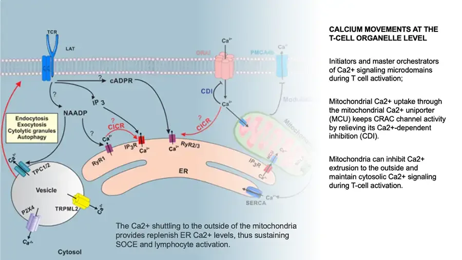
Diagram: calcium movements between T-cell organelles. TCR LAT ζζ Ca²⁺ ORAI PMCA4b Ca²⁺ ? cADPR ? IP 3 NAADP CDI Ca²⁺ Modulation Endocytosis Exocytosis Cytolytic granules Autophagy ? ? CICR Ca²⁺ IP₃R Ca²⁺ RyR1 Ca²⁺ RyR2/3 ? CICR Mitochondria Ca²⁺ TPC1/2 ER IP₃R MCU Ca²⁺ Ca²⁺ Vesicle TRPML2 Ca²⁺ P2X4 Ca²⁺ SERCA Ca²⁺ Cytosol The Ca2+ shuttling to the outside of the mitochondria provides replenish ER Ca2+ levels, thus sustaining SOCE and lymphocyte activation. CALCIUM MOVEMENTS AT THE T-CELL ORGANELLE LEVEL Initiators and master orchestrators of Ca2+ signaling microdomains during T cell activation; Mitochondrial Ca2+ uptake through the mitochondrial Ca2+ uniporter (MCU) keeps CRAC channel activity by relieving its Ca2+-dependent inhibition (CDI). Mitochondria can inhibit Ca2+ extrusion to the outside and maintain cytosolic Ca2+ signaling during T-cell activation.
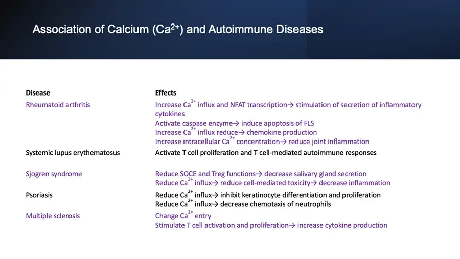
Association of Calcium (Ca²⁺) and Autoimmune Diseases Disease Effects Rheumatoid arthritis Increase Ca²⁺ influx and NFAT transcription→ stimulation of secretion of inflammatory cytokines Activate caspase enzyme→ induce apoptosis of FLS Increase Ca²⁺ influx reduce→ chemokine production Increase intracellular Ca²⁺ concentration→ reduce joint inflammation Systemic lupus erythematosus Activate T cell proliferation and T cell-mediated autoimmune responses Sjogren syndrome Reduce SOCE and Treg functions→ decrease salivary gland secretion Reduce Ca²⁺ influx→ reduce cell-mediated toxicity→ decrease inflammation Psoriasis Reduce Ca²⁺ influx→ inhibit keratinocyte differentiation and proliferation Reduce Ca²⁺ influx→ decrease chemotaxis of neutrophils Multiple sclerosis Change Ca²⁺ entry Stimulate T cell activation and proliferation→ increase cytokine production
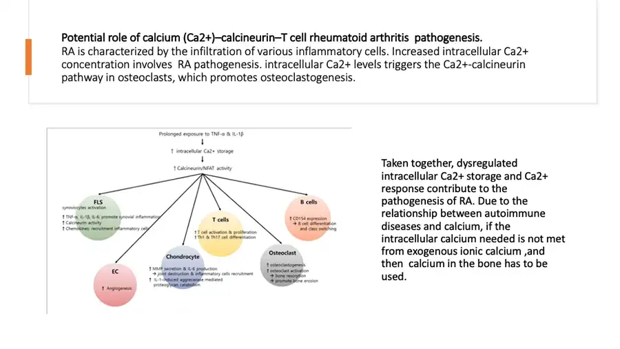
Potential role of calcium (Ca2+)–calcineurin–T cell rheumatoid arthritis pathogenesis. RA is characterized by the infiltration of various inflammatory cells. Increased intracellular Ca2+ concentration involves RA pathogenesis. intracellular Ca2+ levels triggers the Ca2+-calcineurin pathway in osteoclasts, which promotes osteoclastogenesis. Diagram: calcineurin/NFAT activity branching to six cell types. Prolonged exposure to TNF-α & IL-1β ↓ ↑ intracellular Ca2+ storage ↓ ↑ Calcineurin/NFAT activity FLS synoviocytes activation ↑ TNF-α, IL-1β, IL-6: promote synovial inflammation ↑ Calcineurin activity ↑ Chemokines: recruitment inflammatory cells T cells ↑ T cell activation & proliferation ↑ Th1 & Th17 cell differentiation B cells ↑ CD154 expression → B cell differentiation and class switching EC ↑ Angiogenesis Chondrocyte ↑ MMP secretion & IL-6 production → joint destruction & inflammatory cells recruitment ↑ IL-1–induced aggrecanase mediated proteoglycan catabolism Osteoclast ↑ osteoclastogenesis ↑ osteoclast activation → bone resorption → promote bone erosion Taken together, dysregulated intracellular Ca2+ storage and Ca2+ response contribute to the pathogenesis of RA. Due to the relationship between autoimmune diseases and calcium, if the intracellular calcium needed is not met from exogenous ionic calcium ,and then calcium in the bone has to be used.