Notice. The following information is provided for your convenience. ACRI is not directly involved in this research and does not make any medical claims.

AIC THERAPY in Neurodegeneration
Background: glowing blue neuron network. AIC THERAPY IN Neurodegeneration

AIC’s EFFECT on Alzeheimer’s DISEASE
  • AIC’s EFFECT ON ALZHEIMER’S Image: translucent blue brain with glowing red spot.
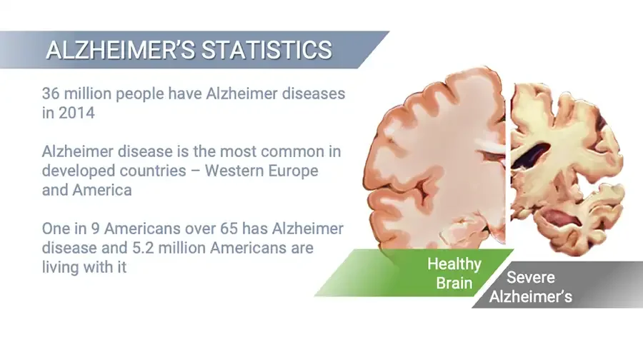
  • ALZHEIMER’S STATISTICS 36 million people have Alzheimer diseases in 2014 Alzheimer disease is the most common in developed countries – Western Europe and America One in 9 Americans over 65 has Alzheimer disease and 5.2 million Americans are living with it Brain cross-sections side by side: healthy and shrunken. Healthy Brain Severe Alzheimer’s
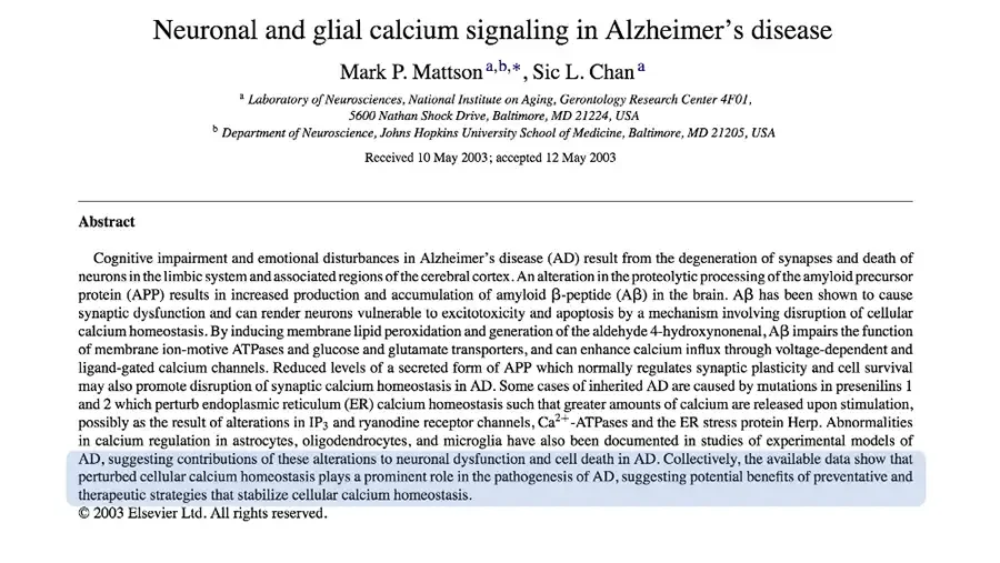
  • Neuronal and glial calcium signaling in Alzheimer’s disease Mark P. Mattson a,b,*, Sic L. Chan a a Laboratory of Neurosciences, National Institute on Aging, Gerontology Research Center 4F01, 5600 Nathan Shock Drive, Baltimore, MD 21224, USA b Department of Neuroscience, Johns Hopkins University School of Medicine, Baltimore, MD 21205, USA Received 10 May 2003; accepted 12 May 2003 Abstract Cognitive impairment and emotional disturbances in Alzheimer’s disease (AD) result from the degeneration of synapses and death of neurons in the limbic system and associated regions of the cerebral cortex. An alteration in the proteolytic processing of the amyloid precursor protein (APP) results in increased production and accumulation of amyloid β-peptide (Aβ) in the brain. Aβ has been shown to cause synaptic dysfunction and can render neurons vulnerable to excitotoxicity and apoptosis by a mechanism involving disruption of cellular calcium homeostasis. By inducing membrane lipid peroxidation and generation of the aldehyde 4-hydroxynonenal, Aβ impairs the function of membrane ion-motive ATPases and glucose and glutamate transporters, and can enhance calcium influx through voltage-dependent and ligand-gated calcium channels. Reduced levels of a secreted form of APP which normally regulates synaptic plasticity and cell survival may also promote disruption of synaptic calcium homeostasis in AD. Some cases of inherited AD are caused by mutations in presenilins 1 and 2 which perturb endoplasmic reticulum (ER) calcium homeostasis such that greater amounts of calcium are released upon stimulation, possibly as the result of alterations in IP3 and ryanodine receptor channels, Ca2+-ATPases and the ER stress protein Herp. Abnormalities in calcium regulation in astrocytes, oligodendrocytes, and microglia have also been documented in studies of experimental models of AD, suggesting contributions of these alterations to neuronal dysfunction and cell death in AD. Collectively, the available data show that perturbed cellular calcium homeostasis plays a prominent role in the pathogenesis of AD, suggesting potential benefits of preventative and therapeutic strategies that stabilize cellular calcium homeostasis. Light blue highlight over the abstract's last three lines. © 2003 Elsevier Ltd. All rights reserved.
  • Background: blue painted brushstroke banner. “Collectively, the available data show that perturbed cellular calcium homeostasis plays a prominent role in the pathogenesis of AD, suggesting potential benefits of preventive and therapeutic strategies that stabilize cellular calcium homeostasis.”
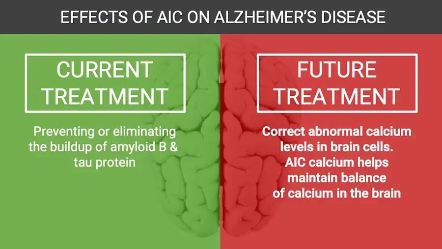
  • EFFECTS OF AIC ON ALZHEIMER’S DISEASE Background: brain illustration, left half green, right half red. CURRENT TREATMENT Preventing or eliminating the buildup of amyloid B & tau protein FUTURE TREATMENT Correct abnormal calcium levels in brain cells. AIC calcium helps maintain balance of calcium in the brain
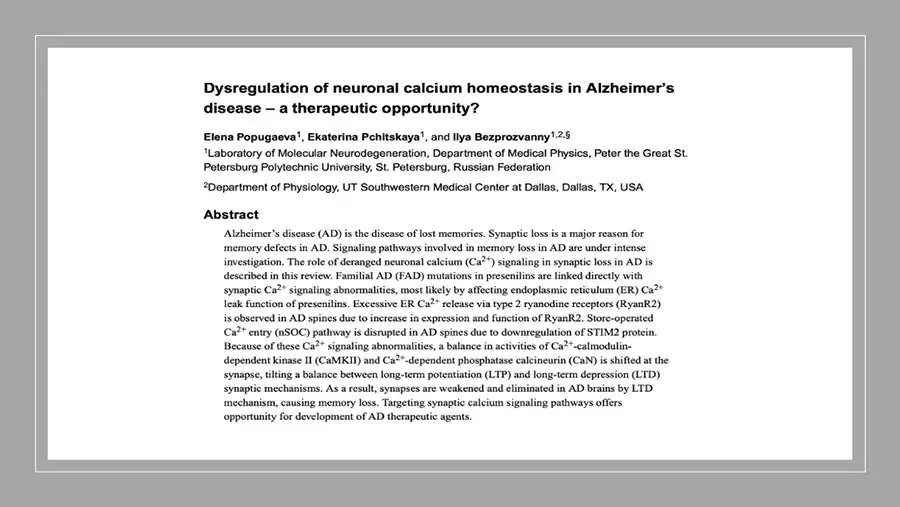
  • Dysregulation of neuronal calcium homeostasis in Alzheimer’s disease – a therapeutic opportunity? Elena Popugaeva1, Ekaterina Pchitskaya1, and Ilya Bezprozvanny1,2,§ 1Laboratory of Molecular Neurodegeneration, Department of Medical Physics, Peter the Great St. Petersburg Polytechnic University, St. Petersburg, Russian Federation 2Department of Physiology, UT Southwestern Medical Center at Dallas, Dallas, TX, USA Abstract Alzheimer’s disease (AD) is the disease of lost memories. Synaptic loss is a major reason for memory defects in AD. Signaling pathways involved in memory loss in AD are under intense investigation. The role of deranged neuronal calcium (Ca2+) signaling in synaptic loss in AD is described in this review. Familial AD (FAD) mutations in presenilins are linked directly with synaptic Ca2+ signaling abnormalities, most likely by affecting endoplasmic reticulum (ER) Ca2+ leak function of presenilins. Excessive ER Ca2+ release via type 2 ryanodine receptors (RyanR2) is observed in AD spines due to increase in expression and function of RyanR2. Store-operated Ca2+ entry (nSOC) pathway is disrupted in AD spines due to downregulation of STIM2 protein. Because of these Ca2+ signaling abnormalities, a balance in activities of Ca2+-calmodulin-dependent kinase II (CaMKII) and Ca2+-dependent phosphatase calcineurin (CaN) is shifted at the synapse, tilting a balance between long-term potentiation (LTP) and long-term depression (LTD) synaptic mechanisms. As a result, synapses are weakened and eliminated in AD brains by LTD mechanism, causing memory loss. Targeting synaptic calcium signaling pathways offers opportunity for development of AD therapeutic agents.
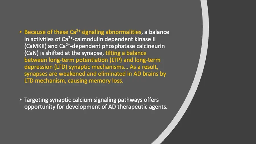
  • Because of these Ca2+ signaling abnormalities, a balance in activities of Ca2+-calmodulin dependent kinase II (CaMKII) and Ca2+-dependent phosphatase calcineurin (CaN) is shifted at the synapse, tilting a balance between long-term potentiation (LTP) and long-term depression (LTD) synaptic mechanisms… As a result, synapses are weakened and eliminated in AD brains by LTD mechanism, causing memory loss. Targeting synaptic calcium signaling pathways offers opportunity for development of AD therapeutic agents.
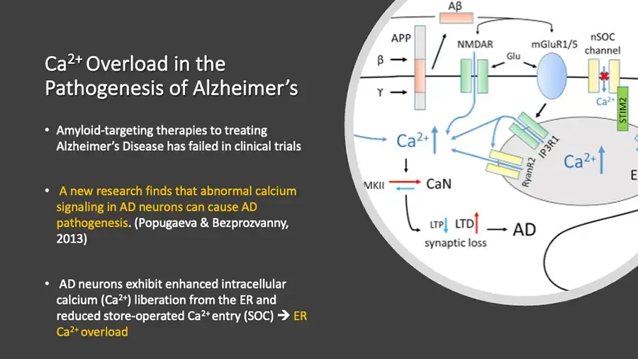
  • Ca2+ Overload in the Pathogenesis of Alzheimer’s Amyloid-targeting therapies to treating Alzheimer’s Disease has failed in clinical trials A new research finds that abnormal calcium signaling in AD neurons can cause AD pathogenesis. (Popugaeva & Bezprozvanny, 2013) AD neurons exhibit enhanced intracellular calcium (Ca2+) liberation from the ER and reduced store-operated Ca2+ entry (SOC) ➔ ER Ca2+ overload Diagram, circular crop: neuron membrane receptors, ER, calcium signaling. Aβ APP NMDAR mGluR1/5 nSOC channel β → Glu ϒ → Ca2+ STIM2 Ca2+ ↑ IP3R1 RyanR2 Ca2+ ↑ E label cut off at image edge. MKII label cut off at circle edge. ⇄ CaN LTP ↓ LTD ↑ → AD synaptic loss
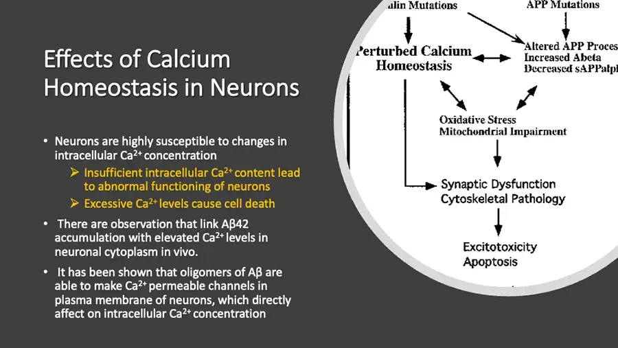
  • Effects of Calcium Homeostasis in Neurons Neurons are highly susceptible to changes in intracellular Ca2+ concentration Insufficient intracellular Ca2+ content lead to abnormal functioning of neurons Excessive Ca2+ levels cause cell death There are observation that link Aβ42 accumulation with elevated Ca2+ levels in neuronal cytoplasm in vivo. It has been shown that oligomers of Aβ are able to make Ca2+ permeable channels in plasma membrane of neurons, which directly affect on intracellular Ca2+ concentration Flow diagram, circular crop: mutations to calcium disruption to apoptosis. ilin Mutations label cut off at left. APP Mutations Perturbed Calcium Homeostasis Altered APP Proces cut off at right. Increased Abeta Decreased sAPPalph cut off at right. Oxidative Stress Mitochondrial Impairment Synaptic Dysfunction Cytoskeletal Pathology Excitotoxicity Apoptosis
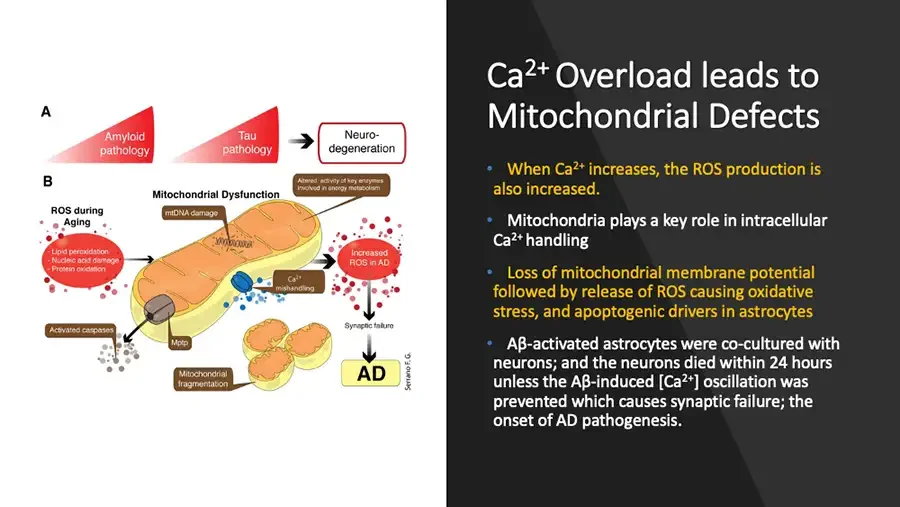
  • Diagram: amyloid and tau pathology, mitochondrial dysfunction leading to AD. A Amyloid pathology Tau pathology Neuro-degeneration B Altered activity of key enzymes involved in energy metabolism Mitochondrial Dysfunction ROS during Aging mtDNA damage Lipid peroxidation Nucleic acid damage Protein oxidation Increased ROS in AD Ca2+ mishandling Activated caspases Mptp Synaptic failure Mitochondrial fragmentation AD Serrano F. G. Ca2+ Overload leads to Mitochondrial Defects When Ca2+ increases, the ROS production is also increased. Mitochondria plays a key role in intracellular Ca2+ handling Loss of mitochondrial membrane potential followed by release of ROS causing oxidative stress, and apoptogenic drivers in astrocytes Aβ-activated astrocytes were co-cultured with neurons; and the neurons died within 24 hours unless the Aβ-induced Ca2+. oscillation was prevented which causes synaptic failure; the onset of AD pathogenesis.
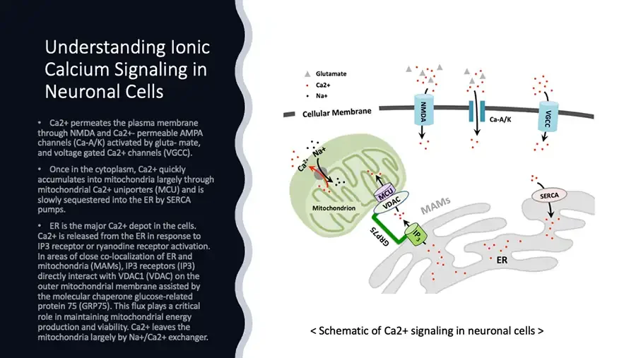
  • Understanding Ionic Calcium Signaling in Neuronal Cells Ca2+ permeates the plasma membrane through NMDA and Ca2+- permeable AMPA channels (Ca-A/K) activated by gluta- mate, and voltage gated Ca2+ channels (VGCC). Once in the cytoplasm, Ca2+ quickly accumulates into mitochondria largely through mitochondrial Ca2+ uniporters (MCU) and is slowly sequestered into the ER by SERCA pumps. ER is the major Ca2+ depot in the cells. Ca2+ is released from the ER in response to IP3 receptor or ryanodine receptor activation. In areas of close co-localization of ER and mitochondria (MAMs), IP3 receptors (IP3) directly interact with VDAC1 (VDAC) on the outer mitochondrial membrane assisted by the molecular chaperone glucose-related protein 75 (GRP75). This flux plays a critical role in maintaining mitochondrial energy production and viability. Ca2+ leaves the mitochondria largely by Na+/Ca2+ exchanger. Diagram: neuronal membrane channels, mitochondrion and ER with calcium flows. Legend symbols: gray triangle, red dot, black dot. Glutamate Ca2+ Na+ Cellular Membrane NMDA Ca-A/K VGCC Ca2+ Na+ MCU VDAC Mitochondrion MAMs SERCA GRP75 IP3 ER < Schematic of Ca2+ signaling in neuronal cells >

AIC’s EFFECT on PARKINSON’s ALS & MS
  • AIC’s EFFECT ON PARKINSON’S ALS & MS Image: translucent blue brain with glowing red spot.
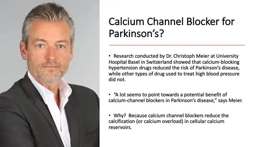
  • Photo: gray-haired man in dark jacket and white shirt. Calcium Channel Blocker for Parkinson’s? Research conducted by Dr. Christoph Meier at University Hospital Basel in Switzerland showed that calcium-blocking hypertension drugs reduced the risk of Parkinson’s disease, while other types of drug used to treat high blood pressure did not. “A lot seems to point towards a potential benefit of calcium-channel blockers in Parkinson’s disease,” says Meier. Why? Because calcium channel blockers reduce the calcification (or calcium overload) in cellular calcium reservoirs.
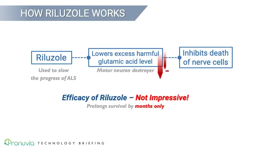
  • HOW RILUZOLE WORKS Flow diagram: three boxes joined by dashed lines. Riluzole Used to slow the progress of ALS Lowers excess harmful glutamic acid level Motor neuron destroyer Red downward arrows with minus sign. Inhibits death of nerve cells Efficacy of Riluzole – Not Impressive! Prolongs survival by months only Pronuvia logo. Pronuvia TECHNOLOGY BRIEFING
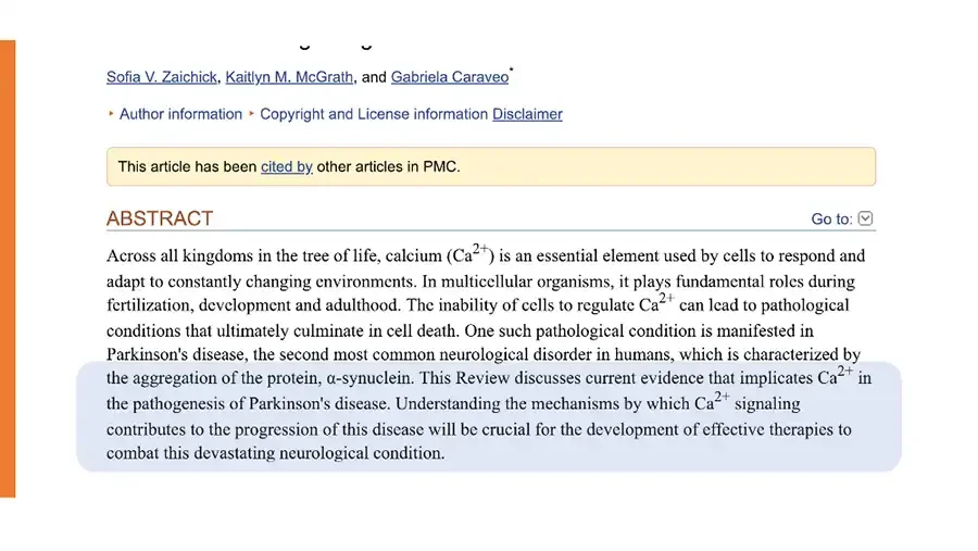
  • Webpage screenshot of journal article; title line cut off at top. Sofia V. Zaichick, Kaitlyn M. McGrath, and Gabriela Caraveo* ▸ Author information ▸ Copyright and License information Disclaimer This article has been cited by other articles in PMC. ABSTRACT Go to: dropdown icon. Across all kingdoms in the tree of life, calcium (Ca2+) is an essential element used by cells to respond and adapt to constantly changing environments. In multicellular organisms, it plays fundamental roles during fertilization, development and adulthood. The inability of cells to regulate Ca2+ can lead to pathological conditions that ultimately culminate in cell death. One such pathological condition is manifested in Parkinson's disease, the second most common neurological disorder in humans, which is characterized by the aggregation of the protein, α-synuclein. This Review discusses current evidence that implicates Ca2+ in the pathogenesis of Parkinson's disease. Understanding the mechanisms by which Ca2+ signaling contributes to the progression of this disease will be crucial for the development of effective therapies to combat this devastating neurological condition. Light blue highlight over the abstract's last four lines.
  • This Review discusses current evidence that implicates Ca2+ in the pathogenesis of Parkinson's disease. Understanding the mechanisms by which Ca2+ signaling contributes to the progression of this disease will be crucial for the development of effective therapies to combat this devastating neurological condition. An emerging, key pathological feature caused by α-synuclein aggregation is the disruption of calcium (Ca2+) homeostasis. Because Ca2+ signaling affects all aspects of neuronal cell biology, cells must tightly regulate Ca2+ levels to avoid uncontrolled responses that could otherwise lead to pathological conditions and cell death. In this Review, we discuss the current evidence that implicates defective Ca2+ homeostasis in the pathogenesis of PD. Elucidating the role of α-synuclein, and of other PD-associated proteins, in Ca2+ homeostasis could provide new opportunities for developing novel therapeutics to treat synucleinopathies.
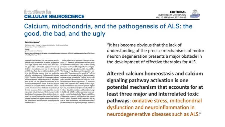
  • frontiers in CELLULAR NEUROSCIENCE EDITORIAL published: 31 October 2013 doi: 10.3389/fncel.2013.00199 Journal logo icon. Calcium, mitochondria, and the pathogenesis of ALS: the good, the bad, and the ugly Small image of the article's first page. Manoj Kumar Jaiswal* Next five lines are very small and blurred; best reading. Department of Anatomy, Physiology, and Genetics, School of Medicine, USUHS, Bethesda, MD, USA *Correspondence: manoj.jaiswal.ctr@usuhs.edu Edited by: Egidio D'Angelo , University of Pavia, Italy Keywords: amyotrophic lateral sclerosis, calcium homeostasis dysregulation, mitochondrial dysfunction, neurodegeneration, calcium buffer, selective vulnerability, motor neuron disease Amyotrophic lateral sclerosis (ALS) is a devastating neurodegenerative disease characterized by the selective and progressive loss of upper and lower motor neurons (MNs) of the brainstem, spinal cord and cerebral cortex. Key discoveries in the field of ALS have exponentially increased since it was first described in 1869 by Jean Martin Charcot, and the identification in 1993 of the first ALS-causing mutations in the gene encoding the well-studied antioxidant enzyme Cu, Zn superoxide dismutase (SOD1). The etiology of sporadic ALS largely remains unknown and the mechanisms of MN degeneration are still being investigated. The only FDA drug approved for the treatment of ALS, riluzole only modestly benefits patients, but multiple drugs are currently in the development pipeline and in human ALS clinical trials. It has become obvious that the lack of understanding of the precise mechanisms of motor neuron degeneration presents a major obstacle in the development of effective therapies for ALS. Altered calcium homeostasis and calcium signaling pathway activation is one potential mechanism that accounts for at least three major and interrelated toxic pathways: oxidative stress, mitochondrial dysfunction and neuroinflammation in neurodegenerative diseases such as ALS. Next line cut off at bottom edge. Further evidence for the involvement of disruption of intracellular Ca2+ homeostasis arises from several studies of cellular and experimental animal models of ALS in which Ca2+ binding proteins such as calbindin-D28K and parvalbumin in MN populations are lost early (hypoglossal, spinal, and lower cranial MNs). These findings are in good agreement with a quantitative comparison of Ca2+ homeostasis where low cytosolic Ca2+ buffering capacity acts as an important risk factor for degeneration and in contrast an increase in cytosolic Ca2+ buffering capacity could protect vulnerable MNs from degeneration both in-vitro and in-vivo. Several lines of evidence support altered Ca2+ homeostasis leading to MN degeneration in ALS, notably disturbance of glutamate neurotransmission and subsequent glutamate triggered Ca2+ entry, increased extracellular glutamate levels probably due to reduced glial glutamate uptake caused by oxidative damage to EAAT2, study of fALS in cell lines and animal mouse models where potential mechanism for Ca2+ disruption is inhibition of glial glutamate transport by mSOD1 similar to those proposed for sALS. Evidence also suggests that ROS generated in MNs can cross the plasma membrane and cause oxidative disruption of glutamate transporters in neighbouring astrocytes. However, in Next line cut off at bottom edge. “It has become obvious that the lack of understanding of the precise mechanisms of motor neuron degeneration presents a major obstacle in the development of effective therapies for ALS. Altered calcium homeostasis and calcium signaling pathway activation is one potential mechanism that accounts for at least three major and interrelated toxic pathways: oxidative stress, mitochondrial dysfunction and neuroinflammation in neurodegenerative diseases such as ALS.”
  • “These findings are in good agreement with a quantitative comparison of Ca2+ homeostasis where low cytosolic Ca2+ buffering capacity acts as an important risk factor for degeneration, and in contrast an increase in cytosolic Ca2+ buffering capacity could protect vulnerable MNs from degeneration both in-vitro and in-vivo."
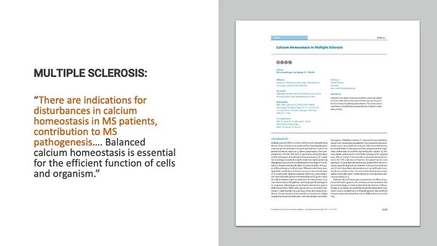
  • MULTIPLE SCLEROSIS: “There are indications for disturbances in calcium homeostasis in MS patients, contribution to MS pathogenesis…. Balanced calcium homeostasis is essential for the efficient function of cells and organism.” Small image of a journal article's first page; most text too small to read. Thieme Calcium Homeostasis in Multiple Sclerosis Creative Commons license icons. Authors Petra Hundehege, Lisa Epping, S. G. Meuth Affiliation Key words Bibliography Correspondence ABSTRACT Introduction E127
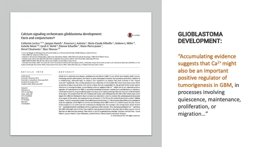
  • Small image of a journal article's first page. Calcium signaling orchestrates glioblastoma development: Facts and conjunctures☆ CrossMark logo. CrossMark Catherine Leclerc a,b,*, Jacques Haiech c, Francisco J. Aulestia a, Marie-Claude Kilhoffer c, Andrew L. Miller d, Isabelle Néant a,b, Sarah E. Webb d, Etienne Schaeffer e, Marie-Pierre Junier f, Hervé Chneiweiss f, Marc Moreau a,b Affiliation lines below are very small and blurred; best reading. a Centre de Biologie du Développement, Université Toulouse 3, 118 route de Narbonne, F31062 Toulouse, Cedex 04, France b CNRS UMR5547 , Toulouse F31062 , France c Laboratoire d'Innovation Thérapeutique, Laboratoire d'Excellence Medalis , UMR 7200 Université de Strasbourg / CNRS, 67412 Illkirch, France d Division of Life Science and State Key Laboratory of Molecular Neuroscience, HKUST, Clear Water Bay, Hong Kong, PR China e IREBS UMR7242 ESBS, Pôle API , Parc d'Innovation d'Illkirch, 67412 Illkirch cedex, France f Sorbonne Universités, UPMC Univ Paris 06, Centre National de la Recherche Scientifique (CNRS), UMR8246 , Institut National de la Santé et de la Recherche Médicale (INSERM), U1130 , Institut de Biologie Paris Seine (IBPS), Neuroscience Paris Seine (NPS), Team Glial Plasticity, 7/9 Quai St Bernard, Paris, France ARTICLE INFO Article history: Received 2 December 2015 Received in revised form 18 January 2016 Accepted 22 January 2016 Available online 28 January 2016 Keywords: Glioblastoma Calcium signaling Cancer stem cells Cell competition Quiescence Proliferation Migration Inflammation ABSTRACT While it is a relatively rare disease, glioblastoma multiform (GBM) is one of the more deadly adult cancers. Following current interventions, the tumor is never eliminated whatever the treatment performed; whether it is radiotherapy, chemotherapy, or surgery. One hypothesis to explain this poor outcome is the “cancer stem cell” hypothesis. This concept proposes that a minority of cells within the tumor mass share many of the properties of adult neural stem cells and it is these that are responsible for the growth of the tumor and its resistance to existing therapies. Accumulating evidence suggests that Ca2+ might also be an important positive regulator of tumorigeness in GBM, in processes involving quiescence, maintenance, proliferation, or migration. Glioblastoma tumors are generally thought to develop by co-opting pathways that are involved in the formation of an organ. We propose that the cells initiating the tumor, and subsequently the cells of the tumor mass, must hijack the different checkpoints that evolution has selected in order to prevent the pathological development of an organ. In this article, two main points are discussed. (i) The first is the establishment of a so-called “cellular society,” which is required to create a favorable microenvironment. (ii) The second is that GBM can be considered to be an organism, which fights to survive and develop. Since GBM evolves in a limited space, its only chance of development is to overcome the evolutionary checkpoints. For example, the deregulation of the normal Ca2+ signaling elements contributes to the progression of the disease. Thus, by manipulating the Ca2+ signaling, the GBM cells might not be killed, but might be reprogrammed toward a new fate that is either easy to cure or that has no aberrant functioning. This article is part of a Special Issue entitled: Calcium and Cell Fate . Guest Editors: Jacques Haiech, Claus Heizmann, Joachim Krebs, Thierry Capiod and Olivier Mignen. © 2016 Elsevier B.V. All rights reserved. GLIOBLASTOMA DEVELOPMENT: “Accumulating evidence suggests that Ca2+ might also be an important positive regulator of tumorigenesis in GBM, in processes involving quiescence, maintenance, proliferation, or migration…”

RECENT WORK ON INTERCELLULAR CALCIUM SIGNALLING IN ASD
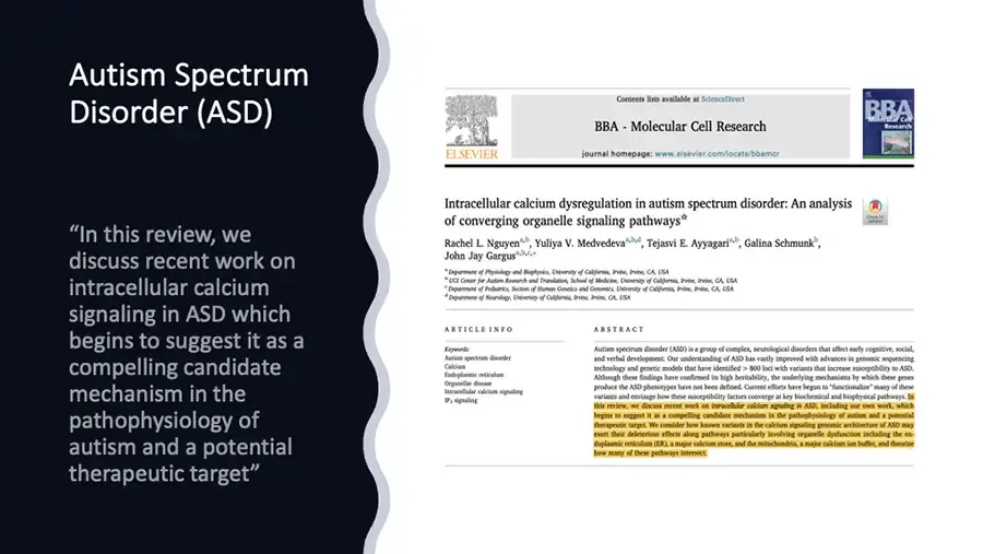
  • Autism Spectrum Disorder (ASD) “In this review, we discuss recent work on intracellular calcium signaling in ASD which begins to suggest it as a compelling candidate mechanism in the pathophysiology of autism and a potential therapeutic target” Screenshot of journal article title page. Elsevier tree logo. ELSEVIER Contents lists available at ScienceDirect BBA - Molecular Cell Research journal homepage: www.elsevier.com/locate/bbamcr Journal cover image. BBA Molecular Cell Research Intracellular calcium dysregulation in autism spectrum disorder: An analysis of converging organelle signaling pathways☆ Check for updates icon. Rachel L. Nguyena,b, Yuliya V. Medvedevaa,b,d, Tejasvi E. Ayyagaria,b, Galina Schmunkb, John Jay Gargusa,b,c,* a Department of Physiology and Biophysics, University of California, Irvine, Irvine, CA, USA b UCI Center for Autism Research and Translation, School of Medicine, University of California, Irvine, Irvine, CA, USA c Department of Pediatrics, Section of Human Genetics and Genomics, University of California, Irvine, Irvine, CA, USA d Department of Neurology, University of California, Irvine, Irvine, CA, USA ARTICLE INFO Keywords: Autism spectrum disorder Calcium Endoplasmic reticulum Organellar disease Intracellular calcium signaling IP3 signaling ABSTRACT Autism spectrum disorder (ASD) is a group of complex, neurological disorders that affect early cognitive, social, and verbal development. Our understanding of ASD has vastly improved with advances in genomic sequencing technology and genetic models that have identified > 800 loci with variants that increase susceptibility to ASD. Although these findings have confirmed its high heritability, the underlying mechanisms by which these genes produce the ASD phenotypes have not been defined. Current efforts have begun to “functionalize” many of these variants and envisage how these susceptibility factors converge at key biochemical and biophysical pathways. In this review, we discuss recent work on intracellular calcium signaling in ASD, including our own work, which begins to suggest it as a compelling candidate mechanism in the pathophysiology of autism and a potential therapeutic target. We consider how known variants in the calcium signaling genomic architecture of ASD may exert their deleterious effects along pathways particularly involving organelle dysfunction including the endoplasmic reticulum (ER), a major calcium store, and the mitochondria, a major calcium ion buffer, and theorize how many of these pathways intersect. Yellow highlight from "In this review" to the end of the abstract.
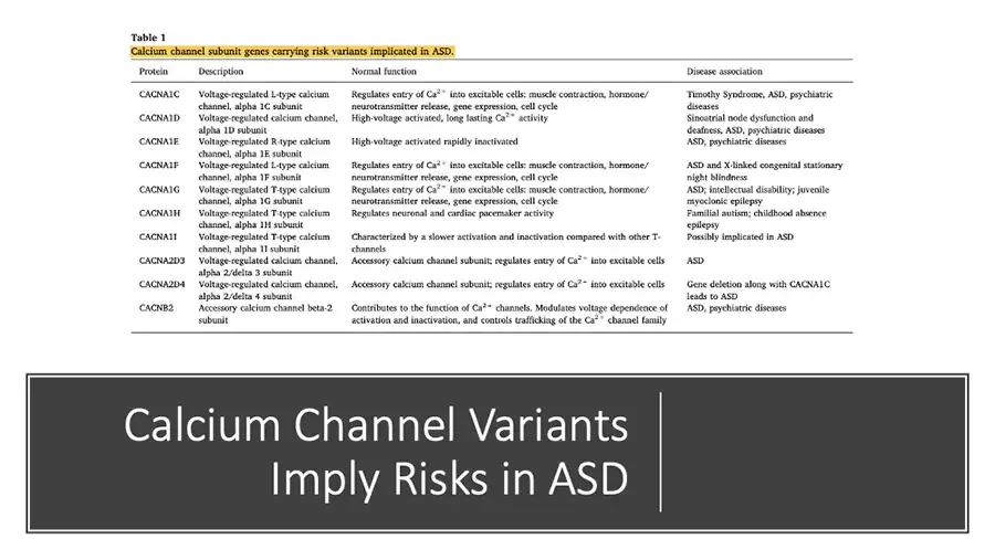
  • Table 1 Calcium channel subunit genes carrying risk variants implicated in ASD. Yellow highlight on table caption. Protein Description Normal function Disease association CACNA1C Voltage-regulated L-type calcium channel, alpha 1C subunit Regulates entry of Ca2+ into excitable cells: muscle contraction, hormone/neurotransmitter release, gene expression, cell cycle Timothy Syndrome, ASD, psychiatric diseases CACNA1D Voltage-regulated calcium channel, alpha 1D subunit High-voltage activated, long lasting Ca2+ activity Sinoatrial node dysfunction and deafness, ASD, psychiatric diseases CACNA1E Voltage-regulated R-type calcium channel, alpha 1E subunit High-voltage activated rapidly inactivated ASD, psychiatric diseases CACNA1F Voltage-regulated L-type calcium channel, alpha 1F subunit Regulates entry of Ca2+ into excitable cells: muscle contraction, hormone/neurotransmitter release, gene expression, cell cycle ASD and X-linked congenital stationary night blindness CACNA1G Voltage-regulated T-type calcium channel, alpha 1G subunit Regulates entry of Ca2+ into excitable cells: muscle contraction, hormone/neurotransmitter release, gene expression, cell cycle ASD; intellectual disability; juvenile myoclonic epilepsy CACNA1H Voltage-regulated T-type calcium channel, alpha 1H subunit Regulates neuronal and cardiac pacemaker activity Familial autism; childhood absence epilepsy CACNA1I Voltage-regulated T-type calcium channel, alpha 1I subunit Characterized by a slower activation and inactivation compared with other T-channels Possibly implicated in ASD CACNA2D3 Voltage-regulated calcium channel, alpha 2/delta 3 subunit Accessory calcium channel subunit; regulates entry of Ca2+ into excitable cells ASD CACNA2D4 Voltage-regulated calcium channel, alpha 2/delta 4 subunit Accessory calcium channel subunit; regulates entry of Ca2+ into excitable cells Gene deletion along with CACNA1C leads to ASD CACNB2 Accessory calcium channel beta-2 subunit Contributes to the function of Ca2+ channels. Modulates voltage dependence of activation and inactivation, and controls trafficking of the Ca2+ channel family ASD, psychiatric diseases Calcium Channel Variants Imply Risks in ASD
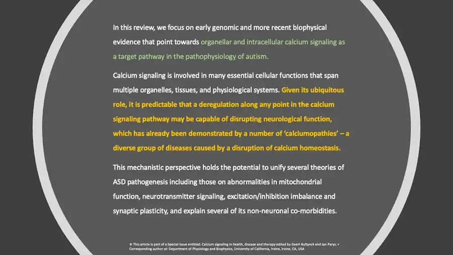
  • In this review, we focus on early genomic and more recent biophysical evidence that point towards organellar and intracellular calcium signaling as a target pathway in the pathophysiology of autism. Calcium signaling is involved in many essential cellular functions that span multiple organelles, tissues, and physiological systems. Given its ubiquitous role, it is predictable that a deregulation along any point in the calcium signaling pathway may be capable of disrupting neurological function, which has already been demonstrated by a number of ‘calciumopathies’ – a diverse group of diseases caused by a disruption of calcium homeostasis. This mechanistic perspective holds the potential to unify several theories of ASD pathogenesis including those on abnormalities in mitochondrial function, neurotransmitter signaling, excitation/inhibition imbalance and synaptic plasticity, and explain several of its non-neuronal co-morbidities. ☆ This article is part of a Special Issue entitled: Calcium signaling in health, disease and therapy edited by Geert Bultynck and Jan Parys. • Corresponding author at: Department of Physiology and Biophysics, University of California, Irvine, Irvine, CA, USA
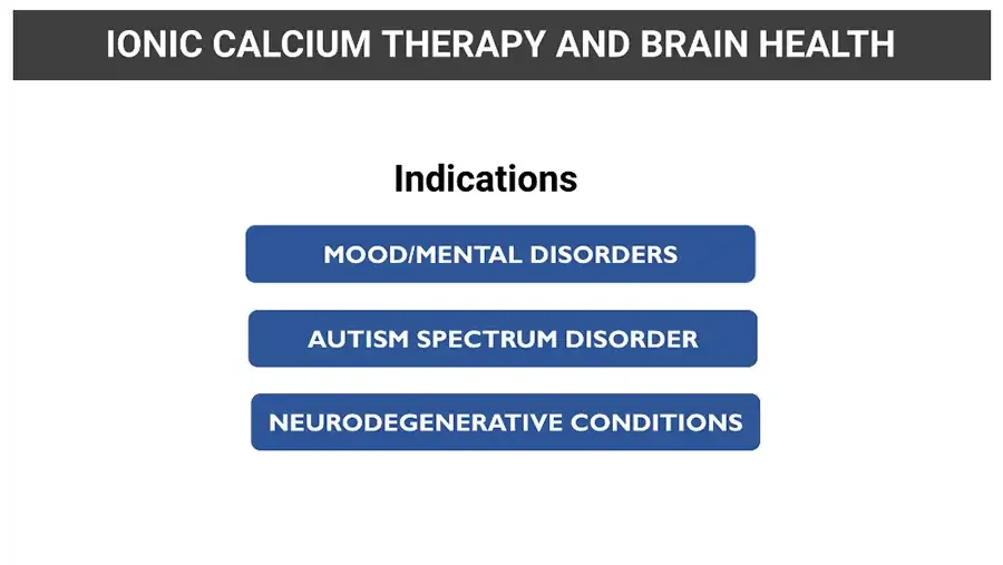
  • IONIC CALCIUM THERAPY AND BRAIN HEALTH Indications MOOD/MENTAL DISORDERS AUTISM SPECTRUM DISORDER NEURODEGENERATIVE CONDITIONS
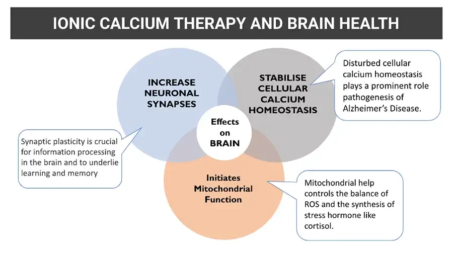
  • IONIC CALCIUM THERAPY AND BRAIN HEALTH Diagram: three overlapping circles around a central circle, with callout boxes. INCREASE NEURONAL SYNAPSES STABILISE CELLULAR CALCIUM HOMEOSTASIS Disturbed cellular calcium homeostasis plays a prominent role pathogenesis of Alzheimer’s Disease. Effects on BRAIN Synaptic plasticity is crucial for information processing in the brain and to underlie learning and memory Initiates Mitochondrial Function Mitochondrial help controls the balance of ROS and the synthesis of stress hormone like cortisol.

CASE STUDY1: DEMENTIA

  • Background: glowing blue network of lines and dots. Case Study 1: Dementia Logo: puzzle pieces; QR code. BRAINWORKS Live your Life without Limits
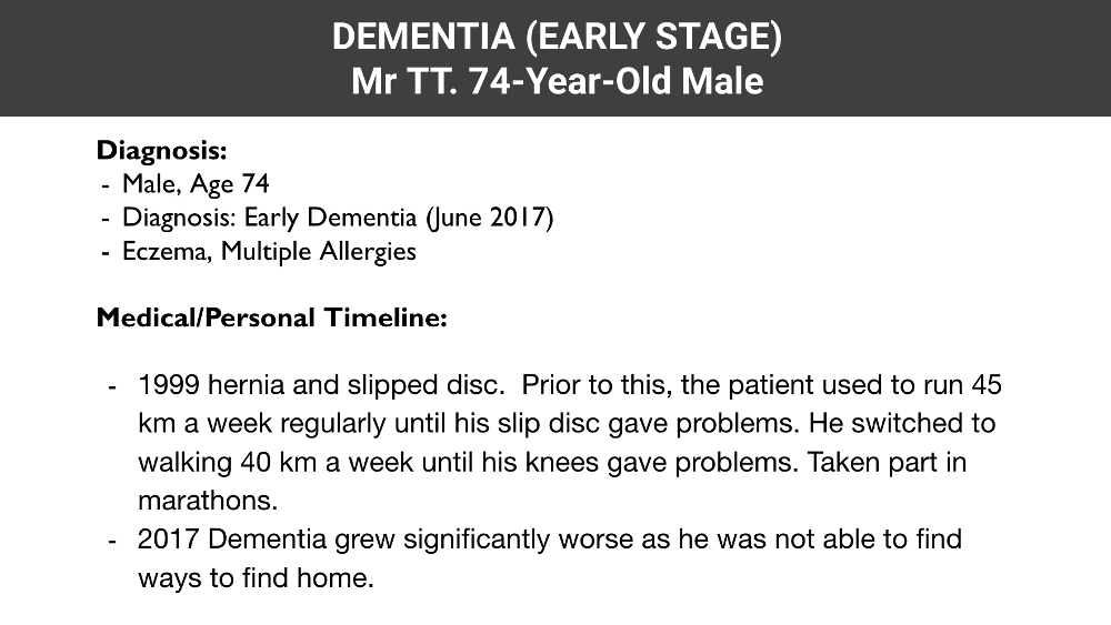
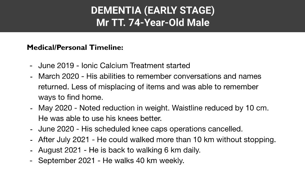
  • DEMENTIA (EARLY STAGE) Mr TT. 74-Year-Old Male Diagnosis: Male, Age 74 Diagnosis: Early Dementia (June 2017) Eczema, Multiple Allergies Medical/Personal Timeline: 1999 hernia and slipped disc. Prior to this, the patient used to run 45 km a week regularly until his slip disc gave problems. He switched to walking 40 km a week until his knees gave problems. Taken part in marathons. 2017 Dementia grew significantly worse as he was not able to find ways to find home.
  • DEMENTIA (EARLY STAGE) Mr TT. 74-Year-Old Male Medical/Personal Timeline: June 2019 - Ionic Calcium Treatment started March 2020 - His abilities to remember conversations and names returned. Less of misplacing of items and was able to remember ways to find home. May 2020 - Noted reduction in weight. Waistline reduced by 10 cm. He was able to use his knees better. June 2020 - His scheduled knee caps operations cancelled. After July 2021 - He could walked more than 10 km without stopping. August 2021 - He is back to walking 6 km daily. September 2021 - He walks 40 km weekly.

CASE STUDY2: DEMENTIA

  • Background: glowing blue network of lines and dots. Case Study 2: Dementia Logo: puzzle pieces; QR code. BRAINWORKS Live your Life without Limits
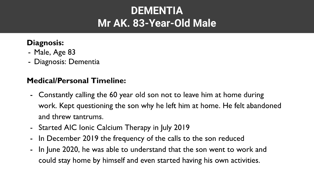
  • DEMENTIA Mr AK. 83-Year-Old Male Diagnosis: Male, Age 83 Diagnosis: Dementia Medical/Personal Timeline: Constantly calling the 60 year old son not to leave him at home during work. Kept questioning the son why he left him at home. He felt abandoned and threw tantrums. Started AIC Ionic Calcium Therapy in July 2019 In December 2019 the frequency of the calls to the son reduced In June 2020, he was able to understand that the son went to work and could stay home by himself and even started having his own activities.

CASE STUDY3: AUTISM WITH EPILEPSY

  • Background: glowing blue network of lines and dots. Case Study 3: Autism with Epilepsy Logo: puzzle pieces; QR code. BRAINWORKS Live your Life without Limits
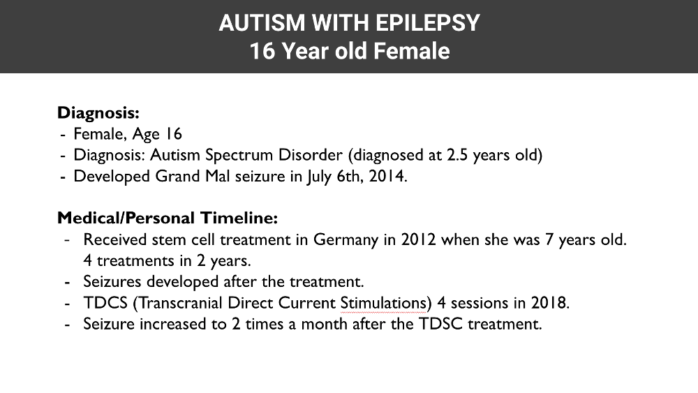
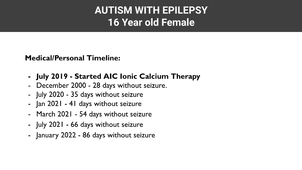
  • AUTISM WITH EPILEPSY 16 Year old Female Diagnosis: Female, Age 16 Diagnosis: Autism Spectrum Disorder (diagnosed at 2.5 years old) Developed Grand Mal seizure in July 6th, 2014. Medical/Personal Timeline: Received stem cell treatment in Germany in 2012 when she was 7 years old. 4 treatments in 2 years. Seizures developed after the treatment. TDCS (Transcranial Direct Current Stimulations) 4 sessions in 2018. Red spell-check underline under "Stimulations". Seizure increased to 2 times a month after the TDSC treatment.
  • AUTISM WITH EPILEPSY 16 Year old Female Medical/Personal Timeline: July 2019 - Started AIC Ionic Calcium Therapy December 2000 - 28 days without seizure. July 2020 - 35 days without seizure Jan 2021 - 41 days without seizure March 2021 - 54 days without seizure July 2021 - 66 days without seizure January 2022 - 86 days without seizure

SAMPLE TREATMENT CASE: AUTISM SPECTRUM DISORDER

  • Background: glowing blue network of lines and dots. Sample Treatment Case Autism Spectrum Disorder Logo: puzzle pieces; QR code. BRAINWORKS Live your Life without Limits
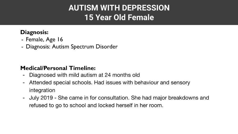
  • AUTISM WITH DEPRESSION 15 Year Old Female Diagnosis: Female, Age 16 Diagnosis: Autism Spectrum Disorder Medical/Personal Timeline: Diagnosed with mild autism at 24 months old Attended special schools. Had issues with behaviour and sensory integration July 2019 - She came in for consultation. She had major breakdowns and refused to go to school and locked herself in her room.
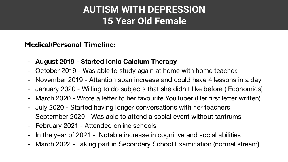
  • AUTISM WITH DEPRESSION 15 Year Old Female Medical/Personal Timeline: August 2019 - Started Ionic Calcium Therapy October 2019 - Was able to study again at home with home teacher. November 2019 - Attention span increase and could have 4 lessons in a day January 2020 - Willing to do subjects that she didn’t like before ( Economics) March 2020 - Wrote a letter to her favourite YouTuber (Her first letter written) July 2020 - Started having longer conversations with her teachers September 2020 - Was able to attend a social event without tantrums February 2021 - Attended online schools In the year of 2021 - Notable increase in cognitive and social abilities March 2022 - Taking part in Secondary School Examination (normal stream)

AIC CLINICAL TESTIMONY

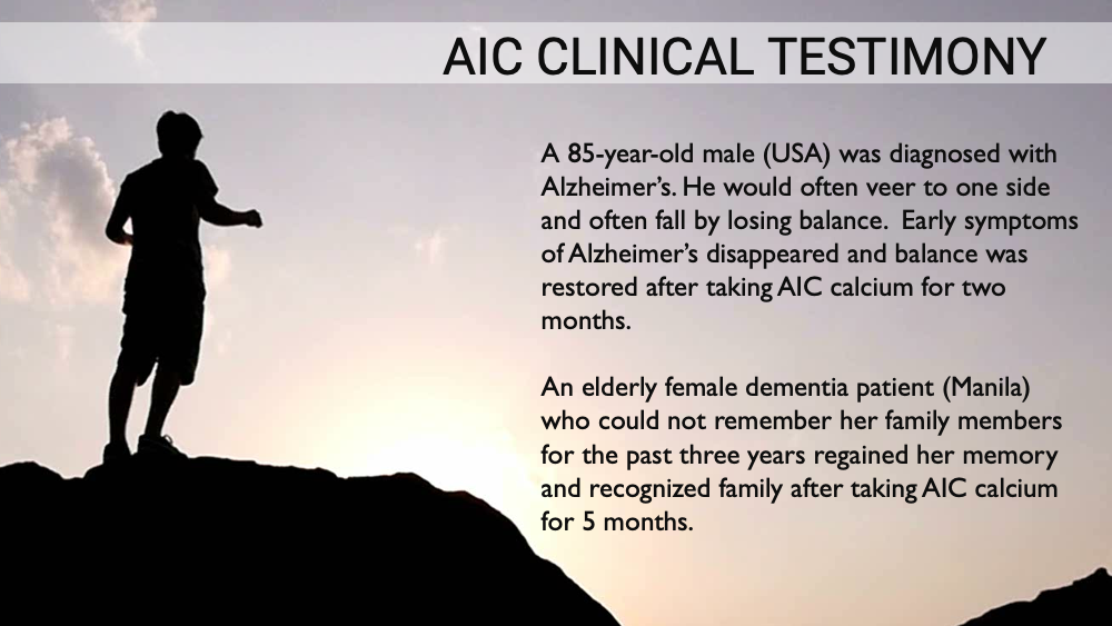
  • AIC CLINICAL TESTIMONY Photo: silhouette of person standing on rocky hilltop at sunset. A 85-year-old male (USA) was diagnosed with Alzheimer’s. He would often veer to one side and often fall by losing balance. Early symptoms of Alzheimer’s disappeared and balance was restored after taking AIC calcium for two months. An elderly female dementia patient (Manila) who could not remember her family members for the past three years regained her memory and recognized family after taking AIC calcium for 5 months.
  • AIC CLINICAL TESTIMONY Photo: silhouette of person standing on rocky hilltop at sunset. A 19 year old male (USA) was diagnosed with early stage ALS. Early symptoms of ALS disappeared after taking AIC calcium for two months. A 65 year old male with late stage ALS was not able to go to the toilet by himself but was able to go to the toilet alone after taking AIC calcium for 3~4 days.
  • AIC CLINICAL TESTIMONY Photo: silhouette of person standing on rocky hilltop at sunset. A 53-year-old male often had swallowing difficulties due to neck muscle weakness. He was able to swallow food easily after taking AIC calcium. A 65-year-old female was diagnosed with MS (multiple sclerosis). After taking AIC calcium for 1 week, she was prescribed lower dose aspirin (from 320mg to 80mg), and her circulation functions were also improved.
  • AIC CLINICAL TESTIMONY Photo: silhouette of person standing on rocky hilltop at sunset. “Dear Dr. Lee, Here is an update of the patient with Parkinson’s disease. I saw him in the church today, and he was ecstatic to see me and wanted to share with me his testimonials. He started out taking three bottles of AIC since October of 2017 without many expectations. However, after others commented that he seems to be doing much better, he was encouraged to take it faithfully through November, twice a day. He said his tremors almost disappeared! He is 75 years old, and his Parkinson’s was not hereditary but caused by severe stress from his work. After the onset of the disease, his life quality was miserable but now lives a new life because of AIC. Though he is not a Christian, he came to my church to share the good news with me! H. Shim”

BONE AS AN ENDOCRINE ORGAN
  • BONE AS AN ENDOCRINE ORGAN A genetics approach has uncovered that bone has more functions than expected. In particular, bone is an endocrine organ that secretes a growing number of hormones. In that context, the discovery of the osteoblast-derived hormone osteocalcin has significantly broadened the field of bone biology because of the number of physiologic processes regulated by this hormone. At present, osteocalcin has been shown to enhance several aspects of energy metabolism, brain development, and cognition. These discoveries shed light on the cross-talk between multiple organs and provide credence to the search for additional endocrine functions of bone
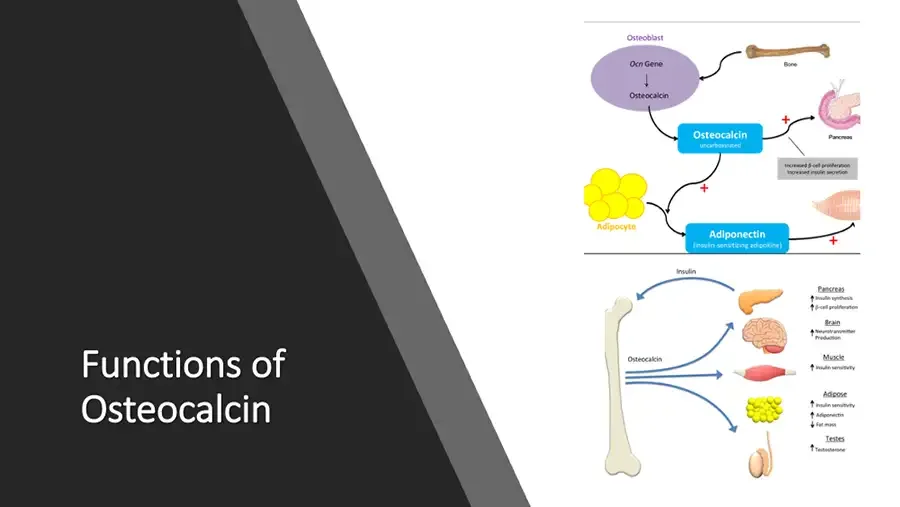
  • Functions of Osteocalcin Diagram: osteocalcin from osteoblasts acting on pancreas, fat cells and muscle. Osteoblast Ocn Gene ↓ Osteocalcin Bone Osteocalcin uncarboxylated + Pancreas Increased β-cell proliferation Increased insulin secretion + Adipocyte Adiponectin (insulin sensitizing adipokine) + Diagram: bone sending osteocalcin to pancreas, brain, muscle, fat and testes. Insulin Osteocalcin Pancreas ↑ Insulin synthesis ↑ β-cell proliferation Brain ↑ Neurotransmitter Production Muscle ↑ Insulin sensitivity Adipose ↑ Insulin sensitivity ↑ Adiponectin ↓ Fat mass Testes ↑ Testosterone
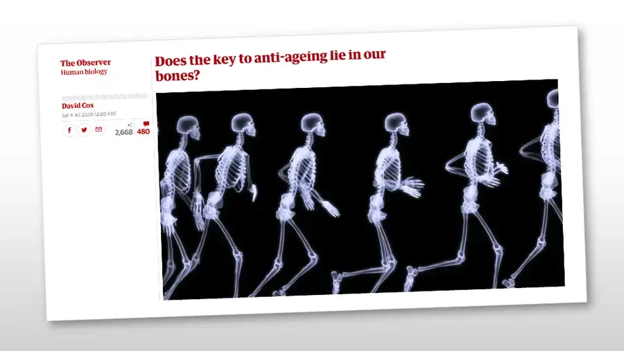
  • Screenshot of a newspaper web article, shown tilted. The Observer Human biology Decorative wavy line. David Cox Sat 4 Jul 2020 12.00 EDT Share icons: Facebook, Twitter, email. < 2,668 Comment icon. 480 Does the key to anti-ageing lie in our bones? Image: X-ray style skeletons running in a row.
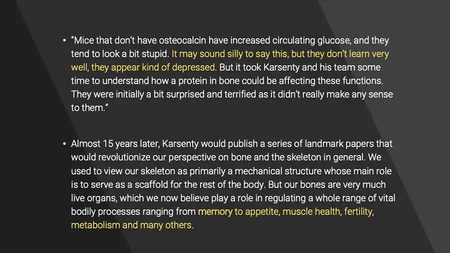
  • “Mice that don’t have osteocalcin have increased circulating glucose, and they tend to look a bit stupid. It may sound silly to say this, but they don’t learn very well, they appear kind of depressed. But it took Karsenty and his team some time to understand how a protein in bone could be affecting these functions. They were initially a bit surprised and terrified as it didn’t really make any sense to them.” Almost 15 years later, Karsenty would publish a series of landmark papers that would revolutionize our perspective on bone and the skeleton in general. We used to view our skeleton as primarily a mechanical structure whose main role is to serve as a scaffold for the rest of the body. But our bones are very much live organs, which we now believe play a role in regulating a whole range of vital bodily processes ranging from memory to appetite, muscle health, fertility, metabolism and many others.
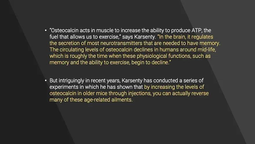
  • “Osteocalcin acts in muscle to increase the ability to produce ATP, the fuel that allows us to exercise,” says Karsenty. “In the brain, it regulates the secretion of most neurotransmitters that are needed to have memory. The circulating levels of osteocalcin declines in humans around mid-life, which is roughly the time when these physiological functions, such as memory and the ability to exercise, begin to decline.” But intriguingly in recent years, Karsenty has conducted a series of experiments in which he has shown that by increasing the levels of osteocalcin in older mice through injections, you can actually reverse many of these age-related ailments.
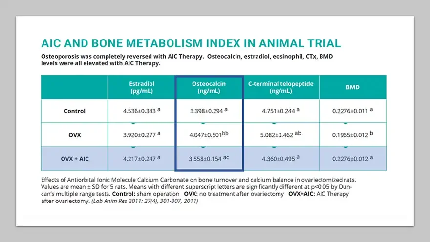
  • AIC AND BONE METABOLISM INDEX IN ANIMAL TRIAL Osteoporosis was completely reversed with AIC Therapy. Osteocalcin, estradiol, eosinophil, CTx, BMD levels were all elevated with AIC Therapy. Estradiol (pg/mL) Osteocalcin (ng/mL) C-terminal telopeptide (ng/mL) BMD Control 4.536±0.343 a 3.398±0.294 a 4.751±0.244 a 0.2276±0.011 a OVX 3.920±0.277 a 4.047±0.501bb 5.082±0.462 ab 0.1965±0.012 b OVX + AIC 4.217±0.247 a 3.558±0.154 ac 4.360±0.495 a 0.2276±0.012 a Osteocalcin column outlined in dark blue; OVX + AIC row shaded light blue. Effects of Antiorbital Ionic Molecule Calcium Carbonate on bone turnover and calcium balance in ovariectomized rats. Values are mean ± SD for 5 rats. Means with different superscript letters are significantly different at p<0.05 by Duncan’s multiple range tests. Control: sham operation OVX: no treatment after ovariectomy OVX+AIC: AIC Therapy after ovariectomy. (Lab Anim Res 2011: 27(4), 301-307, 2011)
  • Background: blue painted circle on white. “Osteocalcin seems to be able to reverse manifestations of ageing in the brain and in muscle,” he says. “What is remarkable is that if you give osteocalcin to old mice, you restore memory and you restore the ability to exercise to the levels seen in a young mouse. That makes it potentially extremely attractive from a medical point of view.” Dr. Gerald Karsenty
Mental & Cognitive Benefits of AIC “After taking AIC, a 17-year-old autistic teenager who had had no conversation with his mother started to make comments like “Your new hair looks good!” when his mother walked in with new hairdo from hair salon. The entire family atmosphere brightened up”. (More expressive) “A first-grade elementary school child with ADHD had never sat at his desk more than five minutes. However, after taking AIC calcium, he reads books for more than half an hour and wanted to continue reading instead of playing outside!” “A woman in her 50’s who was suffering from panic attacks stayed in her room most of the time and did not go out. After taking AIC, she now lives a normal life. Once she visited me with home baked cookies and thanked me." Underlined in image: "autistic teenager", "ADHD", "panic attacks".
Benefits of AIC for Sleep A 60-year-old male had suffered from insomnia for more than 50 years. Ever since his childhood years, strangely he could not fall asleep and continued to turn on his bed all night, keeping his parents up as well. His lifelong wish was to sleep sound just for one full hour. He had tried every treatment available from what western to eastern medicine have to offer in treating insomnia but to no avail. Then from his shoulders down to his arms, he experienced numbing like paralysis. Only after pounding and massaging arms he could gain some senses. His head felt foggy all the time, and to ease the misery he constantly drank liquors which aggravated his conditions more. One day, a good friend of his introduced him to AIC therapy. After taking the first dose, in his head, he felt like a big ice block melting in strong sun rays and then found himself waking up refreshed the next morning. He said he never slept like that all his life and never felt that good waking up refreshed after sleeping. His depression subsided with the treatment and he felt as if he was living a new life. It’s been 30 days already and he still sleeps well, experiencing a total renewal in his life. He told me, “I still cannot believe this is really happening to me. All my friends are amazed, too. I think God sent me an angel. Even my shoulders and arms have regained senses. I have written thousands of poems through those sleepless nights, but now I am looking forward to how my poems will change after sleeping through the full nights. Underlined in image: "insomnia" (first mention); "He said he never slept like that all his life and never felt that good waking up refreshed after sleeping".