Notice. The following information is provided for your convenience. ACRI is not directly involved in this research and does not make any medical claims.

Ionic Calcium in Oncology
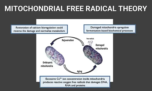
MITOCHONDRIAL FREE RADICAL THEORY Restoration of calcium bioregulation could reverse the damage and normalize metabolism Damaged mitochondria upregulate fermentation-based biochemical processes Diagram: embryonic and damaged mitochondria in an aging/rejuvenation cycle. Rejuvenation free radicals Damaged mitochondria Embryonic mitochondria Aging Excessive Ca²⁺ ion concentration inside mitochondria produces reactive oxygen free radicals that damages DNA, RNA and proteins

Calcium Dysfunction Leads to Cellular Mutation

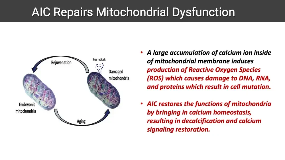
  • A large accumulation of calcium ions inside mitochondrial membranes induces the production of Reactive Oxygen Species (ROS), which causes damage to DNA, RNA, and proteins, resulting in cell mutation, the main driving force of aging and cancer.
  • Mitochondrial damage forces cells to undergo fermentation which upregulates genes to support fermentation, which is cancer cells’ preferred form of metabolism.
  • DNA repair ability is reduced in a variety of pathologic conditions. In addition, in some of these diseases, a disturbance in cellular Ca2+ homeostasis occurs, or cytosolic Ca2+ responses to various stimuli are impaired.
  • These results emphasize the importance of precise cellular Ca2+ level regulation for the optimal DNA repair process.
  • Disturbed Ca2+ metabolism and defective response to Ca2+ stimulators have been reported to exist in some patients with DNA repair deficiency… Thus impaired DNA repairability may result in part from a disturbed Ca2+ homeostasis.
Journal article first page inside a curved decorative frame. Mitochondria as sensors and regulators of calcium signalling Rosario Rizzuto, Diego De Stefani, Anna Raffaello and Cristina Mammucari
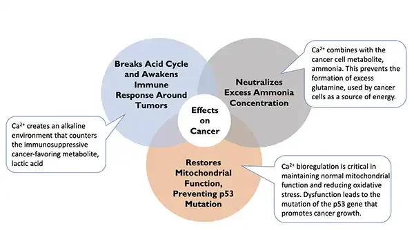
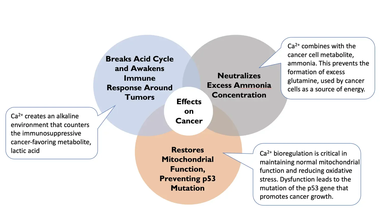
Venn diagram: three overlapping circles with callout boxes. Breaks Acid Cycle and Awakens Immune Response Around Tumors Neutralizes Excess Ammonia Concentration Ca²⁺ combines with the cancer cell metabolite, ammonia. This prevents the formation of excess glutamine, used by cancer cells as a source of energy. Ca²⁺ creates an alkaline environment that counters the immunosuppressive cancer-favoring metabolite, lactic acid Effects on Cancer Restores Mitochondrial Function, Preventing p53 Mutation Ca²⁺ bioregulation is critical in maintaining normal mitochondrial function and reducing oxidative stress. Dysfunction leads to the mutation of the p53 gene that promotes cancer growth.

Ionic Calcium Fights Cancer on Multiple Fronts

  • “The development of malignant tumors results from deregulated proliferation or an inability of cells to undergo apoptotic cell death.
  • Experimental works of the past decade have highlighted the importance of calcium (Ca2+) in the regulation of apoptosis.
  • Several studies indicate that the Ca2+ content of the endoplasmic reticulum (ER) determines the cell’s sensitivity to apoptotic stress and perturbation of ER Ca2+ homeostasis appears to be a key component in the development of several pathological situations. Sensitivity to apoptosis depends on the ability of cells to transfer Ca2+ from the ER to the mitochondria.”
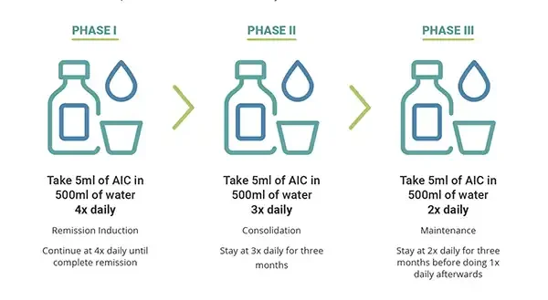
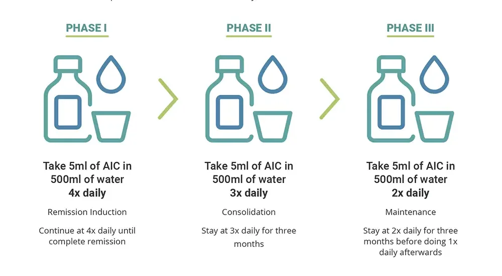
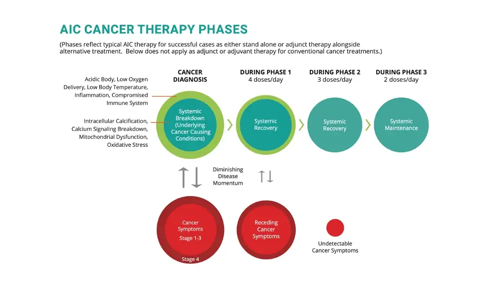
PHASE I Icons: bottle, drop and cup. Take 5ml of AIC in 500ml of water 4x daily Remission Induction Continue at 4x daily until complete remission Arrow. PHASE II Icons: bottle, drop and cup. Take 5ml of AIC in 500ml of water 3x daily Consolidation Stay at 3x daily for three months Arrow. PHASE III Icons: bottle, drop and cup. Take 5ml of AIC in 500ml of water 2x daily Maintenance Stay at 2x daily for three months before doing 1x daily afterwards

AIC Dosing for ALL Stage Cancers

Because AIC Therapy is non-toxic and building healing momentum is important, ACRI recommends minimum 4 doses of AIC therapy daily for a minimum of 3 months to see the benefits of AIC therapy to be convinced to continue. Please take AIC on an empty stomach at least 30 minutes before a meal. Take one dose after waking up, one at 30 minutes before lunch, one at 30 minutes before dinner, and one at 2 hours before bed. (Ionic calcium could trigger an urge to urinate. Blood tests are recommended for every 2-4 weeks to monitor progress. DXA/ultrasound bone scans are also recommended at the beginning of each phase to measure bone health progress. Reaching the minimum T score of -1.0 (normal) is recommended before moving into Phase 3 if the patient started with low bone density.

AIC Ionic Calcium Therapy for Oncology

MULTIPLE MYELOMA #1
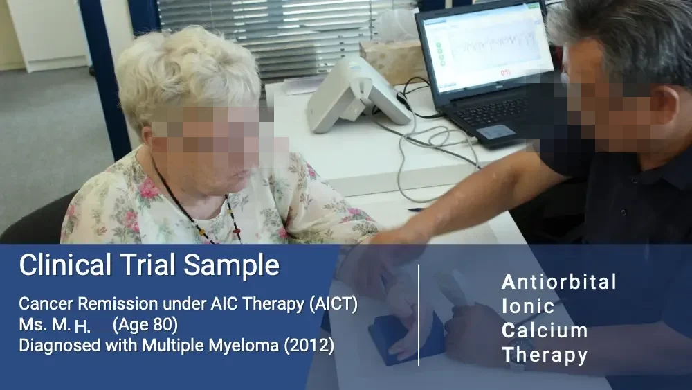
  • Photo: elderly woman next to man at laptop. 0% Clinical Trial Sample Cancer Remission under AIC Therapy (AICT) Ms. M. H. (Age 80) Diagnosed with Multiple Myeloma (2012) Antiorbital Ionic Calcium Therapy
  • Venn diagram: three overlapping circles with callout boxes. Breaks Acid Cycle and Awakens Immune Response Around Tumors Neutralizes Excess Ammonia Concentration Ca²⁺ combines with the cancer cell metabolite, ammonia. This prevents the formation of excess glutamine, used by cancer cells as a source of energy. Ca²⁺ creates an alkaline environment that counters the immunosuppressive cancer-favoring metabolite, lactic acid Effects on Cancer Restores Mitochondrial Function, Preventing p53 Mutation Ca²⁺ bioregulation is critical in maintaining normal mitochondrial function and reducing oxidative stress. Dysfunction leads to the mutation of the p53 gene that promotes cancer growth.
  • PHASE I Icons: bottle, drop and cup. Take 5ml of AIC in 500ml of water 4x daily Remission Induction Continue at 4x daily until complete remission Arrow. PHASE II Icons: bottle, drop and cup. Take 5ml of AIC in 500ml of water 3x daily Consolidation Stay at 3x daily for three months Arrow. PHASE III Icons: bottle, drop and cup. Take 5ml of AIC in 500ml of water 2x daily Maintenance Stay at 2x daily for three months before doing 1x daily afterwards
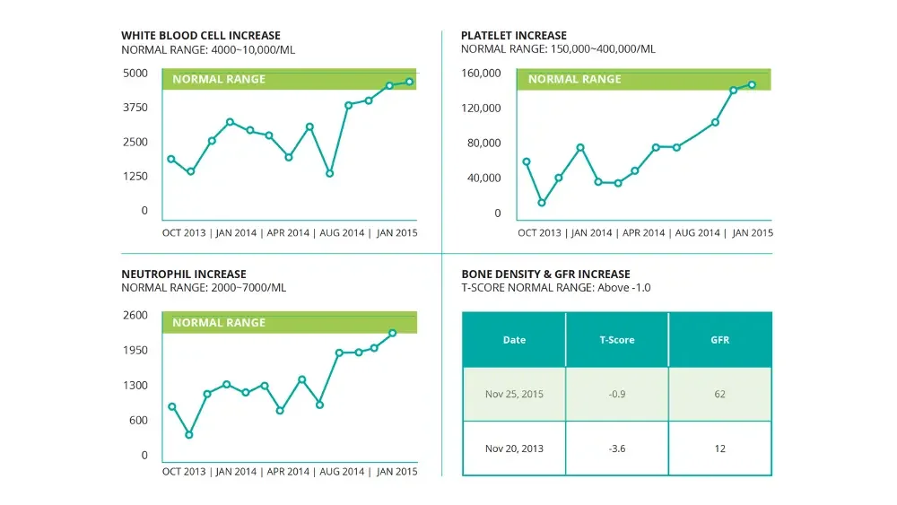
  • WHITE BLOOD CELL INCREASE NORMAL RANGE: 400010,000/ML Line chart. 5000 3750 2500 1250 0 NORMAL RANGE OCT 2013 | JAN 2014 | APR 2014 | AUG 2014 | JAN 2015 PLATELET INCREASE NORMAL RANGE: 150,000400,000/ML Line chart. 160,000 120,000 80,000 40,000 0 NORMAL RANGE OCT 2013 | JAN 2014 | APR 2014 | AUG 2014 | JAN 2015 NEUTROPHIL INCREASE NORMAL RANGE: 2000~7000/ML Line chart. 2600 1950 1300 600 0 NORMAL RANGE OCT 2013 | JAN 2014 | APR 2014 | AUG 2014 | JAN 2015 BONE DENSITY & GFR INCREASE T-SCORE NORMAL RANGE: Above -1.0 Date T-Score GFR Nov 25, 2015 -0.9 62 Nov 20, 2013 -3.6 12
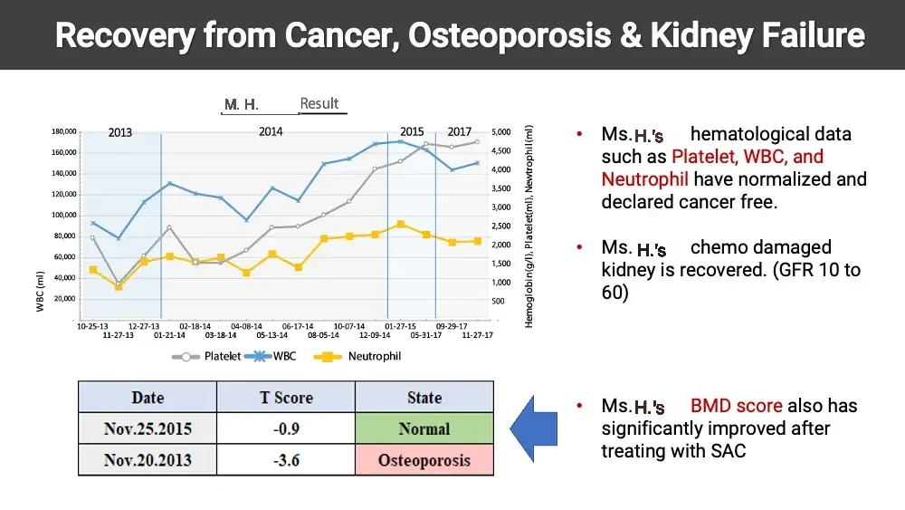
  • Recovery from Cancer, Osteoporosis & Kidney Failure M. H. Result Line chart: Platelet, WBC and Neutrophil over time. 2013 2014 2015 2017 180,000 160,000 140,000 120,000 100,000 80,000 60,000 40,000 20,000 WBC (ml) 5,000 4,500 4,000 3,500 3,000 2,500 2,000 1,500 1,000 500 Hemoglobin(g/l), Platelet(ml), Newtrophil(ml) 10-25-13 11-27-13 12-27-13 01-21-14 02-18-14 03-18-14 04-08-14 05-13-14 06-17-14 08-05-14 10-07-14 12-09-14 01-27-15 05-31-17 09-29-17 11-27-17 Platelet WBC Neutrophil Date T Score State Nov.25.2015 -0.9 Normal Nov.20.2013 -3.6 Osteoporosis Arrow pointing left toward the table. Ms. H.’s hematological data such as Platelet, WBC, and Neutrophil have normalized and declared cancer free. Ms. H.’s chemo damaged kidney is recovered. (GFR 10 to 60) Ms. H.’s BMD score also has significantly improved after treating with SAC
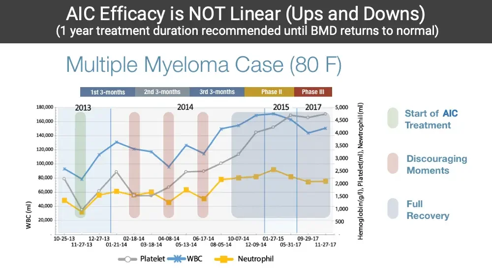
  • AIC Efficacy is NOT Linear (Ups and Downs) (1 year treatment duration recommended until BMD returns to normal) Multiple Myeloma Case (80 F) 1st 3-months 2nd 3-months 3rd 3-months Phase II Phase III Line chart: Platelet, WBC and Neutrophil with shaded periods. 2013 2014 2015 2017 180,000 160,000 140,000 120,000 100,000 80,000 60,000 40,000 20,000 WBC (ml) 5,000 4,500 4,000 3,500 3,000 2,500 2,000 1,500 1,000 500 Hemoglobin(g/l), Platelet(ml), Newtrophil(ml) 10-25-13 11-27-13 12-27-13 01-21-14 02-18-14 03-18-14 04-08-14 05-13-14 06-17-14 08-05-14 10-07-14 12-09-14 01-27-15 05-31-17 09-29-17 11-27-17 Platelet WBC Neutrophil Green marker. Start of AIC Treatment Red marker. Discouraging Moments Gray-blue marker. Full Recovery

MULTIPLE MYELOMA #2

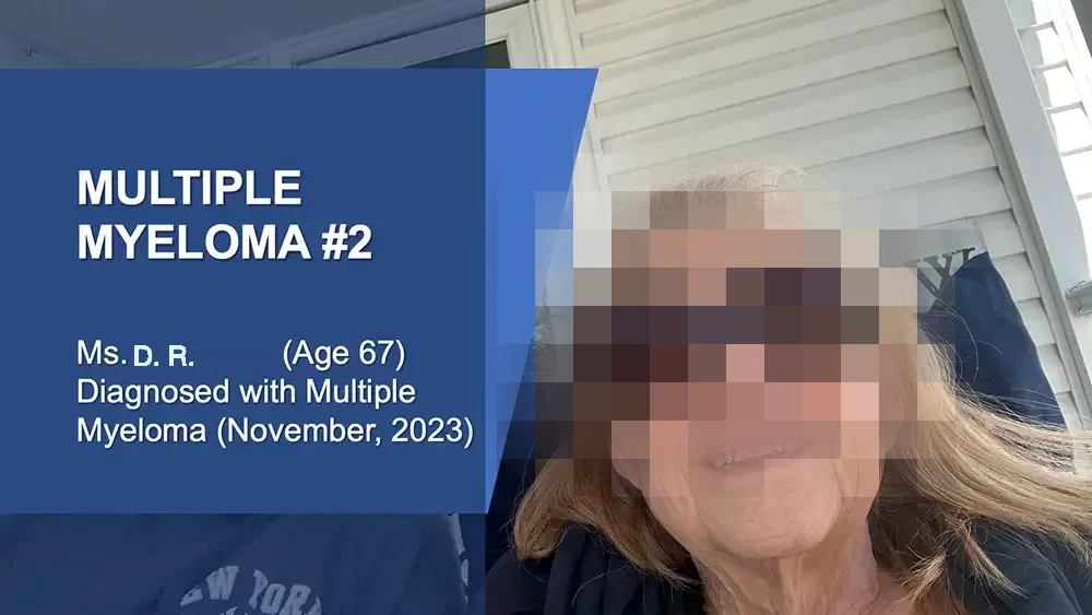
  • Photo: smiling older woman in sunglasses outdoors, selfie. MULTIPLE MYELOMA #2 Ms. D. R. (Age 67) Diagnosed with Multiple Myeloma (November, 2023) Shirt print, partly hidden. EW YORK Sign behind her head, letters partly hidden.
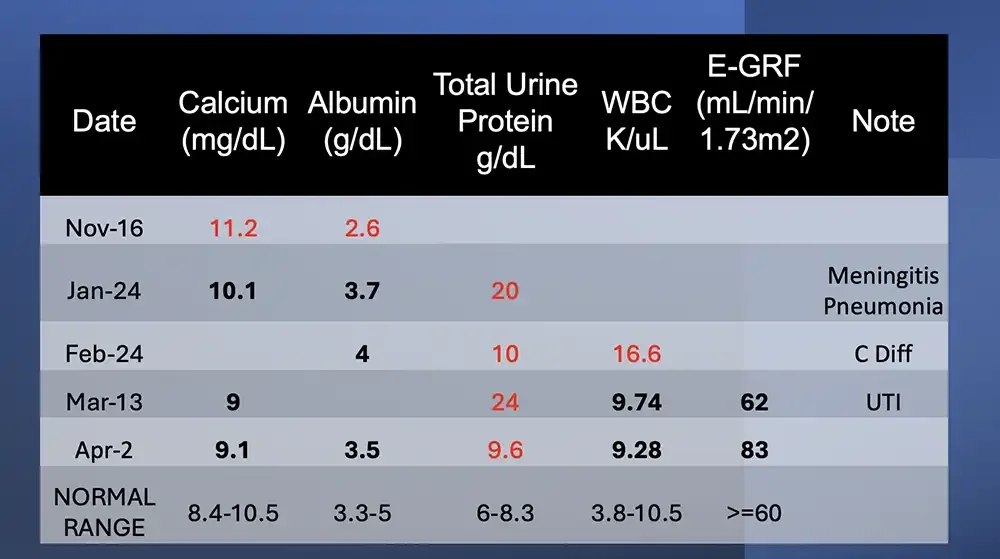
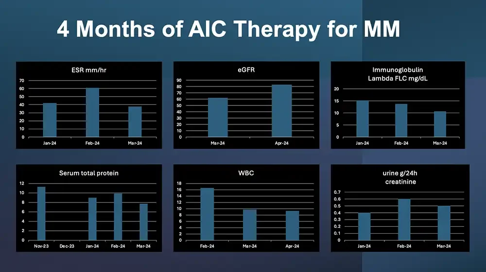
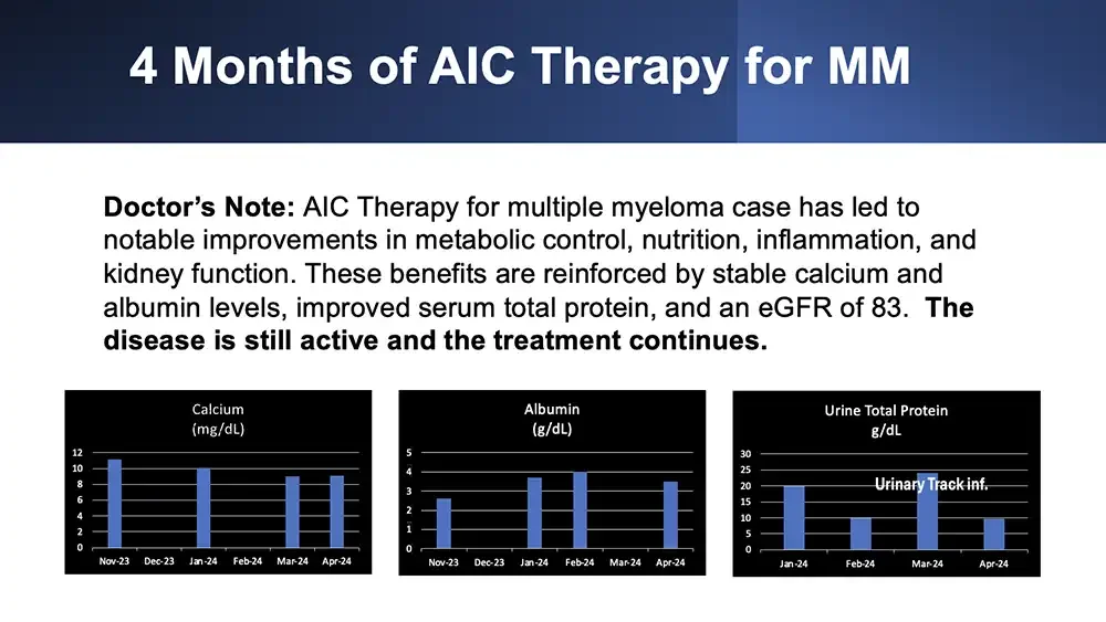
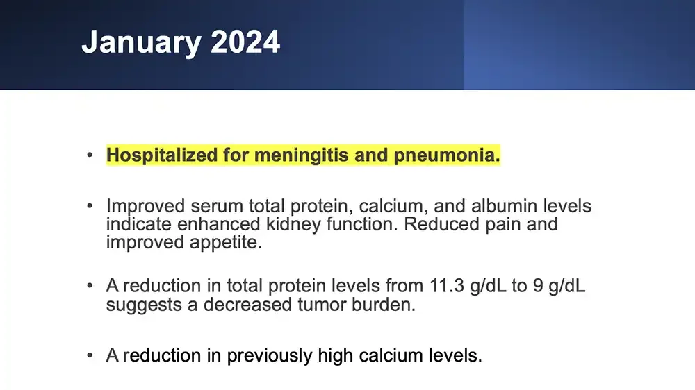
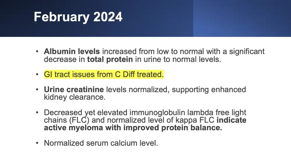
  • Date Calcium (mg/dL) Albumin (g/dL) Total Urine Protein g/dL WBC K/uL E-GRF (mL/min/1.73m2) Note Nov-16 11.2 2.6 Jan-24 10.1 3.7 20 Meningitis Pneumonia Feb-24 4 10 16.6 C Diff Mar-13 9 24 9.74 62 UTI Apr-2 9.1 3.5 9.6 9.28 83 NORMAL RANGE 8.4-10.5 3.3-5 6-8.3 3.8-10.5 >=60
  • 4 Months of AIC Therapy for MM ESR mm/hr Bar chart. 70 60 50 40 30 20 10 0 Jan-24 Feb-24 Mar-24 eGFR Bar chart. 90 80 70 60 50 40 30 20 10 0 Mar-24 Apr-24 Immunoglobulin Lambda FLC mg/dL Bar chart. 20 15 10 5 0 Jan-24 Feb-24 Mar-24 Serum total protein Bar chart. 12 10 8 6 4 2 0 Nov-23 Dec-23 Jan-24 Feb-24 Mar-24 WBC Bar chart. 18 16 14 12 10 8 6 4 2 0 Feb-24 Mar-24 Apr-24 urine g/24h creatinine Bar chart. 0.7 0.6 0.5 0.4 0.3 0.2 0.1 0 Jan-24 Feb-24 Mar-24
  • 4 Months of AIC Therapy for MM Doctor’s Note: AIC Therapy for multiple myeloma case has led to notable improvements in metabolic control, nutrition, inflammation, and kidney function. These benefits are reinforced by stable calcium and albumin levels, improved serum total protein, and an eGFR of 83. The disease is still active and the treatment continues. Calcium (mg/dL) Bar chart. 12 10 8 6 4 2 0 Nov-23 Dec-23 Jan-24 Feb-24 Mar-24 Apr-24 Albumin (g/dL) Bar chart. 5 4 3 2 1 0 Nov-23 Dec-23 Jan-24 Feb-24 Mar-24 Apr-24 Urine Total Protein g/dL Bar chart. 30 25 20 15 10 5 0 Urinary Track inf. Jan-24 Feb-24 Mar-24 Apr-24
  • January 2024 Hospitalized for meningitis and pneumonia. Improved serum total protein, calcium, and albumin levels indicate enhanced kidney function. Reduced pain and improved appetite. A reduction in total protein levels from 11.3 g/dL to 9 g/dL suggests a decreased tumor burden. A reduction in previously high calcium levels.
  • February 2024 Albumin levels increased from low to normal with a significant decrease in total protein in urine to normal levels. GI tract issues from C Diff treated. Urine creatinine levels normalized, supporting enhanced kidney clearance. Decreased yet elevated immunoglobulin lambda free light chains (FLC) and normalized level of kappa FLC indicate active myeloma with improved protein balance. Normalized serum calcium level.
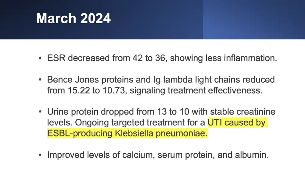
  • March 2024 ESR decreased from 42 to 36, showing less inflammation. Bence Jones proteins and Ig lambda light chains reduced from 15.22 to 10.73, signaling treatment effectiveness. Urine protein dropped from 13 to 10 with stable creatinine levels. Ongoing targeted treatment for a UTI caused by ESBL-producing Klebsiella pneumoniae. Improved levels of calcium, serum protein, and albumin.
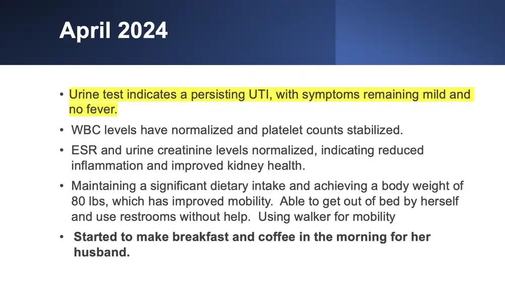
  • April 2024 Urine test indicates a persisting UTI, with symptoms remaining mild and no fever. WBC levels have normalized and platelet counts stabilized. ESR and urine creatinine levels normalized, indicating reduced inflammation and improved kidney health. Maintaining a significant dietary intake and achieving a body weight of 80 lbs, which has improved mobility. Able to get out of bed by herself and use restrooms without help. Using walker for mobility Started to make breakfast and coffee in the morning for her husband.
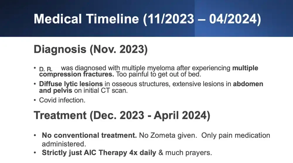
  • Medical Timeline (11/2023 – 04/2024) Diagnosis (Nov. 2023) D. R. was diagnosed with multiple myeloma after experiencing multiple compression fractures. Too painful to get out of bed. Diffuse lytic lesions in osseous structures, extensive lesions in abdomen and pelvis on initial CT scan. Covid infection. Treatment (Dec. 2023 - April 2024) No conventional treatment. No Zometa given. Only pain medication administered. Strictly just AIC Therapy 4x daily & much prayers.

PROSTATE CANCER

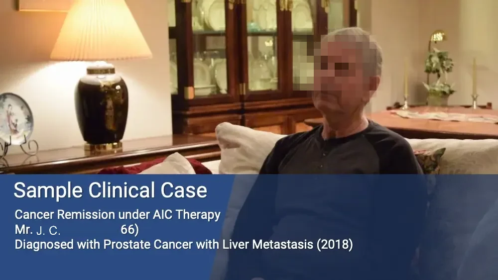
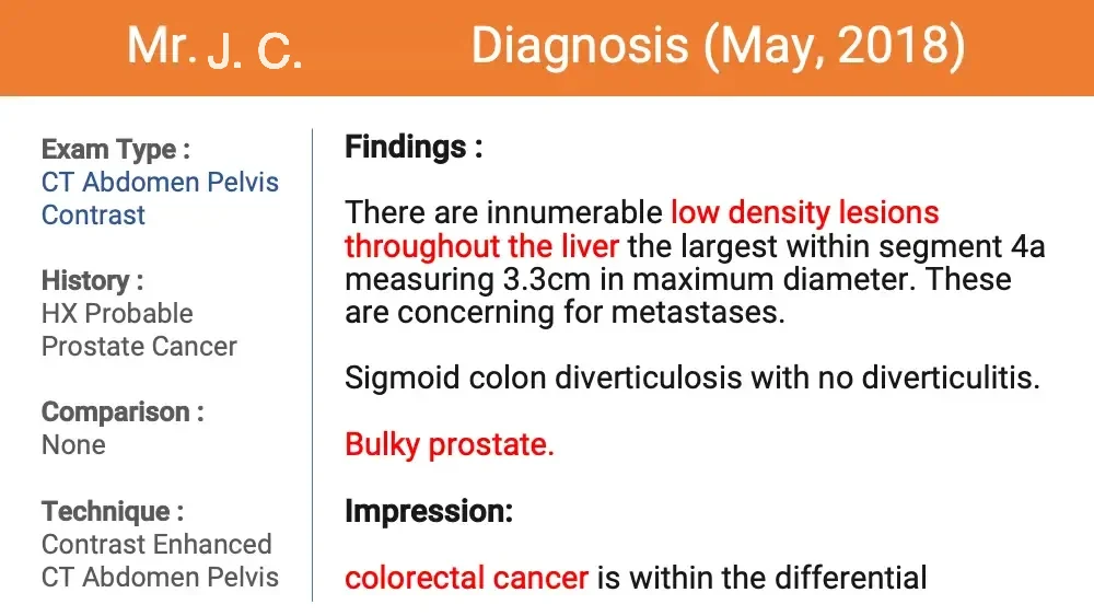
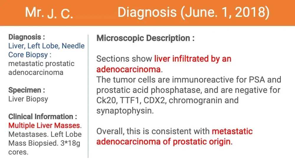
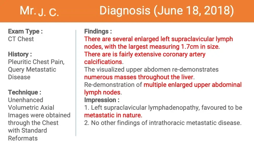
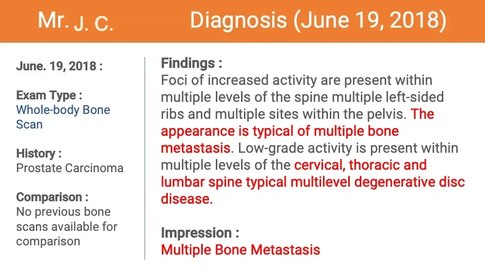
  • Photo: older man seated on a sofa in a lamplit living room. Sample Clinical Case Cancer Remission under AIC Therapy Mr. J. C. (Age 66) Diagnosed with Prostate Cancer with Liver Metastasis (2018)
  • Mr. J. C.’s Diagnosis (May, 2018) Exam Type : CT Abdomen Pelvis Contrast History : HX Probable Prostate Cancer Comparison : None Technique : Contrast Enhanced CT Abdomen Pelvis Findings : There are innumerable low density lesions throughout the liver the largest within segment 4a measuring 3.3cm in maximum diameter. These are concerning for metastases. Sigmoid colon diverticulosis with no diverticulitis. Bulky prostate. Impression: colorectal cancer is within the differential
  • Mr. J. C.’s Diagnosis (June. 1, 2018) Diagnosis : Liver, Left Lobe, Needle Core Biopsy : metastatic prostatic adenocarcinoma Specimen : Liver Biopsy Clinical Information : Multiple Liver Masses. Metastases. Left Lobe Mass Biopsied. 3*18g cores. Microscopic Description : Sections show liver infiltrated by an adenocarcinoma. The tumor cells are immunoreactive for PSA and prostatic acid phosphatase, and are negative for Ck20, TTF1, CDX2, chromogranin and synaptophysin. Overall, this is consistent with metastatic adenocarcinoma of prostatic origin.
  • Mr. J. C.’s Diagnosis (June 18, 2018) Exam Type : CT Chest History : Pleuritic Chest Pain, Query Metastatic Disease Technique : Unenhanced Volumetric Axial Images were obtained through the Chest with Standard Reformats Findings : There are several enlarged left supraclavicular lymph nodes, with the largest measuring 1.7cm in size. There are is fairly extensive coronary artery calcifications. The visualized upper abdomen re-demonstrates numerous masses throughout the liver. Re-demonstration of multiple enlarged upper abdominal lymph nodes. Impression : Left supraclavicular lymphadenopathy, favoured to be metastatic in nature. No other findings of intrathoracic metastatic disease.
  • Mr. J. C.’s Diagnosis (June 19, 2018) June. 19, 2018 : Exam Type : Whole-body Bone Scan History : Prostate Carcinoma Comparison : No previous bone scans available for comparison Findings : Foci of increased activity are present within multiple levels of the spine multiple left-sided ribs and multiple sites within the pelvis. The appearance is typical of multiple bone metastasis. Low-grade activity is present within multiple levels of the cervical, thoracic and lumbar spine typical multilevel degenerative disc disease. Impression : Multiple Bone Metastasis
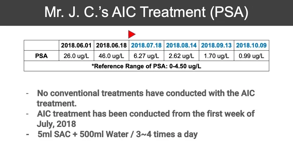
  • Mr. J. C.’s AIC Treatment (PSA) Red triangle marker above the 2018.07.18 column. 2018.06.01 2018.06.18 2018.07.18 2018.08.14 2018.09.13 2018.10.09 PSA 26.0 ug/L 46.0 ug/L 6.27 ug/L 2.62 ug/L 1.70 ug/L 0.99 ug/L *Reference Range of PSA: 0-4.50 ug/L No conventional treatments have conducted with the AIC treatment. AIC treatment has been conducted from the first week of July, 2018 5ml SAC + 500ml Water / 3~4 times a day
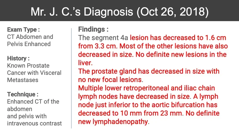
  • Mr. J. C.’s Diagnosis (Oct 26, 2018) Exam Type : CT Abdomen and Pelvis Enhanced History : Known Prostate Cancer with Visceral Metastases Technique : Enhanced CT of the abdomen and pelvis with intravenous contrast Findings : The segment 4a lesion has decreased to 1.6 cm from 3.3 cm. Most of the other lesions have also decreased in size. No definite new lesions in the liver. The prostate gland has decreased in size with no new focal lesions. Multiple lower retroperitoneal and iliac chain lymph nodes have decreased in size. A lymph node just inferior to the aortic bifurcation has decreased to 10 mm from 23 mm. No definite new lymphadenopathy.
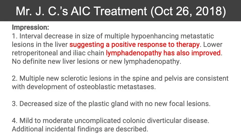
  • Mr. J. C.’s AIC Treatment (Oct 26, 2018) Impression: Interval decrease in size of multiple hypoenhancing metastatic lesions in the liver suggesting a positive response to therapy. Lower retroperitoneal and iliac chain lymphadenopathy has also improved. No definite new liver lesions or new lymphadenopathy. Multiple new sclerotic lesions in the spine and pelvis are consistent with development of osteoblastic metastases. Decreased size of the plastic gland with no new focal lesions. Mild to moderate uncomplicated colonic diverticular disease. Additional incidental findings are described.

OVARY CANCER (STAGE4)

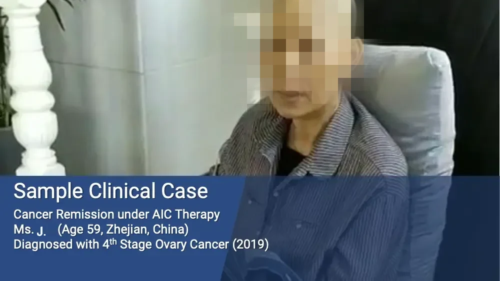
  • Photo: woman with shaved head seated in an armchair. Sample Clinical Case Cancer Remission under AIC Therapy Ms. J. (Age 59, Zhejian, China) Diagnosed with 4th Stage Ovary Cancer (2019)
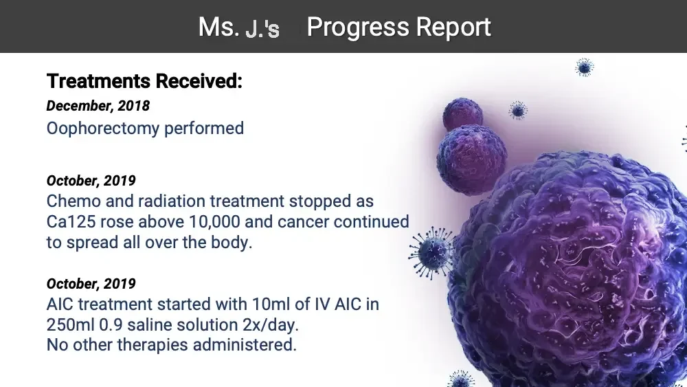
  • Ms. J.’s Progress Report Treatments Received: December, 2018 Oophorectomy performed October, 2019 Chemo and radiation treatment stopped as Ca125 rose above 10,000 and cancer continued to spread all over the body. October, 2019 AIC treatment started with 10ml of IV AIC in 250ml 0.9 saline solution 2x/day. No other therapies administered. Illustration: purple cancer cells and virus-like particles.
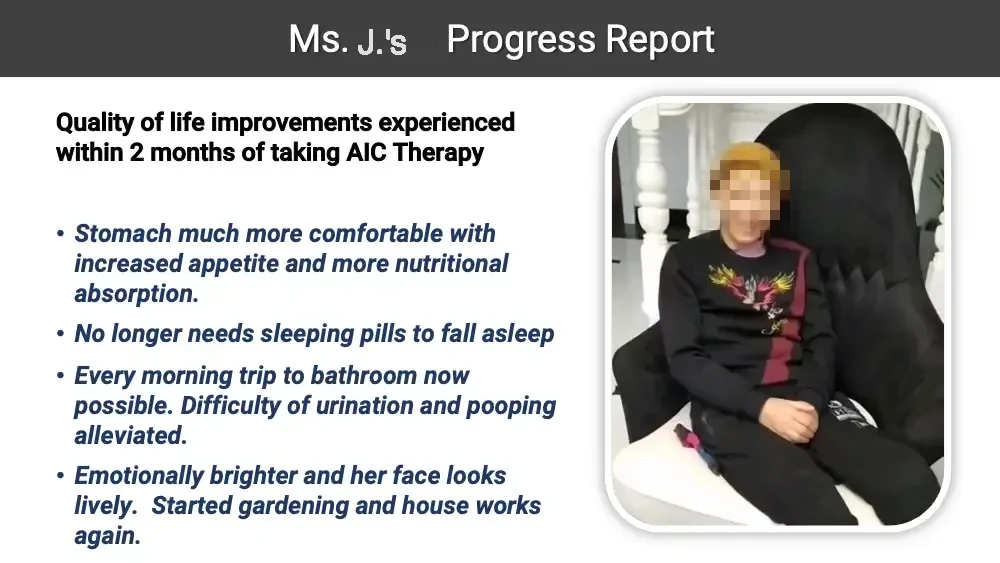
  • Ms. J.’s Progress Report Quality of life improvements experienced within 2 months of taking AIC Therapy Stomach much more comfortable with increased appetite and more nutritional absorption. No longer needs sleeping pills to fall asleep Every morning trip to bathroom now possible. Difficulty of urination and pooping alleviated. Emotionally brighter and her face looks lively. Started gardening and house works again. Photo: smiling woman in a knit hat seated in a chair.
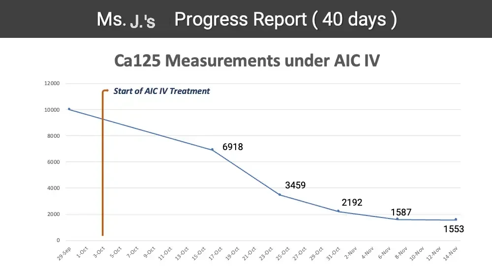
  • Ms. J.’s Progress Report ( 40 days ) Ca125 Measurements under AIC IV Line chart. 12000 10000 8000 6000 4000 2000 0 Start of AIC IV Treatment 6918 3459 2192 1587 1553 29-Sep 1-Oct 3-Oct 5-Oct 7-Oct 9-Oct 11-Oct 13-Oct 15-Oct 17-Oct 19-Oct 21-Oct 23-Oct 25-Oct 27-Oct 29-Oct 31-Oct 2-Nov 4-Nov 6-Nov 8-Nov 10-Nov 12-Nov 14-Nov

CERVICAL CANCER

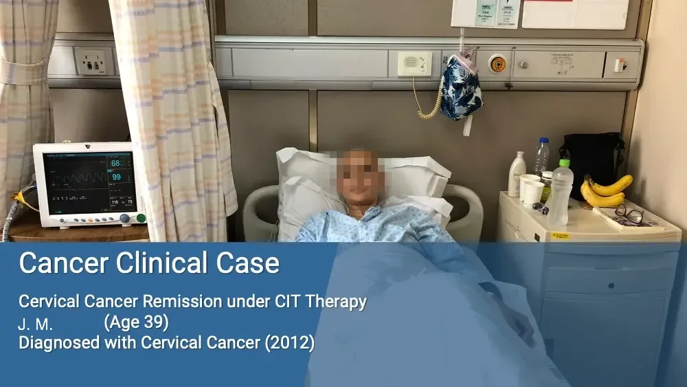
  • Photo: smiling woman with shaved head in a hospital bed. 68 99 116 79 Cancer Clinical Case Cervical Cancer Remission under CIT Therapy J. M. (Age 39) Diagnosed with Cervical Cancer (2012)
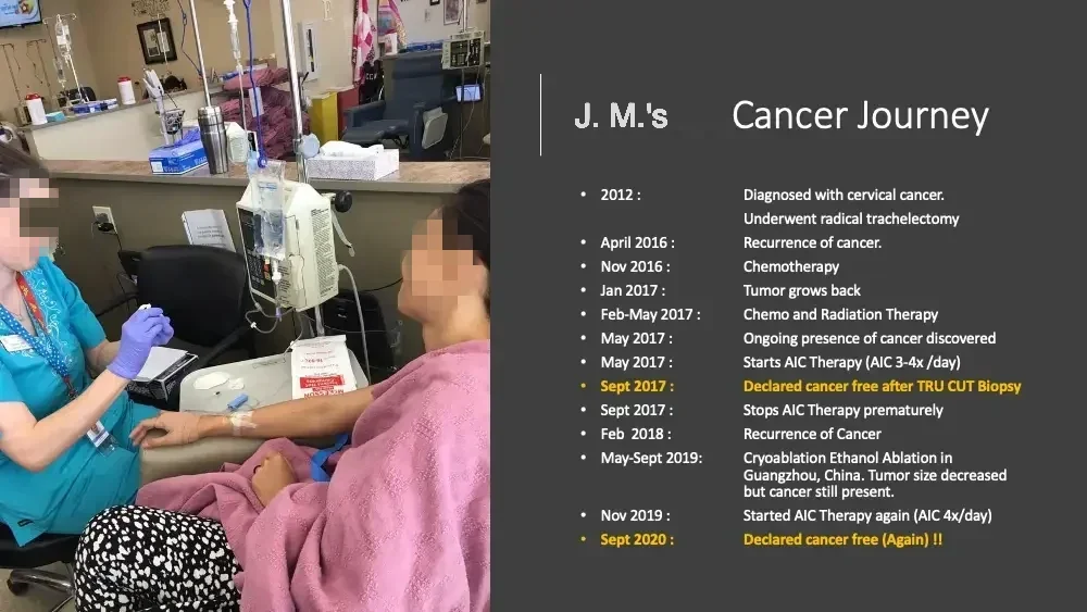
  • Photo: nurse attending a patient receiving an IV infusion. Upside-down packaging print. MCKESSON J. M.’s Cancer Journey 2012 : Diagnosed with cervical cancer. Underwent radical trachelectomy April 2016 : Recurrence of cancer. Nov 2016 : Chemotherapy Jan 2017 : Tumor grows back Feb-May 2017 : Chemo and Radiation Therapy May 2017 : Ongoing presence of cancer discovered May 2017 : Starts AIC Therapy (AIC 3-4x /day) Sept 2017 : Declared cancer free after TRU CUT Biopsy Sept 2017 : Stops AIC Therapy prematurely Feb 2018 : Recurrence of Cancer May-Sept 2019: Cryoablation Ethanol Ablation in Guangzhou, China. Tumor size decreased but cancer still present. Nov 2019 : Started AIC Therapy again (AIC 4x/day) Sept 2020 : Declared cancer free (Again) !!
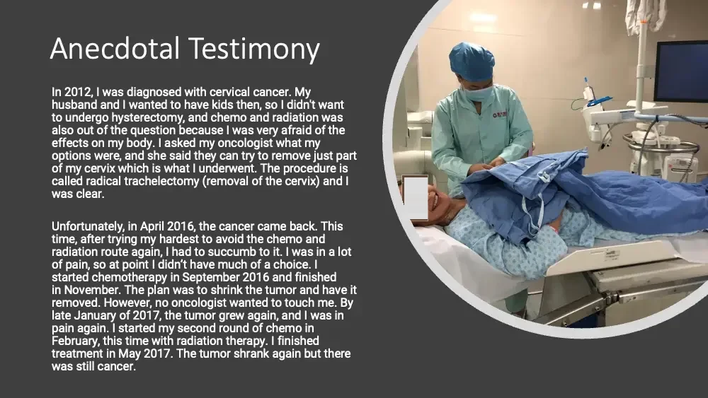
  • Anecdotal Testimony In 2012, I was diagnosed with cervical cancer. My husband and I wanted to have kids then, so I didn't want to undergo hysterectomy, and chemo and radiation was also out of the question because I was very afraid of the effects on my body. I asked my oncologist what my options were, and she said they can try to remove just part of my cervix which is what I underwent. The procedure is called radical trachelectomy (removal of the cervix) and I was clear. Unfortunately, in April 2016, the cancer came back. This time, after trying my hardest to avoid the chemo and radiation route again, I had to succumb to it. I was in a lot of pain, so at point I didn’t have much of a choice. I started chemotherapy in September 2016 and finished in November. The plan was to shrink the tumor and have it removed. However, no oncologist wanted to touch me. By late January of 2017, the tumor grew again, and I was in pain again. I started my second round of chemo in February, this time with radiation therapy. I finished treatment in May 2017. The tumor shrank again but there was still cancer. Photo: smiling patient on a procedure table, masked nurse beside her.
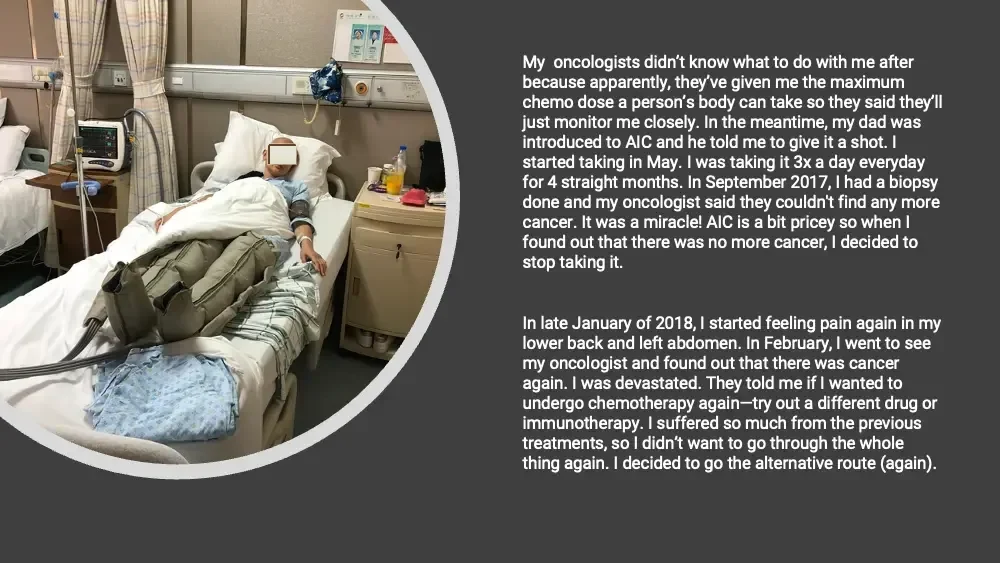
  • Photo: woman with shaved head resting in a hospital bed. My oncologists didn’t know what to do with me after because apparently, they’ve given me the maximum chemo dose a person’s body can take so they said they’ll just monitor me closely. In the meantime, my dad was introduced to AIC and he told me to give it a shot. I started taking in May. I was taking it 3x a day everyday for 4 straight months. In September 2017, I had a biopsy done and my oncologist said they couldn't find any more cancer. It was a miracle! AIC is a bit pricey so when I found out that there was no more cancer, I decided to stop taking it. In late January of 2018, I started feeling pain again in my lower back and left abdomen. In February, I went to see my oncologist and found out that there was cancer again. I was devastated. They told me if I wanted to undergo chemotherapy again—try out a different drug or immunotherapy. I suffered so much from the previous treatments, so I didn’t want to go through the whole thing again. I decided to go the alternative route (again).
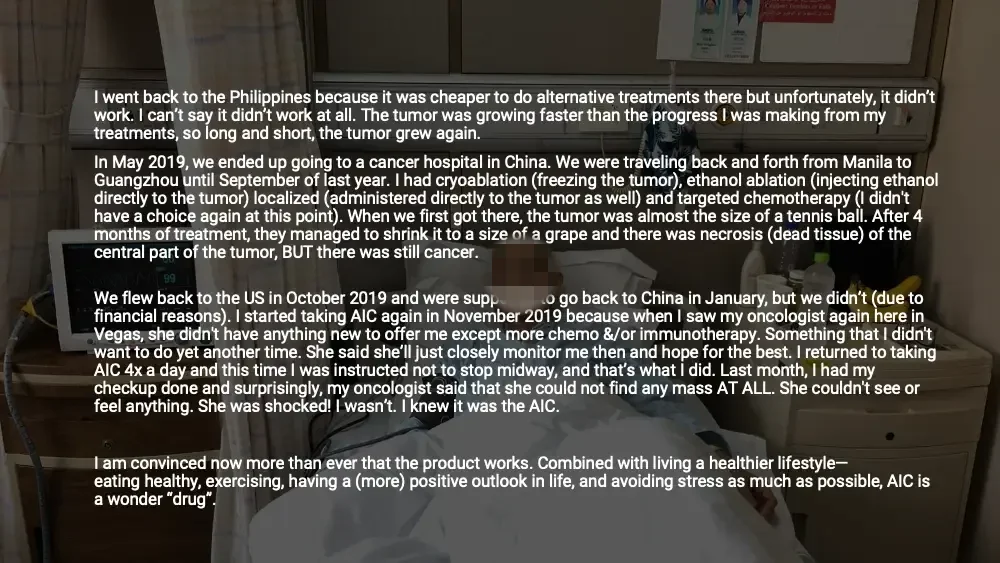
  • Darkened photo: woman with shaved head in a hospital bed. I went back to the Philippines because it was cheaper to do alternative treatments there but unfortunately, it didn’t work. I can’t say it didn’t work at all. The tumor was growing faster than the progress I was making from my treatments, so long and short, the tumor grew again. In May 2019, we ended up going to a cancer hospital in China. We were traveling back and forth from Manila to Guangzhou until September of last year. I had cryoablation (freezing the tumor), ethanol ablation (injecting ethanol directly to the tumor) localized (administered directly to the tumor as well) and targeted chemotherapy (I didn't have a choice again at this point). When we first got there, the tumor was almost the size of a tennis ball. After 4 months of treatment, they managed to shrink it to a size of a grape and there was necrosis (dead tissue) of the central part of the tumor, BUT there was still cancer. Patient monitor in background. 99 We flew back to the US in October 2019 and were supposed to go back to China in January, but we didn’t (due to financial reasons). I started taking AIC again in November 2019 because when I saw my oncologist again here in Vegas, she didn't have anything new to offer me except more chemo &/or immunotherapy. Something that I didn't want to do yet another time. She said she’ll just closely monitor me then and hope for the best. I returned to taking AIC 4x a day and this time I was instructed not to stop midway, and that’s what I did. Last month, I had my checkup done and surprisingly, my oncologist said that she could not find any mass AT ALL. She couldn't see or feel anything. She was shocked! I wasn’t. I knew it was the AIC. I am convinced now more than ever that the product works. Combined with living a healthier lifestyle—eating healthy, exercising, having a (more) positive outlook in life, and avoiding stress as much as possible, AIC is a wonder “drug”.
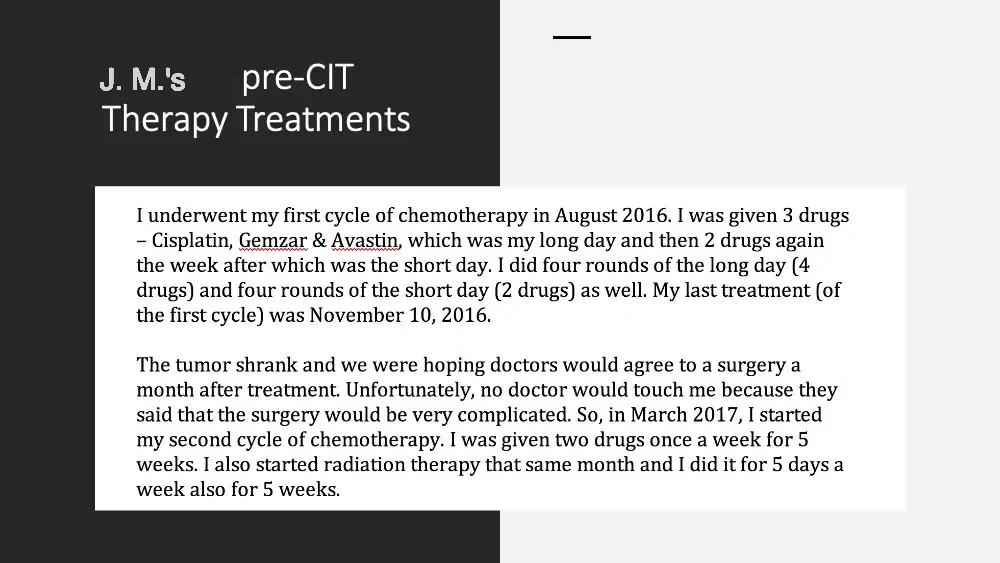
  • J. M.’s pre-CIT Therapy Treatments Short decorative line. I underwent my first cycle of chemotherapy in August 2016. I was given 3 drugs – Cisplatin, Gemzar & Avastin, which was my long day and then 2 drugs again the week after which was the short day. I did four rounds of the long day (4 drugs) and four rounds of the short day (2 drugs) as well. My last treatment (of the first cycle) was November 10, 2016. The tumor shrank and we were hoping doctors would agree to a surgery a month after treatment. Unfortunately, no doctor would touch me because they said that the surgery would be very complicated. So, in March 2017, I started my second cycle of chemotherapy. I was given two drugs once a week for 5 weeks. I also started radiation therapy that same month and I did it for 5 days a week also for 5 weeks.
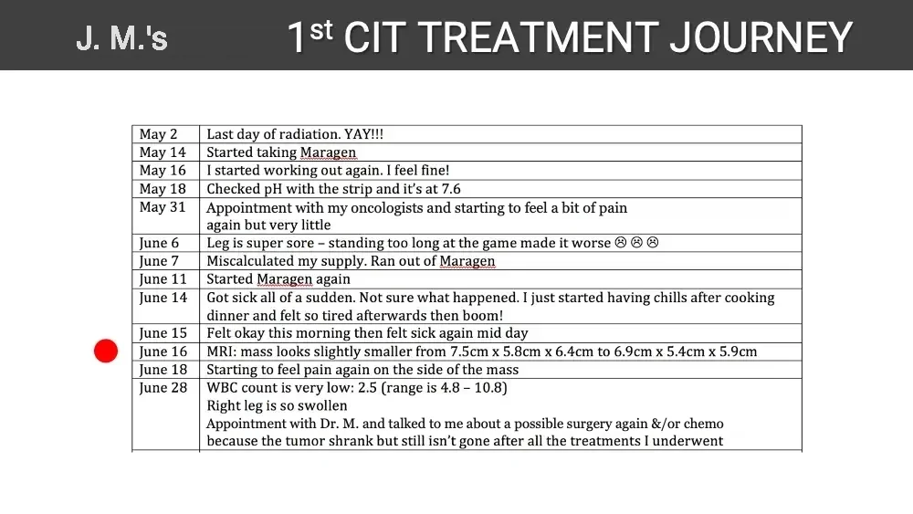
  • J. M.’s 1st CIT TREATMENT JOURNEY May 2 Last day of radiation. YAY!!! May 14 Started taking Maragen May 16 I started working out again. I feel fine! May 18 Checked pH with the strip and it’s at 7.6 May 31 Appointment with my oncologists and starting to feel a bit of pain again but very little June 6 Leg is super sore – standing too long at the game made it worse ☹ ☹ ☹ June 7 Miscalculated my supply. Ran out of Maragen June 11 Started Maragen again June 14 Got sick all of a sudden. Not sure what happened. I just started having chills after cooking dinner and felt so tired afterwards then boom! June 15 Felt okay this morning then felt sick again mid day June 16 MRI: mass looks slightly smaller from 7.5cm x 5.8cm x 6.4cm to 6.9cm x 5.4cm x 5.9cm June 18 Starting to feel pain again on the side of the mass June 28 WBC count is very low: 2.5 (range is 4.8 – 10.8) Right leg is so swollen Appointment with Dr. M. and talked to me about a possible surgery again &/or chemo because the tumor shrank but still isn’t gone after all the treatments I underwent Red dot marker beside the June 16 row.
  • 2021 Photo: woman in a red sweatshirt. Sweatshirt lettering, lower half cropped. ST CLOUD 2022 Photo: woman in flight attendant uniform in a living room, with a suitcase. 2023 Photo: smiling woman wearing a headset in an aircraft cockpit.
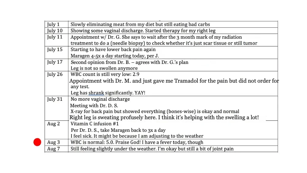
  • July 1 Slowly eliminating meat from my diet but still eating bad carbs July 10 Showing some vaginal discharge. Started therapy for my right leg July 11 Appointment w/ Dr. G. She says to wait after the 3 month mark of my radiation treatment to do a (needle biopsy) to check whether it’s just scar tissue or still tumor July 15 Starting to have lower back pain again Maragen 4-5x a day starting today, per J. July 17 Second opinion from Dr. B. – agrees with Dr. G.’s plan Leg is not so swollen anymore July 26 WBC count is still very low: 2.9 Appointment with Dr. M. and just gave me Tramadol for the pain but did not order for any test. Leg has shrank significantly. YAY! July 31 No more vaginal discharge Meeting with Dr. D. S. X-ray for back pain but showed everything (bones-wise) is okay and normal Right leg is sweating profusely here. I think it’s helping with the swelling a lot! Aug 2 Vitamin C infusion #1 Per Dr. D. S., take Maragen back to 3x a day I feel sick. It might be because I am adjusting to the weather Aug 3 WBC is normal: 5.0. Praise God! I have a fever today, though Aug 7 Still feeling slightly under the weather. I’m okay but still a bit of joint pain Red dot marker beside the Aug 3 row.
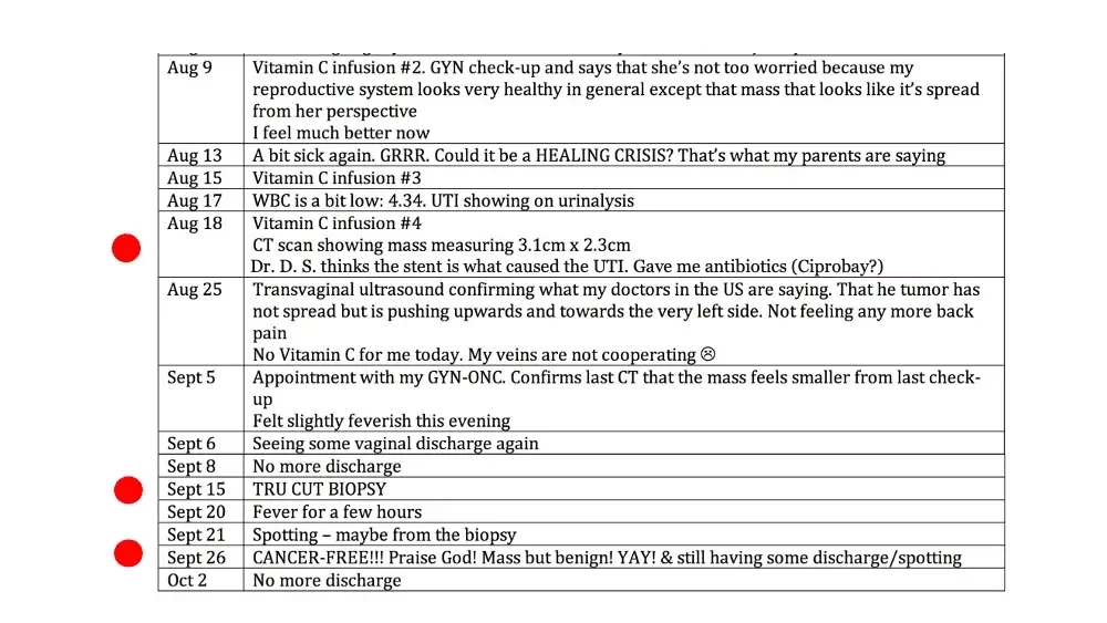
  • Aug 9 Vitamin C infusion #2. GYN check-up and says that she’s not too worried because my reproductive system looks very healthy in general except that mass that looks like it’s spread from her perspective I feel much better now Aug 13 A bit sick again. GRRR. Could it be a HEALING CRISIS? That’s what my parents are saying Aug 15 Vitamin C infusion #3 Aug 17 WBC is a bit low: 4.34. UTI showing on urinalysis Aug 18 Vitamin C infusion #4 CT scan showing mass measuring 3.1cm x 2.3cm Dr. D. S. thinks the stent is what caused the UTI. Gave me antibiotics (Ciprobay?) Aug 25 Transvaginal ultrasound confirming what my doctors in the US are saying. That he tumor has not spread but is pushing upwards and towards the very left side. Not feeling any more back pain No Vitamin C for me today. My veins are not cooperating ☹ Sept 5 Appointment with my GYN-ONC. Confirms last CT that the mass feels smaller from last check-up Felt slightly feverish this evening Sept 6 Seeing some vaginal discharge again Sept 8 No more discharge Sept 15 TRU CUT BIOPSY Sept 20 Fever for a few hours Sept 21 Spotting – maybe from the biopsy Sept 26 CANCER-FREE!!! Praise God! Mass but benign! YAY! & still having some discharge/spotting Oct 2 No more discharge Red dots beside the Aug 18, Sept 15 and Sept 26 rows.
  • “Celebrating my birthday this month. And a few weeks after found out no more cancer. Best birthday gift ever 🥳🙏🏼” J. M. (Oct 2020) Photo: smiling woman holding plate of two cupcakes with number candles. 39
  • Testimony of J. M. SELFLOVE

ADENOID CANCER
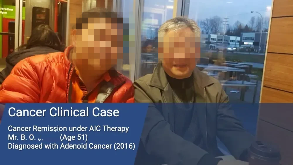
  • Photo: two men seated together at a café table. peration 0pm Partial sign text, cut off at photo edge. Cancer Clinical Case Cancer Remission under AIC Therapy Mr. B. O. J. (Age 51) Diagnosed with Adenoid Cancer (2016)
  • “AIC saved me from dangerous radiotherapy in my head and took away all the traces of cancer!” I am 51 years old and have worked as a manager of a motel a long time. The stress from my work and constant heavy drinking after the work took the toll on my body. Then one day, I noticed a growth inside my left cheek and had to rush to see my doctor. The test revealed that the mass was cancerous. The tumor had a diameter of 3.5 cm and was growing where nerve and blood vessels were intricately intertwined. My oncologist recommended me for surgery because radiation therapy was not deemed helpful then. Last September of 2016, I heard about AIC through my brother but decided to follow the recommendation of the oncologist and surgically removed the big tumor. Many smaller tumors where surgical removal was not possible were left alone. Right after the surgery, I started to take AIC. I immediately experienced my body temperature rising and felt much stronger even after exhaustive chores from my work. I began to have both wonder and hope.
  • “AIC saved me from dangerous radiotherapy in my head and took away all the traces of cancer!” About twenty days after the surgery, the hospital called me and tried to convince me to receive 33 scheduled treatments of radiation therapy. When I inquired and learned about the terrible side effects of the therapy, I could not muster the courage to go ahead with it. I decided not to take the radiation therapy and rely on AIC all the way since I was already experiencing manifold positive effects from it. Honestly, the side effects of the recommended radiotherapy, namely, dry mouth, hot flush, losing taste buds, and possible brain hemorrhage sounded scarier than the cancer itself. Now, I am so glad that I bypassed the radiation therapy and stayed with AIC. AIC removed all the traces of remaining cancer, even in the most difficult places. (March 2017)
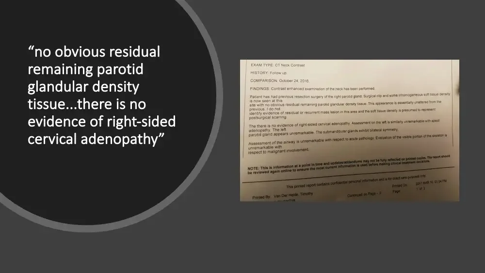
  • “no obvious residual remaining parotid glandular density tissue...there is no evidence of right-sided cervical adenopathy” Photo: printed CT report page. EXAM TYPE: CT Neck Contrast HISTORY: Follow up. COMPARISON: October 24, 2016. FINDINGS: Contrast enhanced examination of the neck has been performed. Patient has had previous resection surgery of the right parotid gland. Surgical clip and some inhomogeneous soft tissue density is now seen at this site with no obvious residual remaining parotid glandular density tissue. This appearance is essentially unaltered from the previous. I do not identify evidence of residual or recurrent mass lesion in this area and the soft tissue density is presumed to represent postsurgical scarring. The there is no evidence of right-sided cervical adenopathy. Assessment on the left is similarly unremarkable with apect adenopathy. The left parotid gland appears unremarkable. The submandibular glands exhibit bilateral symmetry. Assessment of the airway is unremarkable with respect to acute pathology. Evaluation of the visible portion of the skeleton is unremarkable with respect to malignant involvement. NOTE: This is information at a point in time and updates/addendums may not be fully reflected on printed copies. The report should be reviewed again online to ensure the most current information is used before making clinical treatment decisions. This printed report contains confidential personal information and is for direct care purposes only. Printed By: Van Der Heide, Timothy Continued on Page - 2 Printed On: 2017 MAR 10 01:04 PM Page: 1 of 2
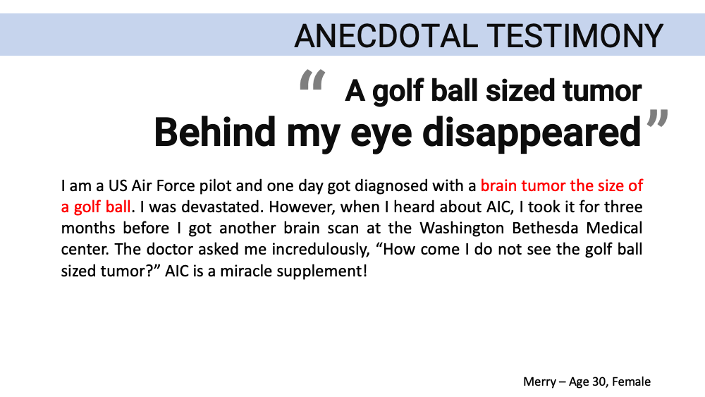
ANECDOTAL TESTIMONY “A golf ball sized tumor Behind my eye disappeared” I am a US Air Force pilot and one day got diagnosed with a brain tumor the size of a golf ball. I was devastated. However, when I heard about AIC, I took it for three months before I got another brain scan at the Washington Bethesda Medical center. The doctor asked me incredulously, “How come I do not see the golf ball sized tumor?” AIC is a miracle supplement! Merry – Age 30, Female
THE RELATIONSHIP BETWEEN CANCER AND CALCIUM (The Calcium Factor by Dr. Carl J. Reich, M.D. and Robert R. Barefoot) Thus, he considered cancer as the ultimate adaptation to ionic calcium deficiency, "tailor-made" to survive and to thrive in an ionic calcium deficiency environment. Dr. Reich found that the cancer in many of his patients seemed to go into remission once their calcium deficiency was rectified (corrected), by a change of lifestyle including diet and with mineral and vitamin supplements that raised the pH of their cellular fluids. Their associated ionic calcium deficiency diseases were also suppressed.
THE RELATIONSHIP BETWEEN CANCER AND CALCIUM (The Calcium Factor by Dr. Carl J. Reich, M.D. and Robert R. Barefoot) In the healthy, calcium buffered, slightly alkaline cell environment, the conditions required for the propagation of cancer do not exist. It therefore remains dormant, or dies. Dr. Reich noted that his cancer patients demonstrated: Lifestyle defects responsible for deficiency of one or both calcium and vitamin D. Symptoms and physical signs of ionic calcium deficiency syndrome. A greater than normal incidence of these ionic calcium deficiency diseases.
CANCER PROBLEM: PHYSICAL & FINANCIAL Suffering cause by the disease itself and its treatment is dreaded by every patients. It takes about $80,000-100,000 in the USA to die with cancer by receiving treatments. Immunotherapy could costs you $150,000 / year. (Ex. Jimmy Carter) IS THERE AN ALTERNATIVE SOLUTION ?
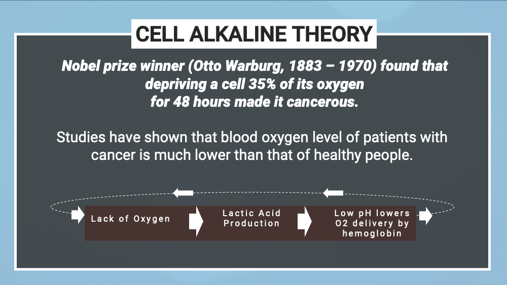
CELL ALKALINE THEORY Nobel prize winner (Otto Warburg, 1883 – 1970) found that depriving a cell 35% of its oxygen for 48 hours made it cancerous. Studies have shown that blood oxygen level of patients with cancer is much lower than that of healthy people. Cycle diagram: three boxes linked by arrows in a loop. Lack of Oxygen Lactic Acid Production Low pH lowers O2 delivery by hemoglobin
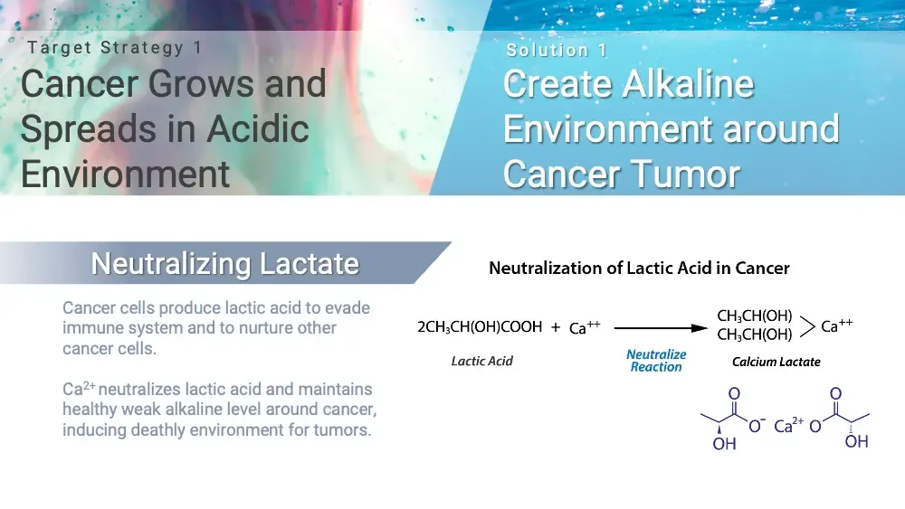
Target Strategy 1 Cancer Grows and Spreads in Acidic Environment Solution 1 Create Alkaline Environment around Cancer Tumor Neutralizing Lactate Cancer cells produce lactic acid to evade immune system and to nurture other cancer cells. Ca²⁺ neutralizes lactic acid and maintains healthy weak alkaline level around cancer, inducing deathly environment for tumors. Neutralization of Lactic Acid in Cancer 2CH₃CH(OH)COOH + Ca⁺⁺ → CH₃CH(OH) CH₃CH(OH) > Ca⁺⁺ Two CH₃CH(OH) groups stacked, bracketed to Ca⁺⁺. Lactic Acid Neutralize Reaction Calcium Lactate Skeletal formula of calcium lactate. O O O⁻ Ca²⁺ O OH OH
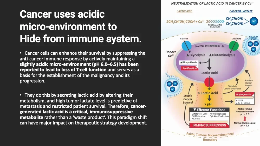
Cancer uses acidic micro-environment to Hide from immune system. Cancer cells can enhance their survival by suppressing the anti-cancer immune response by actively maintaining a slightly acidic micro-environment (pH 6.0–6.5) has been reported to lead to loss of T-cell function and serves as a basis for the establishment of the malignancy and its progression. They do this by secreting lactic acid by altering their metabolism, and high tumor lactate level is predictive of metastasis and restricted patient survival. Therefore, cancer-generated lactic acid is a critical, immunosuppressive metabolite rather than a ‘waste product’. This paradigm shift can have major impact on therapeutic strategy development. Diagram: cancer cell in acidic tumour microenvironment, calcium neutralizing lactic acid. NEUTRALIZATION OF LACTIC ACID IN CANCER BY Ca⁺⁺ LACTIC ACID CALCIUM LACTATE 2CH₃CH(OH)COOH + Ca⁺⁺ NEUTRALIZING REACTION CH₃CH(OH) CH₃CH(OH) > Ca²⁺ Glucose Glutamine CALCIUM IONS Ca²⁺ Ca²⁺ Ca²⁺ Cancer Cell Normal Intracellular pH ▲Glycolysis ▲Glutaminolysis ▲Biosynthesis Proliferation Lactic Acid Ca²⁺ Ca²⁺ Ca²⁺ Promote Cancer Growth Stimulate Cancer Metastasis Enable Cancer Survival ▲Lactic Acid Angiogenesis ▲ VEGF ▲ IL-8 / CXCL8 ▼pH ▼ Effector Functions Cytotoxic T Cells DC Maturation NK Cells M1 Macrophages Cytokine Production Acidic Tumour Microenvironment pH ~ 6.5 Normal Physiological pH = 7.4 IMMUNOSUPPRESSION Acidic Tumour Microenvironment Boundary
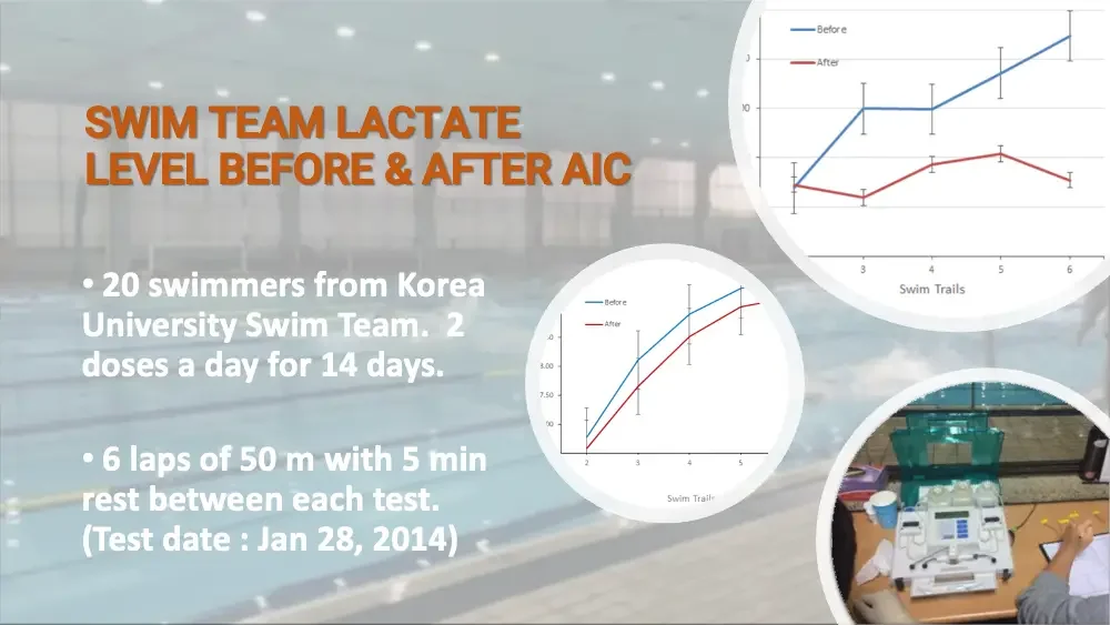
Background photo: indoor swimming pool. SWIM TEAM LACTATE LEVEL BEFORE & AFTER AIC 20 swimmers from Korea University Swim Team. 2 doses a day for 14 days. 6 laps of 50 m with 5 min rest between each test. (Test date : Jan 28, 2014) Line chart with error bars, cropped in a circle. Before After y-axis labels cut off by crop. 3 4 5 6 Swim Trails Line chart with error bars, cropped in a circle. Before After cut off. 8.00 7.50 cut off. 2 3 4 5 Swim Trails Photo: lactate analyzer on a table during testing.
“A rise of intracellular Ca²⁺ concentrations is also required for efficient CTL and NK cell function and thus for killing their targets, in this case cancer cells.” Screenshot: first page of a journal article (Elsevier). Calcium, cancer and killing: The role of calcium in killing cancer cells by cytotoxic T lymphocytes and natural killer cells☆ Logo: CrossMark. CrossMark Eva C. Schwarz, Bin Qu, Markus Hoth * Department of Biophysics, Saarland University, Homburg, Germany ARTICLE INFO Article history: Received 22 September 2012 Received in revised form 16 November 2012 Accepted 18 November 2012 Available online 3 December 2012 Keywords: Calcium Cancer Killing Cytotoxic T lymphocytes Natural killer cells ORAI calcium channels ABSTRACT © 2012 Elsevier B.V. All rights reserved. Introduction Calcium channels
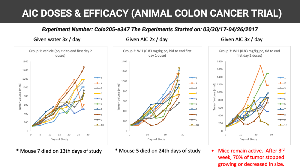
AIC DOSES & EFFICACY (ANIMAL COLON CANCER TRIAL) Experiment Number: Colo205-e347 The Experiments Started on: 03/30/17-04/26/2017 Given water 3x / day Line chart: tumor volume per mouse over time. Group 1: vehicle (po, tid to end first day 2 doses) Tumor Volume (mm3) 0 200 400 600 800 1000 1200 1400 1600 Days of Study 0 5 10 15 20 25 30 1 2 3 4 5 6 7 8 9 10 Mouse 7 died on 13th days of study Given AIC 2x / day Line chart: tumor volume per mouse over time. Group 2: WI1 (0.83 mg/kg,po, bid to end first day 1 dose) Tumor Volume (mm3) 0 200 400 600 800 1000 1200 1400 Days of Study 0 10 20 30 1 2 3 4 5 6 7 8 9 10 Mouse 5 died on 24th days of study Given AIC 3x / day Line chart: tumor volume per mouse over time. Group 3: WI1 (0.83 mg/kg,po, tid to end first day 2 doses) Tumor Volume (mm3) 0 200 400 600 800 1000 1200 1400 1600 1800 Days of Study 0 10 20 30 1 2 3 4 5 6 7 8 9 10 Mice remain active. After 3rd week, 70% of tumor stopped growing or decreased in size.
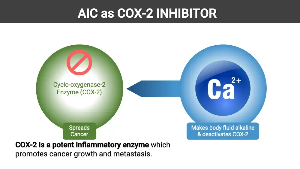
AIC as COX-2 INHIBITOR Diagram: green enzyme sphere with prohibition sign; blue Ca²⁺ sphere arrow pointing at it. Cyclo-oxygenase-2 Enzyme (COX-2) Spreads Cancer Ca²⁺ Makes body fluid alkaline & deactivates COX-2 COX-2 is a potent inflammatory enzyme which promotes cancer growth and metastasis.
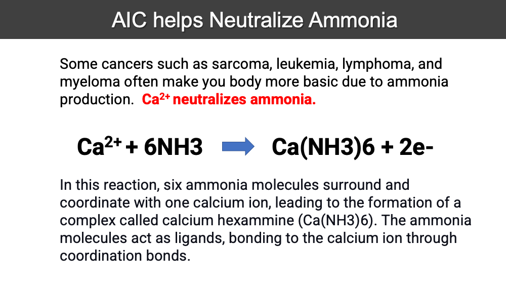
AIC helps Neutralize Ammonia Some cancers such as sarcoma, leukemia, lymphoma, and myeloma often make you body more basic due to ammonia production. Ca²⁺ neutralizes ammonia. Ca²⁺ + 6NH3 → Ca(NH3)6 + 2e- In this reaction, six ammonia molecules surround and coordinate with one calcium ion, leading to the formation of a complex called calcium hexammine (Ca(NH3)6). The ammonia molecules act as ligands, bonding to the calcium ion through coordination bonds.
What is COX-2 Enzyme? COX-2 is a potent inflammatory enzyme which promotes cancer growth and metastasis. Levels of COX-2 were one 100x higher in cancer cells from individuals with head and neck cancers as compared to healthy volunteers. Levels of COX-2 in pancreatic cancer cells to be 60x greater than in normal pancreatic cells. COX-2 fuels cancer growth by stimulating the formation of new blood vessels feeding the tumor. COX-2 increases cancer cell adhesion to the blood vessel walls. COX-2 also enhances the ability of cancer cells to metastasize, as experiments with mice revealed that colon cancer cells expressing high levels of COX-2 metastasized freely to the liver, while colon cancer cells expressing low levels of COX-2 did not metastasize to the liver. (Source: Life Extension Foundation)
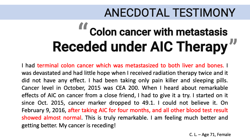
ANECDOTAL TESTIMONY “Colon cancer with metastasis Receded under AIC Therapy” I had terminal colon cancer which was metastasized to both liver and bones. I was devastated and had little hope when I received radiation therapy twice and it did not have any effect. I had been taking only pain killer and sleeping pills. Cancer level in October, 2015 was CEA 200. When I heard about remarkable effects of AIC on cancer from a close friend, I had to give it a try. I started on it since Oct. 2015, cancer marker dropped to 49.1. I could not believe it. On February 9, 2016, after taking AIC for four months, and all other blood test result showed almost normal. This is truly remarkable. I am feeling much better and getting better. My cancer is receding! C. L. – Age 71, Female
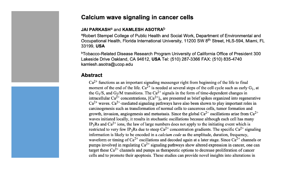
Decorative grey and blue blocks at left. Screenshot: first page of a journal article manuscript. Calcium wave signaling in cancer cells JAI PARKASHᵃ and KAMLESH ASOTRAᵇ ᵃRobert Stempel College of Public Health and Social Work, Department of Environmental and Occupational Health, Florida International University, 11200 SW 8th Street, HLS-594, Miami, FL 33199, USA ᵇTobacco-Related Disease Research Program University of California Office of President 300 Lakeside Drive Oakland, CA 94612, USA Tel: (510) 287-3366 FAX: (510) 835-4740 kamlesh.asotra@ucop.edu Abstract
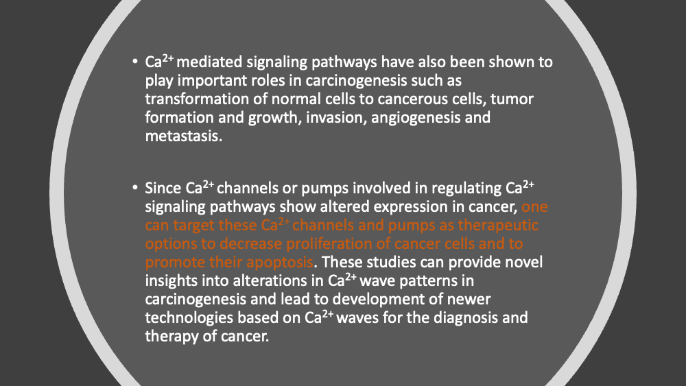
Ca²⁺ mediated signaling pathways have also been shown to play important roles in carcinogenesis such as transformation of normal cells to cancerous cells, tumor formation and growth, invasion, angiogenesis and metastasis. Since Ca²⁺ channels or pumps involved in regulating Ca²⁺ signaling pathways show altered expression in cancer, one can target these Ca²⁺ channels and pumps as therapeutic options to decrease proliferation of cancer cells and to promote their apoptosis. These studies can provide novel insights into alterations in Ca²⁺ wave patterns in carcinogenesis and lead to development of newer technologies based on Ca²⁺ waves for the diagnosis and therapy of cancer.
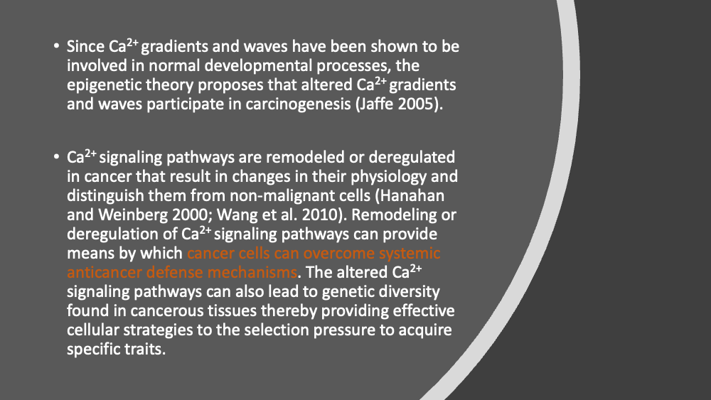
Since Ca²⁺ gradients and waves have been shown to be involved in normal developmental processes, the epigenetic theory proposes that altered Ca²⁺ gradients and waves participate in carcinogenesis (Jaffe 2005). Ca²⁺ signaling pathways are remodeled or deregulated in cancer that result in changes in their physiology and distinguish them from non-malignant cells (Hanahan and Weinberg 2000; Wang et al. 2010). Remodeling or deregulation of Ca²⁺ signaling pathways can provide means by which cancer cells can overcome systemic anticancer defense mechanisms. The altered Ca²⁺ signaling pathways can also lead to genetic diversity found in cancerous tissues thereby providing effective cellular strategies to the selection pressure to acquire specific traits.
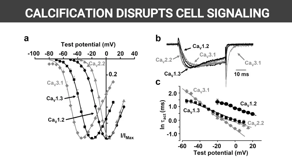
CALCIFICATION DISRUPTS CELL SIGNALING Scientific figure with three panels: current-voltage curves and traces. a Test potential (mV) -100 -80 -60 -40 -20 0 20 40 CaV2.2 0.2 CaV3.1 CaV1.3 CaV1.2 I/IMax b CaV1.2 CaV2.2 CaV3.1 CaV1.3 CaV3.1 10 ms c CaV3.1 CaV1.3 CaV1.2 CaV2.2 ln τact (ms) 2.0 1.0 0.0 -1.0 -60 -40 -20 0 20 Test potential (mV)
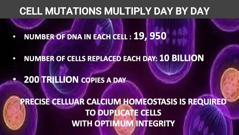
CELL MUTATIONS MULTIPLY DAY BY DAY Background: microscope image of purple dividing cells. NUMBER OF DNA IN EACH CELL : 19, 950 NUMBER OF CELLS REPLACED EACH DAY: 10 BILLION 200 TRILLION COPIES A DAY PRECISE CELLUAR CALCIUM HOMEOSTASIS IS REQUIRED TO DUPLICATE CELLS WITH OPTIMUM INTEGRITY
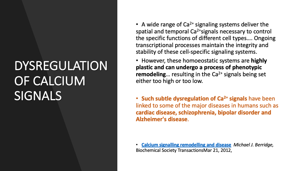
DYSREGULATION OF CALCIUM SIGNALS A wide range of Ca²⁺ signaling systems deliver the spatial and temporal Ca²⁺signals necessary to control the specific functions of different cell types…. Ongoing transcriptional processes maintain the integrity and stability of these cell-specific signaling systems. However, these homoeostatic systems are highly plastic and can undergo a process of phenotypic remodeling… resulting in the Ca²⁺ signals being set either too high or too low. Such subtle dysregulation of Ca²⁺ signals have been linked to some of the major diseases in humans such as cardiac disease, schizophrenia, bipolar disorder and Alzheimer's disease. Calcium signalling remodelling and disease Michael J. Berridge, Biochemical Society TransactionsMar 21, 2012,
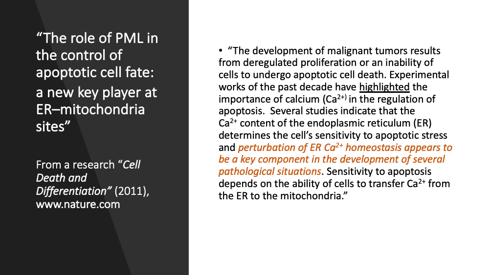
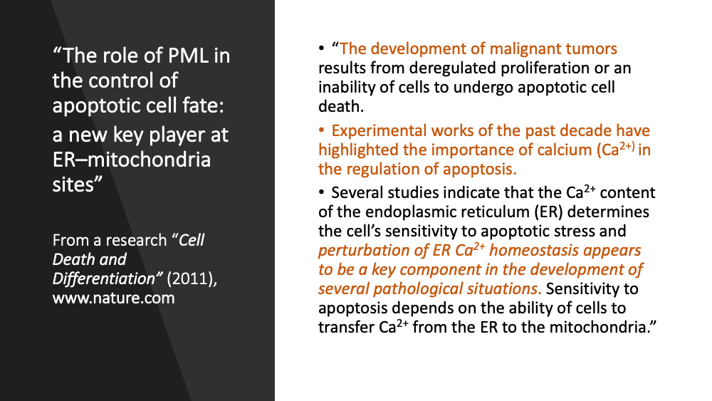
“The role of PML in the control of apoptotic cell fate: a new key player at ER–mitochondria sites” From a research “Cell Death and Differentiation” (2011), www.nature.com “The development of malignant tumors results from deregulated proliferation or an inability of cells to undergo apoptotic cell death. Experimental works of the past decade have highlighted the importance of calcium (Ca²⁺) in the regulation of apoptosis. Several studies indicate that the Ca²⁺ content of the endoplasmic reticulum (ER) determines the cell’s sensitivity to apoptotic stress and perturbation of ER Ca²⁺ homeostasis appears to be a key component in the development of several pathological situations. Sensitivity to apoptosis depends on the ability of cells to transfer Ca²⁺ from the ER to the mitochondria.”
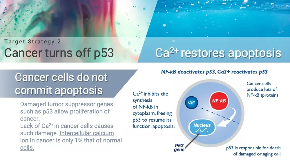
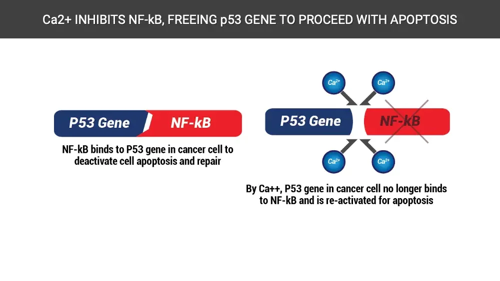
Target Strategy 2 Cancer turns off p53 Ca²⁺ restores apoptosis Cancer cells do not commit apoptosis Damaged tumor suppressor genes such as p53 allow proliferation of cancer. Lack of Ca²⁺ in cancer cells causes such damage. Intercellular calcium ion in cancer is only 1% that of normal cells. NF-kB deactivates p53, Ca2+ reactivates p53 Ca²⁺ inhibits the synthesis of NF-kB in cytoplasm, freeing p53 to resume its function, apoptosis. Cancer cells produce lots of NF-kB (protein) Diagram: cell with Ca²⁺ ion, NF-kB and nucleus. Ca²⁺ NF-kB Nucleus P53 gene p53 is responsible for death of damaged or aging cell
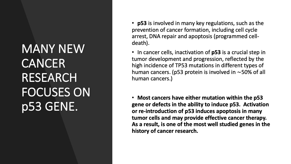
MANY NEW CANCER RESEARCH FOCUSES ON p53 GENE. p53 is involved in many key regulations, such as the prevention of cancer formation, including cell cycle arrest, DNA repair and apoptosis (programmed cell-death). In cancer cells, inactivation of p53 is a crucial step in tumor development and progression, reflected by the high incidence of TP53 mutations in different types of human cancers. (p53 protein is involved in ~50% of all human cancers.) Most cancers have either mutation within the p53 gene or defects in the ability to induce p53. Activation or re-introduction of p53 induces apoptosis in many tumor cells and may provide effective cancer therapy. As a result, is one of the most well studied genes in the history of cancer research.
“The role of PML in the control of apoptotic cell fate: a new key player at ER–mitochondria sites” From a research “Cell Death and Differentiation” (2011), www.nature.com “The development of malignant tumors results from deregulated proliferation or an inability of cells to undergo apoptotic cell death. Experimental works of the past decade have highlighted the importance of calcium (Ca²⁺) in the regulation of apoptosis. Several studies indicate that the Ca²⁺ content of the endoplasmic reticulum (ER) determines the cell’s sensitivity to apoptotic stress and perturbation of ER Ca²⁺ homeostasis appears to be a key component in the development of several pathological situations. Sensitivity to apoptosis depends on the ability of cells to transfer Ca²⁺ from the ER to the mitochondria.”
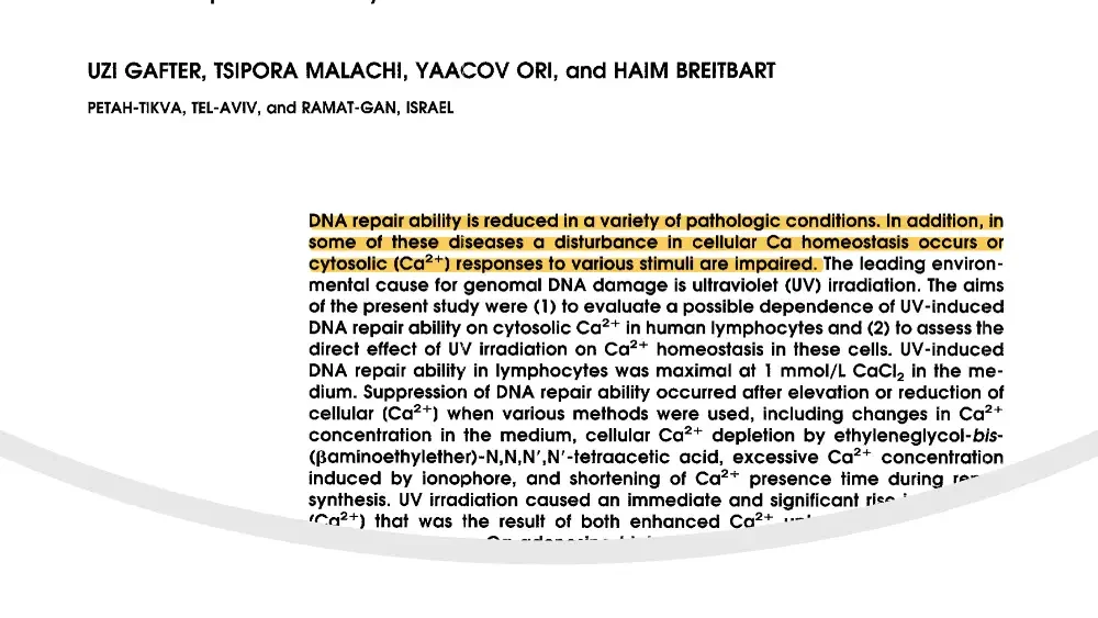
Screenshot: part of a journal article page; lower part covered by white arc. UZI GAFTER, TSIPORA MALACHI, YAACOV ORI, and HAIM BREITBART PETAH-TIKVA, TEL-AVIV, and RAMAT-GAN, ISRAEL
DNA repair ability is reduced in a variety of pathologic conditions. In addition, in some of these diseases a disturbance in cellular Ca homeostasis occurs or cytosolic Ca²⁺ responses to various stimuli are impaired. These results emphasize the importance of a precise cellular Ca²⁺ level regulation for the optimal DNA repair process. Disturbed Ca²⁺ metabolism and a defective response to Ca²⁺ stimulators have been reported to exist in some of the patients with DNA repair deficiency… Thus an impaired DNA repairability may result in part from a disturbed Ca²⁺ homeostasis. Icon: blue DNA double helix in a white circle.
Ca2+ INHIBITS NF-kB, FREEING p53 GENE TO PROCEED WITH APOPTOSIS Diagram: P53 Gene bar joined to NF-kB bar. P53 Gene NF-kB NF-kB binds to P53 gene in cancer cell to deactivate cell apoptosis and repair Diagram: four Ca²⁺ ions separate P53 Gene from crossed-out NF-kB. Ca²⁺ Ca²⁺ P53 Gene NF-kB Ca²⁺ Ca²⁺ By Ca++, P53 gene in cancer cell no longer binds to NF-kB and is re-activated for apoptosis
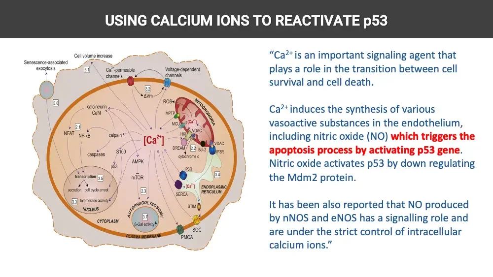
USING CALCIUM IONS TO REACTIVATE p53 Diagram: calcium signaling pathways in a cell, numbered boxes. Cell volume increase Senescence-associated exocytosis 3.1 Ca²⁺-permeable channels Voltage-dependent channels 3.2 ΔVm 3.6 ROS MITOCHONDRIA MPTP calcineurin CaM MCU Ca²⁺. VDAC HKI 2.1 NFAT NF-κB calpain Ca²⁺. DREAM 2.2 Bcl-2 VDAC IP3R cytochrome c caspases S100 p53 AMPK mTOR IP3R 3.4 transcription 3.5 secretion cell cycle arrest Ca²⁺. ENDOPLASMIC RETICULUM SERCA 3.3 telomerase activity NUCLEUS 2.3 STIM AUTOPHAGOLYSOSOME CYTOPLASM 3.7 β-Gal activity▲ SOC PLASMA MEMBRANE PMCA “Ca²⁺ is an important signaling agent that plays a role in the transition between cell survival and cell death. Ca²⁺ induces the synthesis of various vasoactive substances in the endothelium, including nitric oxide (NO) which triggers the apoptosis process by activating p53 gene. Nitric oxide activates p53 by down regulating the Mdm2 protein. It has been also reported that NO produced by nNOS and eNOS has a signalling role and are under the strict control of intracellular calcium ions.”
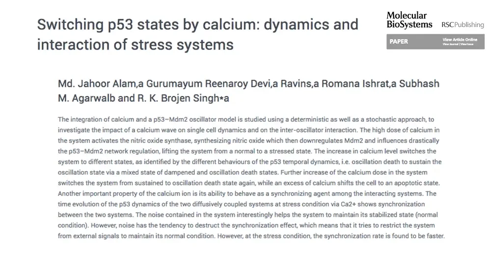
Switching p53 states by calcium: dynamics and interaction of stress systems Logo: Molecular BioSystems journal header. Molecular BioSystems RSCPublishing PAPER View Article Online View Journal | View Issue Md. Jahoor Alam,a Gurumayum Reenaroy Devi,a Ravins,a Romana Ishrat,a Subhash M. Agarwalb and R. K. Brojen Singh*a
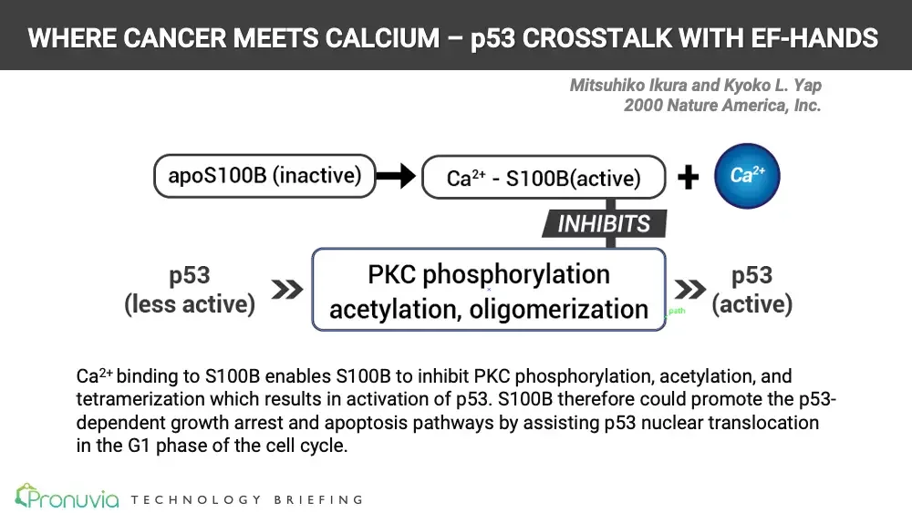
WHERE CANCER MEETS CALCIUM – p53 CROSSTALK WITH EF-HANDS Mitsuhiko Ikura and Kyoko L. Yap 2000 Nature America, Inc. Flow diagram: S100B activation by calcium inhibiting PKC steps. apoS100B (inactive) Ca²⁺ - S100B(active) Ca²⁺ INHIBITS p53 (less active) PKC phosphorylation acetylation, oligomerization p53 (active) path Tiny green editing label beside the box. Ca²⁺ binding to S100B enables S100B to inhibit PKC phosphorylation, acetylation, and tetramerization which results in activation of p53. S100B therefore could promote the p53-dependent growth arrest and apoptosis pathways by assisting p53 nuclear translocation in the G1 phase of the cell cycle. Logo: Pronuvia. Pronuvia TECHNOLOGY BRIEFING
AIC Repairs Mitochondrial Dysfunction Diagram: cycle between embryonic and damaged mitochondria. Rejuvenation free radicals Damaged mitochondria Embryonic mitochondria Aging A large accumulation of calcium ion inside of mitochondrial membrane induces production of Reactive Oxygen Species (ROS) which causes damage to DNA, RNA, and proteins which result in cell mutation. AIC restores the functions of mitochondria by bringing in calcium homeostasis, resulting in decalcification and calcium signaling restoration.
AIC CANCER THERAPY PHASES (Phases reflect typical AIC therapy for successful cases as either stand alone or adjunct therapy alongside alternative treatment. Below does not apply as adjunct or adjuvant therapy for conventional cancer treatments.) Diagram: green phase circles above red cancer-symptom circles. Acidic Body, Low Oxygen Delivery, Low Body Temperature, Inflammation, Compromised Immune System Intracellular Calcification, Calcium Signaling Breakdown, Mitochondrial Dysfunction, Oxidative Stress CANCER DIAGNOSIS Systemic Breakdown (Underlying Cancer Causing Conditions) DURING PHASE 1 4 doses/day Systemic Recovery DURING PHASE 2 3 doses/day Systemic Recovery DURING PHASE 3 2 doses/day Systemic Maintenance Diminishing Disease Momentum Cancer Symptoms Stage 1-3 Stage 4 Receding Cancer Symptoms Undetectable Cancer Symptoms