Notice. The following information is provided for your convenience. ACRI is not directly involved in this research and does not make any medical claims.
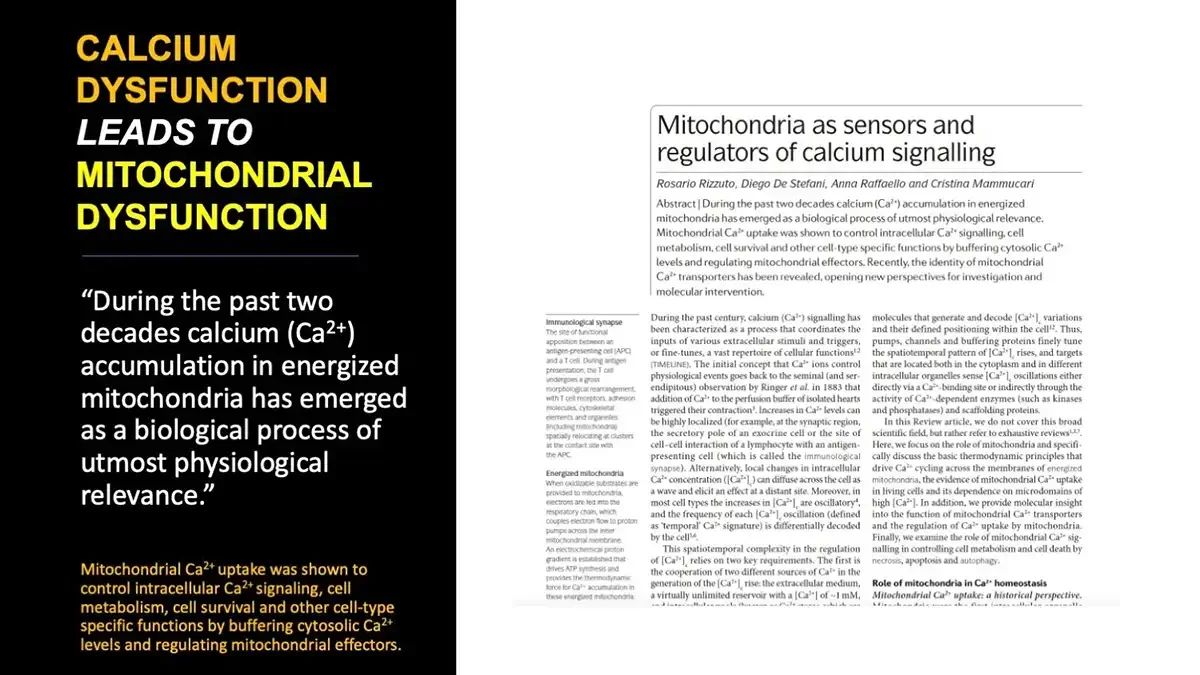
CALCIUM DYSFUNCTION LEADS TO MITOCHONDRIAL DYSFUNCTION “During the past two decades calcium (Ca²⁺) accumulation in energized mitochondria has emerged as a biological process of utmost physiological relevance.” Mitochondrial Ca²⁺ uptake was shown to control intracellular Ca²⁺ signaling, cell metabolism, cell survival and other cell-type specific functions by buffering cytosolic Ca²⁺ levels and regulating mitochondrial effectors. Screenshot of journal review article first page; bottom edge cut off. Mitochondria as sensors and regulators of calcium signalling Rosario Rizzuto, Diego De Stefani, Anna Raffaello and Cristina Mammucari Abstract | During the past two decades calcium (Ca²⁺) accumulation in energized mitochondria has emerged as a biological process of utmost physiological relevance. Mitochondrial Ca²⁺ uptake was shown to control intracellular Ca²⁺ signalling, cell metabolism, cell survival and other cell-type specific functions by buffering cytosolic Ca²⁺ levels and regulating mitochondrial effectors. Recently, the identity of mitochondrial Ca²⁺ transporters has been revealed, opening new perspectives for investigation and molecular intervention. Immunological synapse The site of functional apposition between an antigen-presenting cell (APC) and a T cell. During antigen presentation, the T cell undergoes a gross morphological rearrangement, with T cell receptors, adhesion molecules, cytoskeletal elements and organelles (including mitochondria) spatially relocating at clusters at the contact site with the APC. Energized mitochondria When oxidizable substrates are provided to mitochondria, electrons are fed into the respiratory chain, which couples electron flow to proton pumps across the inner mitochondrial membrane. An electrochemical proton gradient is established that drives ATP synthesis and provides the thermodynamic force for Ca²⁺ accumulation in these energized mitochondria. During the past century, calcium (Ca²⁺) signalling has been characterized as a process that coordinates the inputs of various extracellular stimuli and triggers, or fine-tunes, a vast repertoire of cellular functions¹,² (TIMELINE). The initial concept that Ca²⁺ ions control physiological events goes back to the seminal (and serendipitous) observation by Ringer et al. in 1883 that addition of Ca²⁺ to the perfusion buffer of isolated hearts triggered their contraction³. Increases in Ca²⁺ levels can be highly localized (for example, at the synaptic region, the secretory pole of an exocrine cell or the site of cell–cell interaction of a lymphocyte with an antigen-presenting cell (which is called the immunological synapse). Alternatively, local changes in intracellular Ca²⁺ concentration (Ca²⁺.c) can diffuse across the cell as a wave and elicit an effect at a distant site. Moreover, in most cell types the increases in Ca²⁺.c are oscillatory⁴, and the frequency of each Ca²⁺.c oscillation (defined as 'temporal' Ca²⁺ signature) is differentially decoded by the cell⁵,⁶ . This spatiotemporal complexity in the regulation of Ca²⁺.c relies on two key requirements. The first is the cooperation of two different sources of Ca²⁺ in the generation of the Ca²⁺.c rise: the extracellular medium, a virtually unlimited reservoir with a Ca²⁺. of ~1 mM, molecules that generate and decode Ca²⁺.c variations and their defined positioning within the cell¹,² . Thus, pumps, channels and buffering proteins finely tune the spatiotemporal pattern of Ca²⁺.c rises, and targets that are located both in the cytoplasm and in different intracellular organelles sense Ca²⁺.c oscillations either directly via a Ca²⁺-binding site or indirectly through the activity of Ca²⁺-dependent enzymes (such as kinases and phosphatases) and scaffolding proteins. In this Review article, we do not cover this broad scientific field, but rather refer to exhaustive reviews¹,²,⁷. Here, we focus on the role of mitochondria and specifically discuss the basic thermodynamic principles that drive Ca²⁺ cycling across the membranes of energized mitochondria, the evidence of mitochondrial Ca²⁺ uptake in living cells and its dependence on microdomains of high Ca²⁺.. In addition, we provide molecular insight into the function of mitochondrial Ca²⁺ transporters and the regulation of Ca²⁺ uptake by mitochondria. Finally, we examine the role of mitochondrial Ca²⁺ signalling in controlling cell metabolism and cell death by necrosis, apoptosis and autophagy. Role of mitochondria in Ca²⁺ homeostasis Mitochondrial Ca²⁺ uptake: a historical perspective.
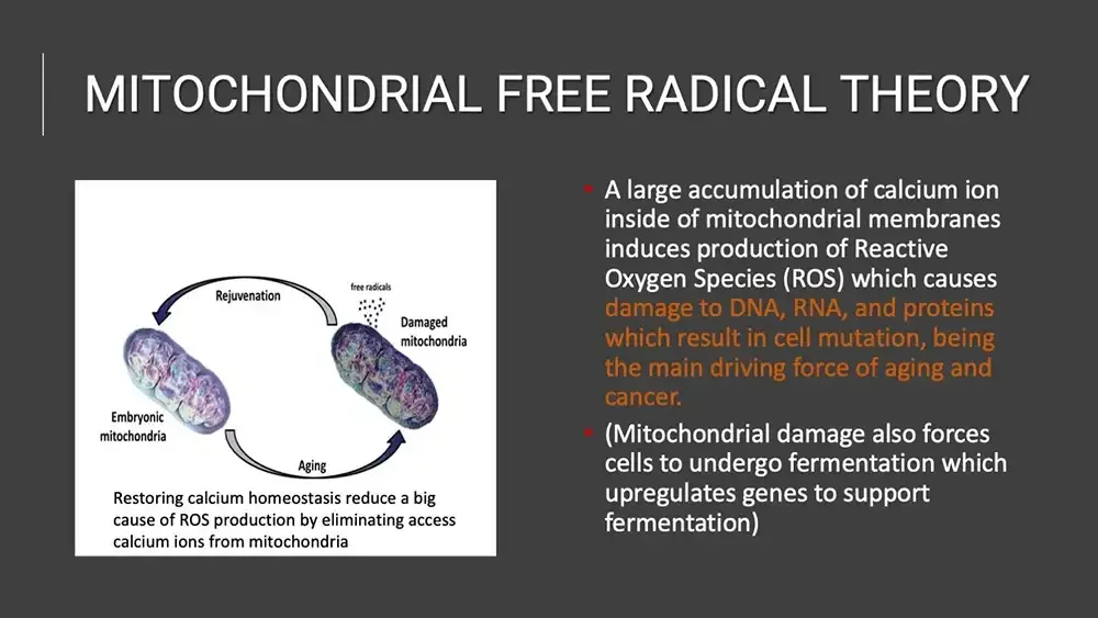
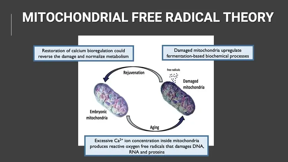
MITOCHONDRIAL FREE RADICAL THEORY Restoration of calcium bioregulation could reverse the damage and normalize metabolism Damaged mitochondria upregulate fermentation-based biochemical processes Cycle diagram: two mitochondria linked by curved arrows. Rejuvenation free radicals Damaged mitochondria Embryonic mitochondria Aging Excessive Ca²⁺ ion concentration inside mitochondria produces reactive oxygen free radicals that damages DNA, RNA and proteins
MITOCHONDRIAL FREE RADICAL THEORY Cycle diagram: two mitochondria linked by curved arrows. Rejuvenation free radicals Damaged mitochondria Embryonic mitochondria Aging Restoring calcium homeostasis reduce a big cause of ROS production by eliminating access calcium ions from mitochondria A large accumulation of calcium ion inside of mitochondrial membranes induces production of Reactive Oxygen Species (ROS) which causes damage to DNA, RNA, and proteins which result in cell mutation, being the main driving force of aging and cancer. (Mitochondrial damage also forces cells to undergo fermentation which upregulates genes to support fermentation)
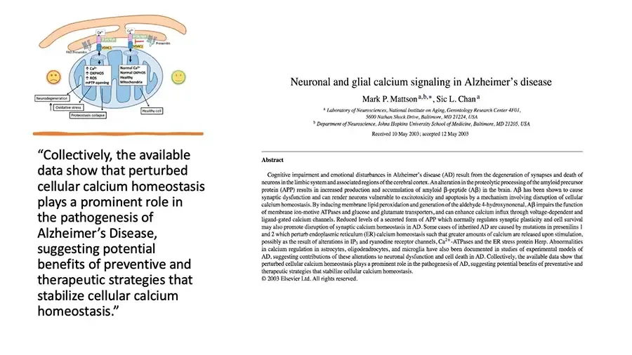
Diagram: ER and mitochondria, sad face left, smiling face right. Ca²⁺ Ca²⁺ VDAC1 VDAC1 FAD Presenilin Presenilin ↑ Ca²⁺ ↑ OXPHOS ↑ ROS mPTP opening Normal Ca²⁺ Normal OXPHOS Healthy Mitochondria Neurodegeneration ↑ Oxidative stress Proteostasis collapse Healthy cell “Collectively, the available data show that perturbed cellular calcium homeostasis plays a prominent role in the pathogenesis of Alzheimer’s Disease, suggesting potential benefits of preventive and therapeutic strategies that stabilize cellular calcium homeostasis.” Screenshot of journal article title page. Neuronal and glial calcium signaling in Alzheimer’s disease Mark P. Mattsonᵃ,ᵇ,*, Sic L. Chanᵃ ᵃ Laboratory of Neurosciences, National Institute on Aging, Gerontology Research Center 4F01, 5600 Nathan Shock Drive, Baltimore, MD 21224, USA ᵇ Department of Neuroscience, Johns Hopkins University School of Medicine, Baltimore, MD 21205, USA Received 10 May 2003; accepted 12 May 2003 Abstract Cognitive impairment and emotional disturbances in Alzheimer’s disease (AD) result from the degeneration of synapses and death of neurons in the limbic system and associated regions of the cerebral cortex. An alteration in the proteolytic processing of the amyloid precursor protein (APP) results in increased production and accumulation of amyloid β-peptide (Aβ) in the brain. Aβ has been shown to cause synaptic dysfunction and can render neurons vulnerable to excitotoxicity and apoptosis by a mechanism involving disruption of cellular calcium homeostasis. By inducing membrane lipid peroxidation and generation of the aldehyde 4-hydroxynonenal, Aβ impairs the function of membrane ion-motive ATPases and glucose and glutamate transporters, and can enhance calcium influx through voltage-dependent and ligand-gated calcium channels. Reduced levels of a secreted form of APP which normally regulates synaptic plasticity and cell survival may also promote disruption of synaptic calcium homeostasis in AD. Some cases of inherited AD are caused by mutations in presenilins 1 and 2 which perturb endoplasmic reticulum (ER) calcium homeostasis such that greater amounts of calcium are released upon stimulation, possibly as the result of alterations in IP₃ and ryanodine receptor channels, Ca²⁺-ATPases and the ER stress protein Herp. Abnormalities in calcium regulation in astrocytes, oligodendrocytes, and microglia have also been documented in studies of experimental models of AD, suggesting contributions of these alterations to neuronal dysfunction and cell death in AD. Collectively, the available data show that perturbed cellular calcium homeostasis plays a prominent role in the pathogenesis of AD, suggesting potential benefits of preventative and therapeutic strategies that stabilize cellular calcium homeostasis. © 2003 Elsevier Ltd. All rights reserved.
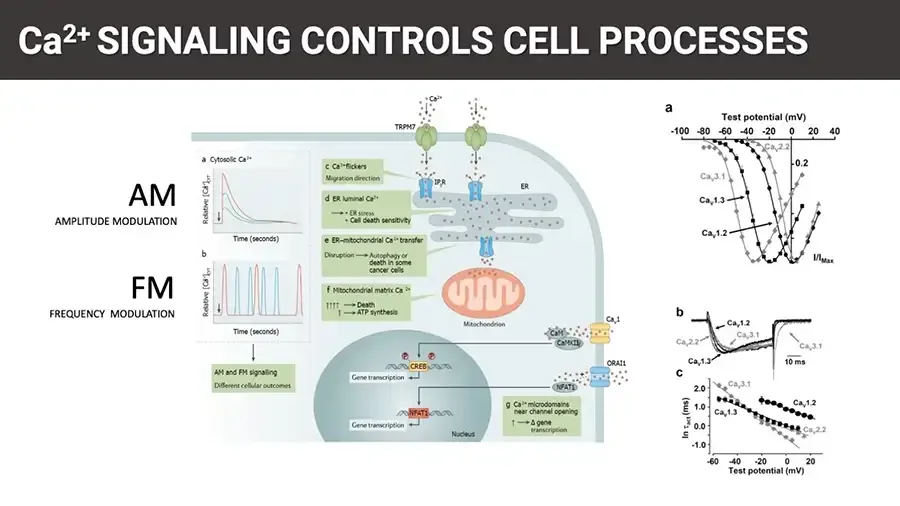
Ca²⁺ SIGNALING CONTROLS CELL PROCESSES AM AMPLITUDE MODULATION FM FREQUENCY MODULATION Figure: cell diagram of calcium signalling pathways, traces at left. a Cytosolic Ca²⁺ Line graph: three transient peaks of different heights. Relative Ca²⁺.CYT Time (seconds) b Line graph: repeated spikes at different frequencies. Relative Ca²⁺.CYT Time (seconds) AM and FM signalling Different cellular outcomes Ca²⁺ TRPM7 IP₃R ER c Ca²⁺ flickers Migration direction d ER luminal Ca²⁺ → • ER stress • Cell death sensitivity e ER–mitochondrial Ca²⁺ transfer Disruption → Autophagy or death in some cancer cells f Mitochondrial matrix Ca²⁺ ↑↑↑↑ → Death ↑ → ATP synthesis Mitochondrion CaM CaMKII Caᵥ1 ORAI1 NFAT1 P P CREB Gene transcription NFAT1 Gene transcription Nucleus g Ca²⁺ microdomains near channel opening ↑ → Δ gene transcription Three electrophysiology graphs at right: a, b, c. a Test potential (mV) X-axis. -100 -80 -60 -40 -20 0 20 40 Caᵥ2.2 0.2 Caᵥ3.1 Caᵥ1.3 Caᵥ1.2 I/I_Max b Caᵥ1.2 Caᵥ3.1 Caᵥ2.2 Caᵥ1.3 Caᵥ3.1 10 ms c ln τ_act (ms) Y-axis. 2.0 1.0 0.0 -1.0 Caᵥ3.1 Caᵥ1.2 Caᵥ1.3 Caᵥ2.2 X-axis. -60 -40 -20 0 20 Test potential (mV)
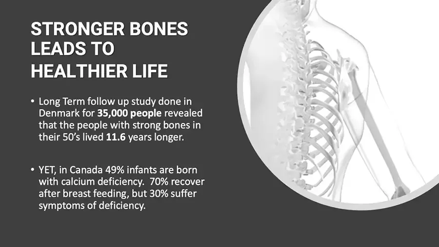
STRONGER BONES LEADS TO HEALTHIER LIFE Long Term follow up study done in Denmark for 35,000 people revealed that the people with strong bones in their 50’s lived 11.6 years longer. YET, in Canada 49% infants are born with calcium deficiency. 70% recover after breast feeding, but 30% suffer symptoms of deficiency. Illustration: translucent upper body showing spine, ribs and arm bone.
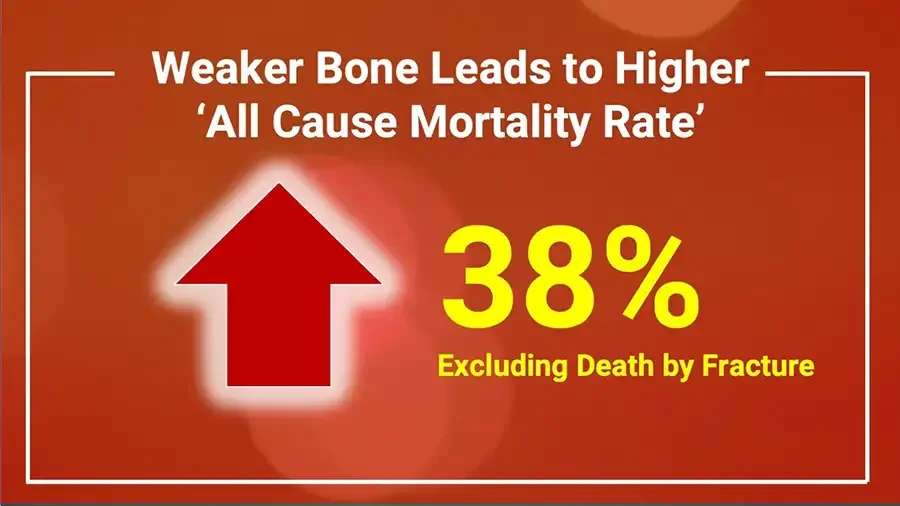
Weaker Bone Leads to Higher ‘All Cause Mortality Rate’ Large red upward arrow. 38% Excluding Death by Fracture
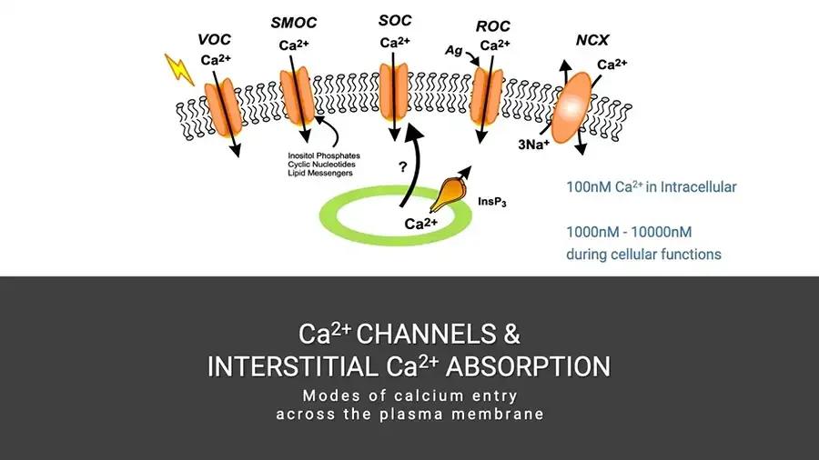
Diagram: calcium channels in a cell membrane, lightning bolt at left. VOC Ca²⁺ SMOC Ca²⁺ SOC Ca²⁺ ROC Ag Ca²⁺ NCX Ca²⁺ 3Na⁺ Inositol Phosphates Cyclic Nucleotides Lipid Messengers ? InsP₃ Ca²⁺ 100nM Ca²⁺ in Intracellular 1000nM - 10000nM during cellular functions Ca²⁺ CHANNELS & INTERSTITIAL Ca²⁺ ABSORPTION Modes of calcium entry across the plasma membrane
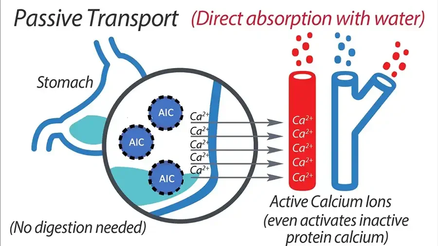
Passive Transport (Direct absorption with water) Illustration: stomach with magnified circle, arrows into blood vessels. Stomach AIC AIC AIC Ca²⁺ Ca²⁺ Ca²⁺ Ca²⁺ Ca²⁺ Ca²⁺ Ca²⁺ Ca²⁺ Ca²⁺ Ca²⁺ (No digestion needed) Active Calcium Ions (even activates inactive protein calcium)
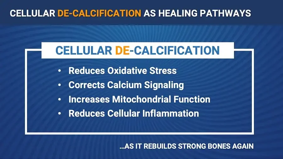
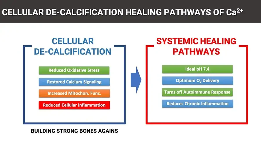
CELLULAR DE-CALCIFICATION AS HEALING PATHWAYS CELLULAR DE-CALCIFICATION Reduces Oxidative Stress Corrects Calcium Signaling Increases Mitochondrial Function Reduces Cellular Inflammation ...AS IT REBUILDS STRONG BONES AGAIN
CELLULAR DE-CALCIFICATION HEALING PATHWAYS OF Ca²⁺ CELLULAR DE-CALCIFICATION Reduced Oxidative Stress Restored Calcium Signaling Increased Mitochon. Func. Reduced Cellular Inflammation BUILDING STRONG BONES AGAINS Blue arrow pointing right. SYSTEMIC HEALING PATHWAYS Ideal pH 7.4 Optimum O₂ Delivery Turns off Autoimmune Response Reduces Chronic Inflammation
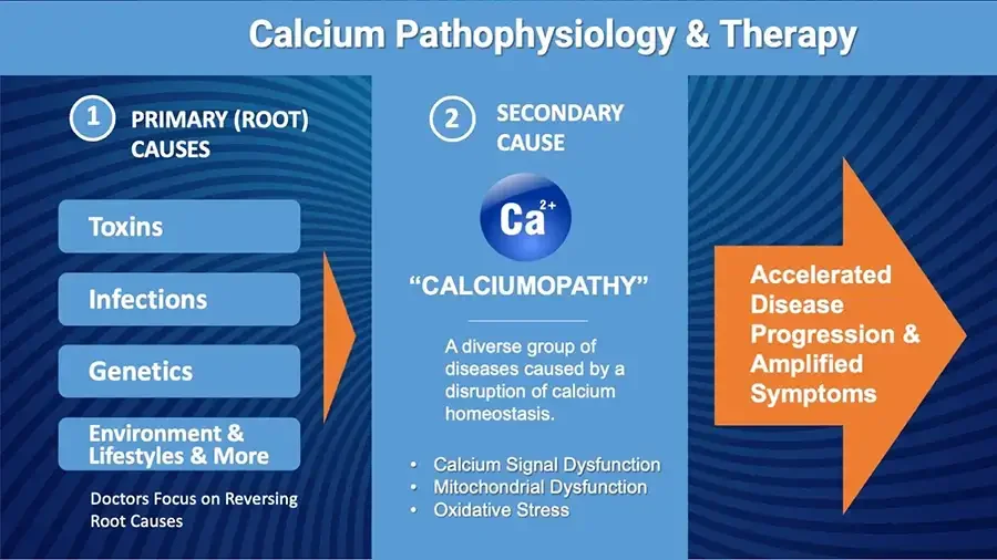
Calcium Pathophysiology & Therapy 1 PRIMARY (ROOT) CAUSES Toxins Infections Genetics Environment & Lifestyles & More Doctors Focus on Reversing Root Causes Orange arrow pointing right. 2 SECONDARY CAUSE Ca²⁺ “CALCIUMOPATHY” A diverse group of diseases caused by a disruption of calcium homeostasis. Calcium Signal Dysfunction Mitochondrial Dysfunction Oxidative Stress Large orange arrow pointing right. Accelerated Disease Progression & Amplified Symptoms
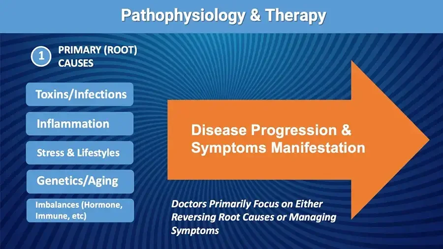
Pathophysiology & Therapy 1 PRIMARY (ROOT) CAUSES Toxins/Infections Inflammation Stress & Lifestyles Genetics/Aging Imbalances (Hormone, Immune, etc) Large orange arrow pointing right. Disease Progression & Symptoms Manifestation Doctors Primarily Focus on Either Reversing Root Causes or Managing Symptoms
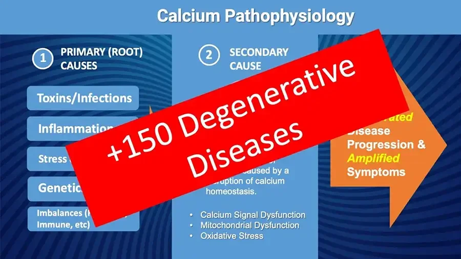
Calcium Pathophysiology Diagonal red banner across slide, hiding parts of the text. +150 Degenerative Diseases 1 PRIMARY (ROOT) CAUSES Toxins/Infections Inflammatio rest hidden by banner. Stress rest hidden by banner. Genetic rest hidden by banner. Imbalances (H rest hidden by banner. Immune, etc) 2 SECONDARY CAUSE hidden by banner. caused by a hidden by banner. ruption of calcium homeostasis. Calcium Signal Dysfunction Mitochondrial Dysfunction Oxidative Stress hidden by banner. rated hidden by banner. isease Progression & Amplified Symptoms
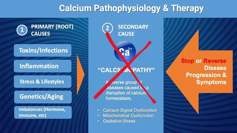
Calcium Pathophysiology & Therapy 1 PRIMARY (ROOT) CAUSES Toxins/Infections Inflammation Stress & Lifestyles Genetics/Aging Imbalances (Hormone, Immune, etc) Orange arrow pointing right. 2 SECONDARY CAUSE Red X drawn over Ca²⁺ icon and text, covering some letters. Ca²⁺ “CALCIUMOPATHY” A diverse group of diseases caused by a disruption of calcium homeostasis. Calcium Signal Dysfunction Mitochondrial Dysfunction Oxidative Stress Large orange arrow pointing left. Stop or Reverse Disease Progression & Symptoms
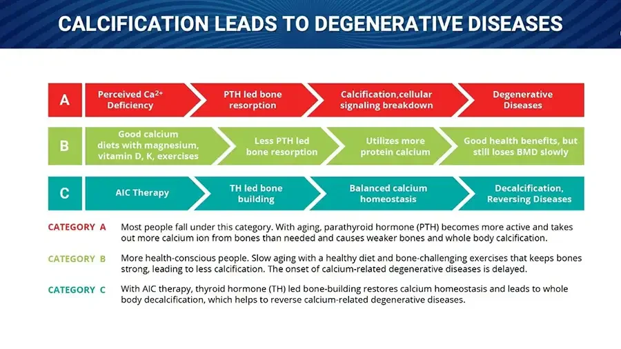
CALCIFICATION LEADS TO DEGENERATIVE DISEASES Three rows of chevron arrow steps, labeled A, B, C. A Perceived Ca²⁺ Deficiency PTH led bone resorption Calcification,cellular signaling breakdown Degenerative Diseases B Good calcium diets with magnesium, vitamin D, K, exercises Less PTH led bone resorption Utilizes more protein calcium Good health benefits, but still loses BMD slowly C AIC Therapy TH led bone building Balanced calcium homeostasis Decalcification, Reversing Diseases CATEGORY A Most people fall under this category. With aging, parathyroid hormone (PTH) becomes more active and takes out more calcium ion from bones than needed and causes weaker bones and whole body calcification. CATEGORY B More health-conscious people. Slow aging with a healthy diet and bone-challenging exercises that keeps bones strong, leading to less calcification. The onset of calcium-related degenerative diseases is delayed. CATEGORY C With AIC therapy, thyroid hormone (TH) led bone-building restores calcium homeostasis and leads to whole body decalcification, which helps to reverse calcium-related degenerative diseases.
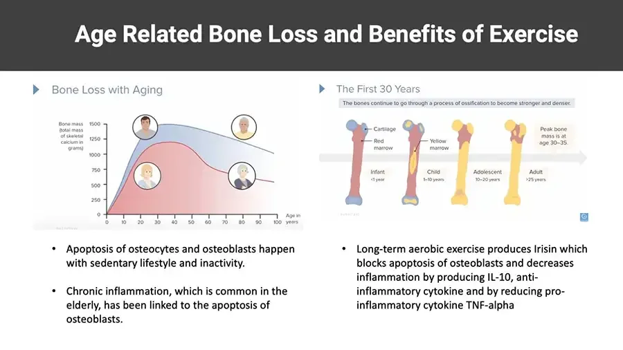
Age Related Bone Loss and Benefits of Exercise Bone Loss with Aging Area chart: bone mass by age, four portrait icons. Bone mass (total mass of skeletal calcium in grams) Y-axis. 1500 1250 1000 750 500 250 0 X-axis. 0 10 20 30 40 50 60 70 80 90 100 Age in years The First 30 Years The bones continue to go through a process of ossification to become stronger and denser. Illustration: four thigh bones at different ages along an arrow. Cartilage Red marrow Yellow marrow Infant <1 year Child 1–10 years Adolescent 10–20 years Adult 25 years Peak bone mass is at age 30–35. Apoptosis of osteocytes and osteoblasts happen with sedentary lifestyle and inactivity. Chronic inflammation, which is common in the elderly, has been linked to the apoptosis of osteoblasts. Long-term aerobic exercise produces Irisin which blocks apoptosis of osteoblasts and decreases inflammation by producing IL-10, anti-inflammatory cytokine and by reducing pro-inflammatory cytokine TNF-alpha
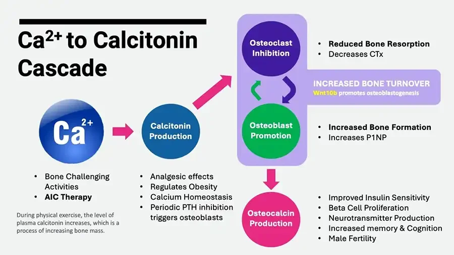
Ca²⁺ to Calcitonin Cascade Flow diagram: circles linked by arrows. Ca²⁺ Bone Challenging Activities AIC Therapy During physical exercise, the level of plasma calcitonin increases, which is a process of increasing bone mass. Calcitonin Production Analgesic effects Regulates Obesity Calcium Homeostasis Periodic PTH inhibition triggers osteoblasts Osteoclast Inhibition Reduced Bone Resorption Decreases CTx INCREASED BONE TURNOVER Wnt10b promotes osteoblastogenesis Osteoblast Promotion Increased Bone Formation Increases P1NP Osteocalcin Production Improved Insulin Sensitivity Beta Cell Proliferation Neurotransmitter Production Increased memory & Cognition Male Fertility
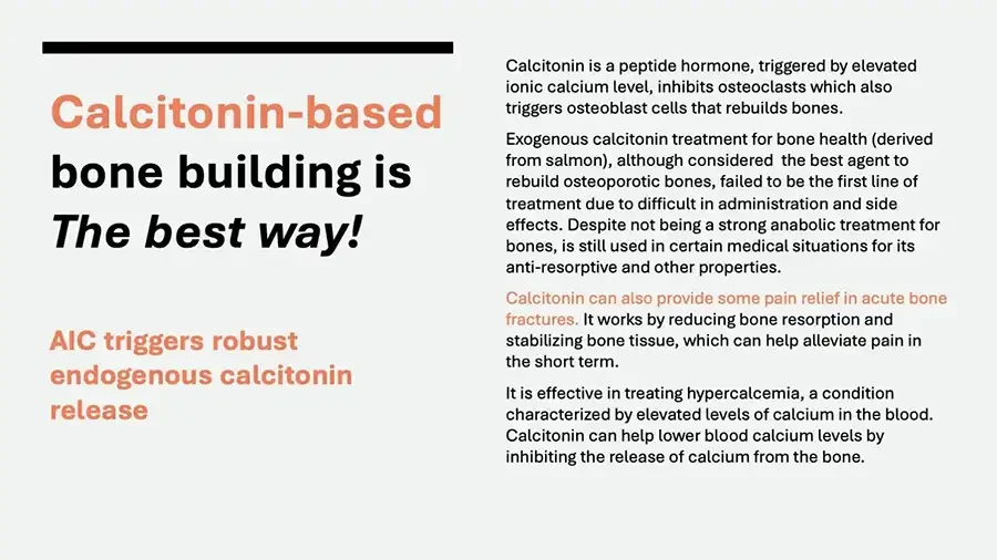
Calcitonin-based bone building is The best way! AIC triggers robust endogenous calcitonin release Calcitonin is a peptide hormone, triggered by elevated ionic calcium level, inhibits osteoclasts which also triggers osteoblast cells that rebuilds bones. Exogenous calcitonin treatment for bone health (derived from salmon), although considered the best agent to rebuild osteoporotic bones, failed to be the first line of treatment due to difficult in administration and side effects. Despite not being a strong anabolic treatment for bones, is still used in certain medical situations for its anti-resorptive and other properties. Calcitonin can also provide some pain relief in acute bone fractures. It works by reducing bone resorption and stabilizing bone tissue, which can help alleviate pain in the short term. It is effective in treating hypercalcemia, a condition characterized by elevated levels of calcium in the blood. Calcitonin can help lower blood calcium levels by inhibiting the release of calcium from the bone.
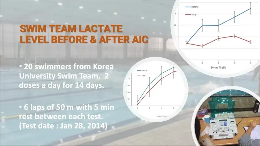
Background photo: indoor swimming pool. SWIM TEAM LACTATE LEVEL BEFORE & AFTER AIC 20 swimmers from Korea University Swim Team. 2 doses a day for 14 days. 6 laps of 50 m with 5 min rest between each test. (Test date : Jan 28, 2014) Line chart in a circular crop, left edge cut off. Before After Y-axis. cut off. 10 X-axis. 3 4 5 6 Swim Trails Line chart in a circular crop, edges cut off. Before After Y-axis. cut off. 8.00 7.50 cut off. X-axis. 2 3 4 5 Swim Trails Photo: lab analyzer on table, hand holding small tubes.