
Calcium Signaling in Erectile Dysfunction
This newsletter reveals why early erectile dysfunction(ED) is often a calcium signaling problem before it becomes a complete loss of erection.
Executive Summary
ED often begins as a disorder of cavernosal calcium handling rather than a simple blood flow failure. Endothelial stress, oxidative injury, and metabolic disease can disrupt nitric oxide signaling, increase contractile calcium activity, and weaken erection stability over time. For that reason, restoring calcium signaling may benefit ED by helping cavernosal smooth muscle relax, improving endothelial-to-smooth muscle communication, and supporting more stable venous trapping.
ED as a Disorder of Cavernosal Calcium Signaling
ED begins as a disorder of cavernosal calcium signaling. Rather than being only a blood flow problem, it often reflects disordered calcium signaling dynamics within penile smooth muscle. In the corpora cavernosa, erection starts when neural and endothelial nitric oxide signaling lowers cytosolic calcium and reduces calcium sensitivity of the contractile apparatus. The review literature shows that penile smooth muscle relaxation depends on reduced cytosolic calcium, activation of potassium currents, and tight coordination between intracellular calcium stores and membrane ion channels. When voltage-dependent calcium entry, calcium-activated chloride activity, transient receptor potential signaling, or Rho kinase-driven calcium sensitization become dominant, the cavernosal state shifts back toward contraction and detumescence.
ED then progresses through endothelial injury, oxidative stress, and structural remodeling. At the tissue level, reduced nitric oxide weakens suppression of smooth muscle calcium loading and limits vasodilation. In diabetes, hypertension, aging, and related vascular conditions, oxidative stress lowers nitric oxide bioavailability, impairs endothelial repair, and reduces cavernosal perfusion. Review data also link ED with cavernosal fibrosis, smooth muscle loss, and collagen remodeling, changes that stiffen corporal tissue and weaken veno-occlusive function. The final organ-level phenotype is failure to sustain coordinated arterial inflow, trabecular relaxation, and venous trapping even when neural initiation remains present.
These changes make restoration of ionic calcium handling directly relevant to cavernosal dysfunction. The central physiological aim is to restore smooth muscle exit from tonic contraction, improve endothelial-to-smooth-muscle signaling, and reduce redox-driven disruption of subcellular calcium handling. Anti-orbital Ionic Calcium Therapy (AIC) restores calcium signaling at membrane channels, endoplasmic reticulum release sites, and the mitochondrial calcium uniporter, helping to limit pathological persistence of cytosolic calcium and improving relaxation stability. Its regulation of cyclooxygenase-2, together with triggered endogenous calcitonin release, supports more organized vascular calcium sensing and less persistent constrictive signaling.
By supporting cellular decalcification, reducing the tendency for ectopic deposition, and improving calcium regulation of the mitochondria and endoplasmic reticulum with reduced oxidative stress, AIC addresses both the contractile defect and the structural drift that sustain ED.
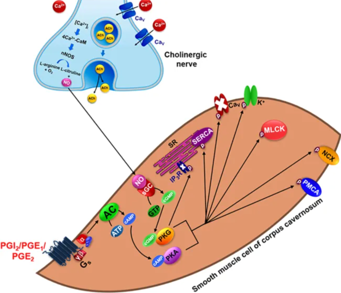
Diniz AFA, Ferreira RC, de Souza ILL, da Silva BA. Ionic Channels as Potential Therapeutic Targets for Erectile Dysfunction: A Review. Front Pharmacol. 2020 Jul 24;11:1120. doi: 10.3389/fphar.2020.01120. PMID: 32848741; PMCID: PMC7396897.https://creativecommons.org/licenses/by/4.0/
This figure explains the physiological mechanism of cavernous smooth muscle relaxation that leads to penile erection. Penile erection occurs when the corpus cavernosum relaxes in response to cholinergic neurotransmission, and nitric oxide is the most important neurotransmitter in this response. The figure also shows that non-adrenergic, non-cholinergic transmitters are present in adrenergic nerves, and that prostacyclin and prostaglandin types 1 and 2 also modulate cavernous smooth muscle relaxation. These mediators act through G protein-coupled receptors, leading to activation of cyclic guanosine monophosphate- and cyclic adenosine monophosphate-dependent protein kinases. Once activated, these protein kinases activate potassium channels and inhibit voltage-dependent calcium channels, supporting smooth muscle relaxation. The figure also includes sarco/endoplasmic reticulum calcium adenosine triphosphatase, myosin light chain kinase, sodium/calcium exchanger, and plasma membrane calcium adenosine triphosphatase as part of the relaxation mechanism shown.
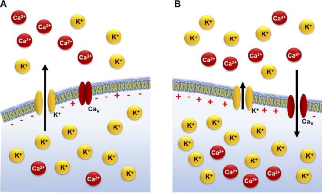
This figure explains how calcium inside the cell starts contraction in cavernous smooth muscle. It shows that this response depends on two types of control: electromechanical coupling and pharmacomechanical coupling. Electromechanical coupling causes contraction when membrane depolarization occurs, either because extracellular potassium rises or because potassium channels are blocked. Although this pathway is important, penile flaccidity primarily depends on pharmacomechanical coupling, which begins when agonists bind to G protein-coupled receptors and activate the inositol signaling pathway. This receptor-driven pathway activates phospholipase C beta 1 and generates signals that cause calcium release from the sarcoplasmic reticulum. The sarcoplasmic reticulum also contains ryanodine receptors, and calcium released through inositol 1,4,5-trisphosphate receptors can activate these channels in a process called calcium-induced calcium release.
de Souza ILL, Ferreira EDS, Vasconcelos LHC, Cavalcante FA, da Silva BA. Erectile Dysfunction: Key Role of Cavernous Smooth Muscle Cells. Front Pharmacol. 2022 Jul 5;13:895044. doi: 10.3389/fphar.2022.895044. PMID: 35865945; PMCID: PMC9294450.https://doi.org/10.3390/ijms24109067.https://creativecommons.org/licenses/by/4.0/
Clinical Tips
1. Sleep apnea can sit quietly behind ED. If a patient mentions snoring, poor sleep, or fewer morning erections, I would not ignore it. ED is sometimes the first clue that sleep quality is a bigger issue than it looks.
2. Testosterone is only one piece of the picture. Some men with ED are actually showing early signs of hidden diabetes, prediabetes, thyroid dysfunction, or high prolactin. A normal testosterone level does not close the case.
3. Simple urinary questions can open the case fast. Nocturia, urgency, weak stream, or incomplete emptying often come with ED. These complaints may reflect the same underlying bladder-prostate-vascular story.
4. The mouth can give us a useful clue. Bleeding gums and chronic periodontal inflammation are easy to miss in an ED work-up. Oral inflammation may be a quiet marker of systemic vascular and inflammatory burden.
Hidden Patterns Behind the Complaint
ED rarely comes alone. In many men, it shows up alongside poor sleep, fewer morning erections, central weight gain, unstable blood sugar patterns, urinary changes, or long-standing gum inflammation. These details matter because they can point to a wider picture of endothelial stress, metabolic strain, and loss of smooth muscle stability. A normal testosterone result does not rule out that deeper pattern. In some cases, ED appears before diabetes, thyroid dysfunction, or other endocrine problems become obvious. In others, the clue is a quieter pattern, such as snoring, nocturia, oral inflammation, or high-normal calcium with symptoms that do not seem connected at first. Looking for these linked signals can make the complaint more clinically meaningful and help explain why ED is often an early whole-body marker, not only a local sexual symptom.
Clinical Practice Snapshot
ED does not always appear as a complete absence of erection. In clinical practice, it often appears as reduced rigidity, delayed full tumescence, brief erection duration, or early loss of firmness during intercourse. These patterns help distinguish impaired erection maintenance from impaired erection initiation.
They also suggest that the functional problem may lie within cavernosal smooth muscle behavior and erection stability rather than desire alone. This distinction matters in patients with diabetes, vascular disease, or an age-related endothelial burden, in whom penile function may become unstable before it is completely lost.
A partial but unsustained erectile response often indicates a system that can initiate tumescence but cannot maintain coordinated corporal function. That clinical pattern fits well with disease states shaped by abnormal ionic regulation within the erectile tissue unit. In this setting, AIC may provide benefit by improving the physiological basis of erection stability.
Conclusion
ED often begins as an early problem of cavernosal stability before it progresses to complete erection loss. Reduced rigidity, slower tumescence, and early loss of firmness suggest that the erectile tissue can still start a response but cannot maintain coordinated relaxation. This kind of instability reflects more than reduced blood flow, because nitric oxide, cyclic nucleotide signaling, potassium channel activity, and controlled calcium movement between membrane channels and intracellular stores all need to stay coordinated for an erection to last.
This perspective also helps connect the mechanism with clinical clues that are easy to miss. When depolarization-driven calcium entry or receptor-driven intracellular calcium release becomes dominant, contraction pathways take over again, oxidative stress rises, and structural remodeling may follow. This helps explain why ED may appear together with poor sleep, fewer morning erections, hidden glycemic or endocrine stress, urinary symptoms, and even chronic oral inflammation before other problems become obvious.
AIC Therapy fits this physiology because ED depends on the orderly handling of calcium across membrane channels, release sites on the endoplasmic reticulum, and mitochondrial control. By helping reduce persistent contractile calcium loading and supporting more stable cavernosal relaxation, AIC Therapy fits ED at both the signaling and tissue levels.
Bring calcium signaling into your practice.
Practitioners can request a consultation to discuss AIC therapy and how these mechanisms apply to specific cases.
Newsletter content is educational and contributed by participating healthcare practitioners. These statements have not been evaluated by the FDA and are not intended to diagnose, treat, cure, or prevent any disease.