When the Brain Misfires: A New Pathway in Autism Relief
This newsletter reveals that restoring calcium flow inside brain cells is a key step toward rebalancing neural circuits, reducing sensory overload, and unlocking meaningful functional improvements in children with autism.
Executive Summary
Autism is increasingly linked to disrupted intracellular flow, affecting how neurons signal, grow, and recover. Calcium plays a central role in this process—when its movement between cellular compartments is impaired, the brain loses its ability to regulate sensory input, sleep, and behavior. By restoring that calcium rhythm, we open a path to more stable neural function and measurable clinical progress.
Beyond Behavior: What the Cells Are Trying to Say
Autism is increasingly understood not as a static neurodevelopmental defect, but as a dynamic disruption of intracellular signaling, especially at the synapse. Dysfunctions in scaffolding proteins, such as SHANK3 and neuroligins, weaken the architecture required for a precise excitatory–inhibitory balance, while epigenetic drift during prenatal development alters the timing of neuronal differentiation and axon guidance. On a metabolic level, mitochondrial insufficiency limits ATP production for synaptogenesis and heightens oxidative stress, contributing to chronic microglial activation and cytokine-driven spine pruning. These combined molecular disruptions desynchronize cortical networks, explaining the social withdrawal, sensory overload, and language delays seen in children like Marquese.
Calcium is not just involved—it’s central. Precise intracellular calcium pulses coordinate a wide range of cellular processes, including synaptic plasticity, gene transcription, and dendritic refinement. Yet, in many autism cases, calcium buffering proteins like parvalbumin are depleted, and TRPC channel variants distort calcium entry, leading to an elevated baseline Ca²⁺ level that accelerates maladaptive pruning.
Clinically, this means you can act by assessing ionized calcium, correcting magnesium and vitamin D deficits, and considering buffered ionic calcium supplementation with intracellular antioxidant support. By restoring calcium rhythm at the cellular level, we open a therapeutic window that aligns molecular correction with behavioral gains.
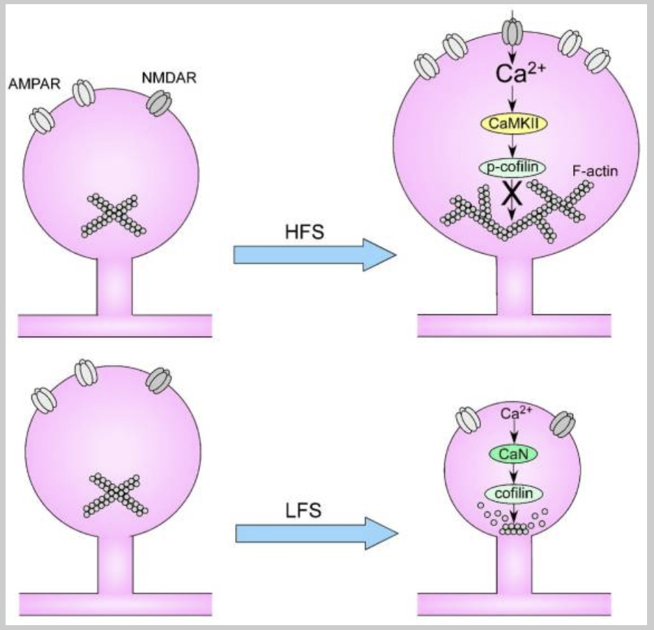
Ugarte G, Piña R, Contreras D, et al. Attention Deficit-Hyperactivity Disorder (ADHD): From Abnormal Behavior to Impairment in Synaptic Plasticity. Biology (Basel). 2023;12(9):1241. Published 2023 Sep 15. doi:10.3390/biology12091241
This figure illustrates how different patterns of stimulation affect the structure of synaptic connections in the brain. When stimulation is intense or frequent, calcium enters the neuron through NMDA-type receptors, triggering the activation of specific enzymes that promote the growth and strengthening of synaptic structures. This process involves not only calcium signaling but also the polymerization of actin filaments, which expand the spine head and enhance the efficiency of neural communication. In contrast, when stimulation is weak or occurs at low frequency, the calcium signal is smaller and activates a different set of enzymes, leading to actin breakdown and resulting in spine shrinkage and reduced synaptic strength. Together, these mechanisms show how neurons adapt structurally, either reinforcing or weakening their connections, based on the nature of the incoming signals.
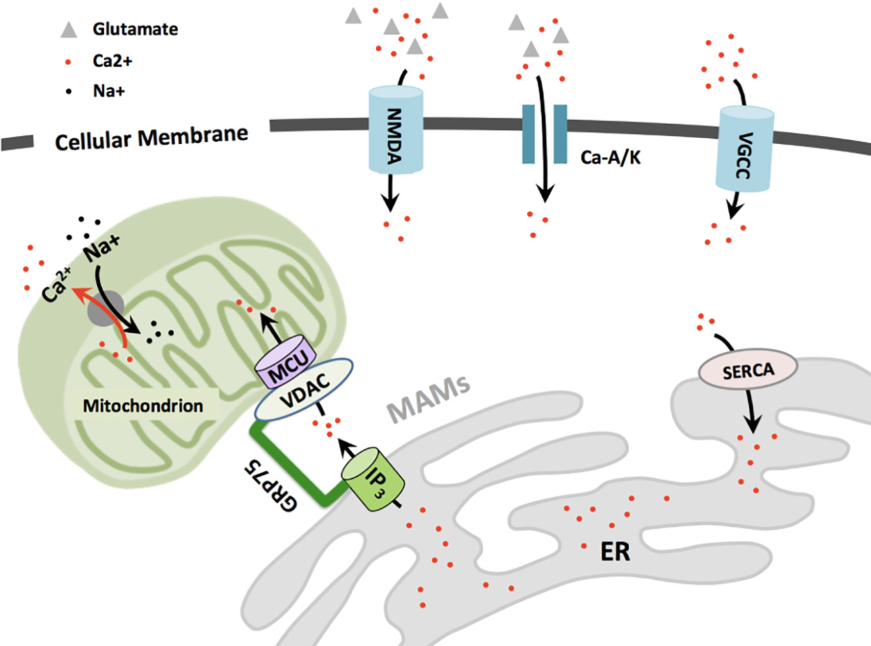
Nguyen RL, Medvedeva YV, Ayyagari TE, Schmunk G, Gargus JJ. Intracellular calcium dysregulation in autism spectrum disorder: An analysis of converging organelle signaling pathways. Biochim Biophys Acta Mol Cell Res. 2018;1865(11 Pt B):1718-1732. doi:10.1016/j.bbamcr.2018.08.003
This figure illustrates how calcium enters and moves within neurons to support essential cellular functions. Calcium first crosses the cell membrane through NMDA and AMPA-type glutamate receptors, as well as voltage-gated calcium channels activated during electrical activity. Once inside the cell, calcium rapidly enters mitochondria through specialized transporters and is also gradually stored in the endoplasmic reticulum, which serves as the main calcium reservoir. At contact points between the ER and mitochondria—called mitochondria-associated membranes—calcium flows directly from the ER into mitochondria, guided by protein complexes that ensure proper energy production. To prevent overload, excess calcium is eventually expelled from the mitochondria through a sodium-calcium exchange system, thereby maintaining cellular balance and viability.
Where AIC Fits Clinically
In many patients with autism, calcium flow inside brain cells is disrupted, leading to problems with energy, signaling, and sensory overload. These shifts often go unnoticed in standard tests but can quietly disturb how neurons communicate and how the brain handles stress.
AIC therapy restores ionic calcium exactly where it's needed, helping cells regain proper rhythm and control. It supports the exchange of calcium between the endoplasmic reticulum and mitochondria, two core systems that maintain neuronal stability. By reactivating this cellular balance, AIC offers a direct and targeted way to support brain regulation from within.
Overlooked Clinical Signals in Autism
1. Texture sensitivity isn’t always about behavior.
Some children reject food not because they’re picky, but because their brain misreads textures as a threat. When calcium signaling in sensory areas is off, even soft foods can feel sharp or “unsafe.”
2. Constipation may start in the nervous system.
Many kids with autism struggle with slow bowels for years. But the issue may not be in the gut itself—it can be a loss of nerve signals, especially when calcium control around the colon breaks down.
3. Sound sensitivity can stem from brain overload, rather than the ears.
If a child covers their ears or panics at normal sounds, it may be due to calcium deposits in the brain’s sound relay centers. Instead of filtering out background noise, the brain turns everything up.
4. Sudden improvement in sleep can signal brain recovery.
When intracellular calcium resets in key brain areas, sleep often returns, sometimes within days. It’s not a coincidence. Deep sleep needs proper calcium timing to switch the brain into rest mode.
Case Reference
This case features a female adolescent with autism and secondary epilepsy who developed worsening seizures following conventional interventions, including stem cell therapy and transcranial brain stimulation. Despite multiple efforts, her seizure frequency progressively increased, showing no sustained neurological relief.
However, after introducing AIC (Anti-orbital Ionic Calcium) therapy, her seizure episodes began decreasing in a clear, stepwise pattern—first stabilizing, then gradually extending seizure-free intervals from weeks to nearly three months. This improvement strongly suggests that AIC therapy helped restore calcium signaling balance, reduce neuronal hyperexcitability, and support intrinsic neuroregulation. This case demonstrates AIC’s capacity to modulate cellular pathways critical to brain resilience, offering a non-invasive, biochemically grounded approach to neurological disorders.
Conclusion
Autism is no longer seen as a fixed disorder—it’s now understood as a condition shaped by disrupted cellular timing and intracellular communication. Cortical imbalance often begins with subtle defects in how neurons handle growth signals, energy flow, and structural adaptation. The figures in this newsletter highlight how the strength or weakness of stimulation shapes the physical growth of synapses, and how calcium moves between compartments to maintain neuronal health.
This patient’s clinical progress—marked by longer seizure-free intervals and improved regulation—began only after restoring ionic calcium dynamics at the cellular level. AIC therapy reestablishes the natural flow between mitochondria and the ER, a process essential for electrical stability and cognitive calm. And overlooked signals like texture refusal, chronic constipation, and sudden sound sensitivity are now better explained by misfiring cellular rhythms rather than behavioral resistance. Together, these insights offer a clear path: when we restore internal flow, the brain regains its ability to regulate from the inside out.
Bring calcium signaling into your practice.
Practitioners can request a consultation to discuss AIC therapy and how these mechanisms apply to specific cases.
Newsletter content is educational and contributed by participating healthcare practitioners. These statements have not been evaluated by the FDA and are not intended to diagnose, treat, cure, or prevent any disease.