Mitochondrial Detox Roadmap, from Exposure Control to Energy Recovery
This newsletter reveals how orderly mitochondrial signaling, redox rebuilding, and measured detox restore ATP coupling by reestablishing brief, well-timed calcium pulses across tissues.
Executive Summary
Toxic metal stress and redox drift trap calcium inside mitochondria, uncoupling ATP and slowing repair across multiple organs, sometimes including cognition, all the while serum values can look normal. Protocols that stabilize the extracellular ionized fraction, ensure magnesium sufficiency, activate Nrf2 for glutathione rebuilding, and recover MCU–NCLX timing tend to outperform antioxidant megadoses alone and quietly point toward ionic strategies such as AIC therapy that support this rhythm.
From Exposure Control to Signal Timing, Rebuilding Mitochondrial Function
Toxic metals such as cadmium, mercury, and lead bind respiratory chain proteins in mitochondria, increase reactive oxygen species, and lower ATP levels, which show up as low energy and slow tissue repair. These metals also disrupt the systems that move calcium across the mitochondrial inner membrane, so calcium builds up inside the organelle even when serum calcium is normal, worsening exercise intolerance and cognitive slowing.
When mitochondrial calcium remains elevated, the permeability transition pore opens more readily, membrane potential collapses, and cells become more sensitive to death signals —a driver of chronic toxicant fatigue and tissue fragility. In practice, mitochondrial detoxification —not just adding generic antioxidants —should reduce metal load and restore calcium flux, because efficient energy coupling depends on brief, well-timed calcium pulses.
The first clinical lever is exposure control, together with rebuilding the glutathione system, since glutathione conjugates metals and stabilizes the redox tone that protects mitochondrial enzymes. Activating the Nrf2 pathway upregulates enzymes involved in glutathione synthesis and phase II conjugation, thereby lowering oxidative triggers that keep calcium channels hyperactive under stress.
Damaged mitochondria are best removed by mitophagy, and restoring this pathway allows healthy organelles to replace dysfunctional ones, thereby stabilizing intracellular calcium buffering. Graded aerobic conditioning acts as mitochondrial medicine by stimulating biogenesis and improving calcium handling during predictable energy demand, so paced post-meal walking is a practical starting step.
Maintaining magnesium sufficiency calms overactive calcium channels in excitable tissues and supports vascular tone, so checking and correcting magnesium is a simple, high-yield step. Nutrition that supports endogenous antioxidant capacity works better than single mega doses, because it strengthens the enzymes that keep mitochondrial redox and calcium signaling in balance.
Before considering chelation, body burden and clinical status should be documented, because chelators can redistribute metals and place excessive stress on mitochondria if used without a clear indication and monitoring. In contrast, AIC Therapy naturally restores mineral equilibrium, activates endogenous calcitonin, and promotes the cellular release of toxic calcium and heavy metals without forcing the body, making it a physiological alternative to aggressive chelation.
Therefore, a cellular longevity detox plan integrates exposure reduction, redox rebuilding, mitophagy support, mineral balance through AIC Therapy, and graded physical activity to gradually restore clean mitochondria and stable calcium signaling over time.
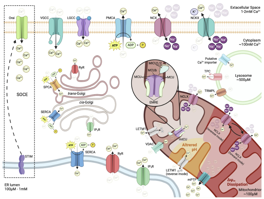
Borbolis F, Ploumi C, Palikaras K. Calcium-mediated regulation of mitophagy: implications in neurodegenerative diseases. NPJ Metab Health Dis. 2025;3(1):4. doi:10.1038/s44324-025-00049-2
This figure illustrates how cells tightly regulate calcium to protect their function and support energy production. Calcium enters the cell through activation channels and is quickly removed or stored to prevent toxicity. The endoplasmic reticulum and Golgi act as main storage sites: they take up calcium when levels rise and release it when the cell needs to activate specific responses. When these stores are depleted, a sensor protein (STIM) signals the Orai channel to bring in more calcium from the extracellular space. Mitochondria also take up calcium to produce ATP, but if calcium becomes excessive, they release it through safety pathways, which is an early sign of cellular stress and potential damage.
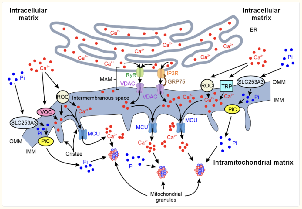
Shapiro IM, Risbud MV, Landis WJ. Toward understanding the cellular control of vertebrate mineralization: The potential role of mitochondria. Bone. 2024;185:117112. doi:10.1016/j.bone.2024.117112
This figure illustrates how calcium is continuously moved through channels, pumps, and exchangers to maintain cellular balance. Calcium enters the cell through activation channels and is then either stored in organelles, such as the endoplasmic reticulum and the Golgi apparatus, or removed by membrane pumps to prevent overload. When intracellular calcium stores become low, a sensing mechanism activates additional calcium entry to sustain essential signaling. Mitochondria temporarily take up calcium to support ATP production, but release it when levels become excessive to protect the cell from damage. This coordinated system ensures stable calcium homeostasis, which is critical for cell survival and function.
Five Common Clinical Misconceptions
Misconception 1: Detox equals chelation.
Clinical angle 1: Chelation is a late tool, not step one. I start with exposure control, bowel and bile flow, hydration, mineral repletion, and redox rebuilding, then document body burden before touching a chelator. This sequence protects mitochondria from redistribution crashes and makes any later chelation both safer and more effective.
Misconception 2: Normal serum calcium means intracellular calcium is fine.
Clinical angle 2: Patients can have mitochondrial matrix calcium overload while serum values appear normal. Address magnesium status, lower phosphate intake, improve sleep and paced activity, and support endothelial nitric oxide to normalize calcium entry and extrusion at the organelle level.
Misconception 3: Antioxidants alone will fix mitochondrial stress.
Clinical angle 3: The core lesion is disordered signaling and organelle turnover, not a simple lack of scavengers. I am biased toward nutrition that strengthens endogenous enzyme systems, Nrf2 activation, and steady mitophagy plus biogenesis through graded aerobic work, so mitochondria can handle calcium transients during real-life demand.
Misconception 4: Faster detox works better.
Clinical angle 4: Rapid mobilization pushes metals and calcium faster than tissues can sequester and excrete, which destabilizes membrane potential and worsens fatigue. A gentle, titrated program with daily movement, fiber-supported elimination, and periodic clinical checks keeps flux directional and symptoms stable.
Misconception 5: Mitochondrial calcium overload is a one-way street.
Clinical angle 5: Calcium handling is dynamic and modifiable. Stabilize the extracellular ionized pool, ensure vitamin K-dependent proteins are active, maintain magnesium sufficiency, and rebuild microcirculation. With those pieces in place, calcium traffic at the mitochondrion becomes orderly again, and energy coupling improves.
Case Reference
A 66-year-old man had biopsy-confirmed metastatic prostate cancer with innumerable liver lesions up to 3.3 cm, a bulky prostate, enlarged abdominal and left supraclavicular nodes, and bone metastases on scan. He began AIC Therapy with no conventional therapy recorded in that window, and PSA fell from 46.0 to 0.99 by early October. Follow-up CT showed the segment 4a liver lesion shrinking to 1.6 cm, most other hepatic lesions and nodal disease were smaller, no new liver lesions, and a smaller prostate, while new sclerotic spine and pelvic foci were described as osteoblastic. Woven into a mitochondrial medicine view, care aimed to steady extracellular ionized calcium and improve redox so Ca²⁺ flux and ATP production could run more normally during recovery.
Conclusion
Toxic metals bind respiratory complexes, raise oxidative stress, and disrupt transport across the inner membrane, so calcium lingers in the matrix even when serum measurements look fine. Persistent matrix calcium lowers membrane potential, sensitizes the permeability transition pore, and heightens susceptibility to cell death signals, which present clinically as fatigue, slow tissue repair, and exercise intolerance. Physiologic balance depends on coordinated entry channels, ER- and Golgi-mediated storage, membrane pumps, and STIM–Orai store-operated entry, while mitochondria buffer brief pulses to support ATP production and release excess as a safety response. Effective care, therefore, starts with exposure reduction, Nrf2-driven glutathione replenishment, magnesium sufficiency, paced aerobic conditioning, and a cautious approach to rapid chelation.
When the extracellular ionized pool is stable and microcirculation improves, calcium pulses regain timing through MCU uptake and NCLX extrusion, and energy coupling recovers with steadier day-to-day function. AIC is helpful because it supports the extracellular ionic fraction and encourages orderly transmembrane flux without pushing total calcium load, which calms overactive channels and restores pulse quality. By promoting timely pulses and relieving aberrant intracellular accumulation, AIC helps normalize ATP production and makes detox progression safer when paired with magnesium, redox rebuilding, and graded activity.
Bring calcium signaling into your practice.
Practitioners can request a consultation to discuss AIC therapy and how these mechanisms apply to specific cases.
Newsletter content is educational and contributed by participating healthcare practitioners. These statements have not been evaluated by the FDA and are not intended to diagnose, treat, cure, or prevent any disease.