This newsletter reveals why calcium signaling is so essential in osteogenesis imperfecta.
Executive Summary
Collagen type I variants initiate most osteogenesis imperfecta phenotypes, yet calcium flux across membrane channels, endoplasmic reticulum release, and mitochondrial uptake shape the severity of the matrix defect. Endoplasmic reticulum stress, oxidative load, and impaired bioenergetics can convert a fixed genetic defect into higher fracture frequency, persistent pain, and unstable turnover signals by degrading collagen processing and secretion efficiency. This rationale supports a protocol that modulates intracellular calcium flux and calcitonin-mediated osteoclast restraint to attenuate ER stress and stabilize collagen processing.
Why Calcium Signaling Matters in Brittle Bone
Clinically, osteogenesis imperfecta presents with recurrent low-trauma fractures, bone deformity, and short stature. Most cases arise from mutations in type I collagen, yet the severity of the phenotype also depends on how bone cells regulate calcium ions during matrix formation and remodeling. Calcium ions function as rapid signals that couple membrane activity, cytoskeletal tension, and collagen secretion from the endoplasmic reticulum. When calcium signaling is mis-timed, osteoblasts mis-handle collagen folding, trafficking, and mineral deposition, even though the causal gene variant remains unchanged. Calcium handling, therefore, becomes a practical mechanistic determinant of brittle bone expression across pediatric and adult forms of osteogenesis imperfecta.
Within the endoplasmic reticulum, calcium supports chaperone binding and the enzymatic steps that modify procollagen before secretion. Variants in intracellular cation channels linked to recessive osteogenesis imperfecta can disrupt calcium flux from the endoplasmic reticulum to the cytosol, altering post-translational processing of type I collagen. Endoplasmic reticulum calcium dysregulation also drives endoplasmic reticulum stress, and stressed osteoblasts tend to generate reactive oxygen species and show weaker differentiation programs. Mitochondria respond to altered calcium transfer with impaired energy handling, which can further reduce osteoblast matrix output and increase remodeling variability. Histology often is consistent with this cell-stress model, with disorganized collagen architecture and bone that fails before it can adequately distribute load.
A treatment rationale starts with restoring ionic calcium dynamics across osteoblasts, osteocytes, and osteoclasts to normalize signal timing during matrix formation and remodeling . The next step is to improve coordination of calcium transfer among membrane channels, endoplasmic reticulum release, and mitochondrial uptake, reducing stress pathway activation and stabilizing collagen secretion. Anti-orbital Ionic Calcium (AIC) Therapy restores calcium signaling and supports coordinated collagen handling in type I collagen-producing cells. AIC Therapy also improves mitochondrial and endoplasmic reticulum calcium regulation and lowers oxidative stress, thereby reducing cellular stress and fragility. AIC regulates cyclooxygenase-2, triggers endogenous calcitonin release, and lowers the tendency for ectopic deposition, supporting remodeling under steadier calcium homeostasis to improve bone matrix mineralization quality and reduce fracture susceptibility in osteogenesis imperfecta.
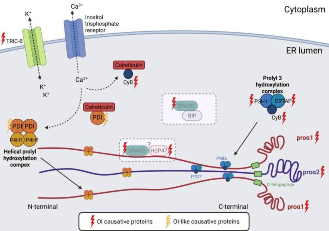
This figure shows that osteogenesis imperfecta can disrupt the processing of type I collagen, leading to either over- or undermodified collagen. The most common cause of too much modification is a point mutation that replaces a glycine in the alpha 1 type I collagen or alpha 2 type I collagen chains, which slows triple helix folding and keeps the chains exposed longer to post-translational modification events, so some overmodified collagen is retained in cells in patients and mouse models.
Excessive modification can also occur when the prolyl 3-hydroxylation complex is defective; this complex includes prolyl 3-hydroxylase 1, cartilage-associated protein, and cyclophilin B. In contrast, mutations in the endoplasmic reticulum trimeric cation channel type B disrupt intracellular calcium flux, altering endoplasmic reticulum enzymes that carry out type I collagen post-translational modifications, leading to insufficient modification. The figure also shows possible collagen “helper” complexes that may act as chaperones, involving secreted protein acidic and rich in cysteine, heat shock protein 47, and binding immunoglobulin protein, and it notes that osteogenesis imperfecta-like protein disulfide isomerase mutations have not been shown to cause collagen overmodification, but more research is needed. This figure has been generated using BioRender.
Cotti S, Pérez Franco W, Forlino A. Molecular drivers of osteogenesis imperfecta: a cellular and extracellular collagen disease. Clin Sci (Lond). 2025 Dec 18;139(24):1733–68. doi: 10.1042/CS20255642. PMID: 41410595; PMCID: PMC12794382.https://creativecommons.org/licenses/by/4.0/
Clinical Tips
- High fracture burden with modest deformity suggests that osteoblast calcium signaling timing failure can amplify collagen gene effects by disrupting secretion kinetics.
- Screen for muscle cramps, fatigue, or palpitations, as some recessive osteogenesis imperfecta subtypes can alter intracellular Ca²⁺ flux in excitable tissues as well as bone.
- Persistent bone pain without a new fracture can reflect osteocyte network stress, where osteocyte calcium signals coordinate population responses to mechanical loading and microdamage cues.
- Variable bone turnover markers can track inflammatory tone, because cyclooxygenase-2-driven prostaglandin signaling intersects osteoclast differentiation and calcium-dependent transcriptional programs.
- When mineralization appears mismatched to collagen quality, prioritize endoplasmic reticulum calcium-dependent collagen processing upstream of mineral deposition rather than focusing only on the mineral phase.
Clinical Practice Snapshot:
Osteogenesis imperfecta is driven by defects in type I collagen, but clinical severity is also shaped by calcium ion handling within osteoblasts and osteocytes. Disrupted endoplasmic reticulum calcium homeostasis promotes endoplasmic reticulum stress, impairs collagen folding and secretion, and lowers matrix quality. Altered calcium transfer between the endoplasmic reticulum and mitochondria increases oxidative stress and restricts cellular energy availability for matrix production and repair. Clinically, this can present as fractures out of proportion to deformity, persistent bone pain without a clear new fracture, fluctuating bone turnover markers, and slower recovery after minor injuries.
AIC Therapy restores calcium signaling across membrane calcium channels, endoplasmic reticulum calcium release, and mitochondrial calcium handling, addressing cellular drivers linked to fragile matrix formation. By improving mitochondrial and endoplasmic reticulum calcium regulation and lowering oxidative stress, it aligns with the osteoblast stress phenotype described in osteogenesis imperfecta and supports remodeling toward a higher-quality matrix state.
Cyclooxygenase-2 regulation connects to prostaglandin signaling that can amplify osteoclastogenesis, making this axis clinically relevant when resorption signals dominate. Triggering endogenous calcitonin release links calcium homeostasis to direct osteoclast restraint, fitting patients with high resorption markers and frequent microfracture activity. Supporting cellular decalcification and reducing the tendency toward ectopic deposition favors physiologic calcium distribution in soft tissues during immobilization, repeated microinjury, or postsurgical periods, which complicate osteogenesis imperfecta care.
Conclusion
Osteogenesis imperfecta is primarily a disorder of type I collagen quantity or quality, but intracellular calcium handling strongly modifies matrix quality and turnover. In dominant disease, glycine substitutions slow triple-helix folding, prolong exposure to collagen-modifying enzymes, and produce overmodified collagen with increased intracellular retention.
Defects in the collagen post-translational modification complex can produce a related processing phenotype by disrupting this pathway. In recessive forms linked to endoplasmic reticulum (ER) ion-channel biology, disturbed ER-to-cytosol cation flux alters modifying-enzyme activity and can shift collagen toward undermodification. Reduced ER luminal calcium also weakens ER chaperone interactions, increases unfolded protein response signaling, and reduces effective secretion of properly processed procollagen. The resulting stress state increases reactive oxygen species and weakens osteoblast differentiation, while altered ER-to-mitochondria calcium transfer impairs ATP availability required for matrix synthesis and repair.
These cellular constraints are consistent with fractures out of proportion to deformity, delayed recovery after minor trauma, persistent bone pain without a new fracture, and variable bone turnover markers. When resorption signaling predominates, calcium-dependent transcription can favor osteoclastogenesis, making the inflammatory context clinically relevant.
Clinically, this is the setting in which matrix quality and remodeling coupling become the main therapeutic limiting factors rather than mineral supply alone. A practical target is the timing of calcium signals across membrane influx, ER release, and mitochondrial uptake, with parallel engagement of endogenous calcitonin to restrain osteoclast output. AIC improves these processes by stabilizing intracellular calcium homeostasis, reducing ER and mitochondrial stress, and shifting remodeling toward more consistent collagen processing and higher matrix quality.
Bring calcium signaling into your practice.
Practitioners can request a consultation to discuss AIC therapy and how these mechanisms apply to specific cases.
Newsletter content is educational and contributed by participating healthcare practitioners. These statements have not been evaluated by the FDA and are not intended to diagnose, treat, cure, or prevent any disease.