
Executive Summary
Hypertension is blood pressure that keeps rising and slowly returns to normal after stress. The real change lies in the arterial wall, where calcium is the signal that triggers muscle contraction. When the wall can no longer clear that calcium, the vessel stays tense and cannot fully relax. Bring calcium handling back into balance, and the pressure eases with it.
How a breakdown in vascular calcium handling raises and sustains blood pressure
Hypertension traces back to a calcium-handling problem in the wall of small arteries, not only to volume load or sympathetic drive. Smooth muscle cells in the vessel wall set vascular tone by regulating intracellular calcium levels. At rest, they maintain very low intracellular calcium and high extracellular calcium, a steep gradient that requires constant energy to maintain. In hypertension that is controlled, slips occur. Calcium entry at the cell surface is higher than it should be. At the same time, the pumps and exchangers that clear calcium fall behind. The vessel then sits closer to its constricted state. As a result, vascular resistance increases and systolic pressure gradually rises before any single alarming reading appears.
The failure is not one channel but several calcium-handling steps shifting in the same direction. The surface channels that allow calcium into smooth muscle cells become more active, so each signal to contract hits harder. The contractile machinery also grows more sensitive to calcium, so vessels stay constricted even when calcium is only modestly raised. Two intracellular enzyme systems drive this sensitization and sustain the contraction signal. Oxidative stress adds another layer. Reactive oxygen species open additional calcium channels at the cell surface and drive a surface exchanger to run in reverse, so it carries calcium in instead of clearing it. The endothelium, which should counter all this by relaxing the vessel, loses that capacity as its calcium signaling becomes disordered and nitric oxide levels fall. The clinical reading is sustained vasoconstriction, arterial stiffness, and blood pressure that is difficult to control.
Slowing this process means restoring calcium handling at every site where it drifted inside the vessel wall. The cell surface, the internal calcium stores, and the mitochondria all have to return to coordinated control. Mitochondria matter here because when they take up calcium poorly, oxidative stress rises and feeds back onto the same calcium routes that started the problem. When calcium entry at the surface returns to the normal range, the pumps and exchangers can keep pace again. As the internal stores and mitochondria regain coordinated handling, the endothelium recovers its capacity to relax the vessel. Anti-orbital Ionic Calcium Therapy (AIC) restores this coordinated calcium handling across those sites. It also lowers the oxidative stress that keeps the cycle running. The clinical reading is steadier blood pressure, a better response when vascular tone is the limiting factor, and a slower progression of arterial stiffening that accompanies long-standing hypertension.
Dai C, Khalil RA. Calcium Signaling Dynamics in Vascular Cells and Their Dysregulation in Vascular Disease. Biomolecules. 2025; 15(6):892. https://doi.org/10.3390/biom15060892
https://creativecommons.org/licenses/by/4.0/
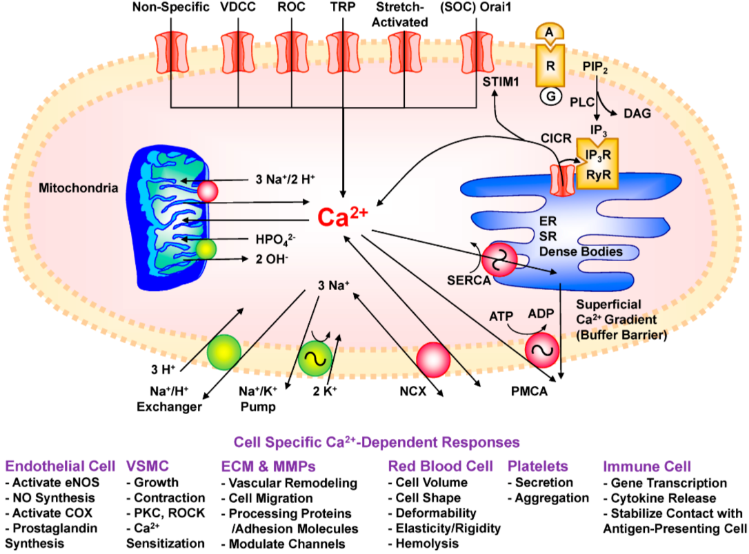
This figure shows how a vascular cell maintains its internal calcium levels by balancing the calcium that enters with the calcium that is removed or stored. When a constrictor signal reaches the cell surface, it releases calcium from the internal stores, and that first release pulls still more calcium out of them. The same signal opens several channels at the cell surface, so calcium also flows in from outside the cell. When the internal stores run low, a sensor triggers extra surface entry to refill them. To lower calcium again, one pump returns it to the internal stores while other pumps and an exchanger push it out of the cell. When calcium levels climb to dangerous levels, the mitochondria take up the excess and release it slowly once the regular pumps can keep pace. The size of this calcium signal then sets how the vascular cell responds.
Dai C, Khalil RA. Calcium Signaling Dynamics in Vascular Cells and Their Dysregulation in Vascular Disease. Biomolecules. 2025; 15(6):892. https://doi.org/10.3390/biom15060892
https://creativecommons.org/licenses/by/4.0/
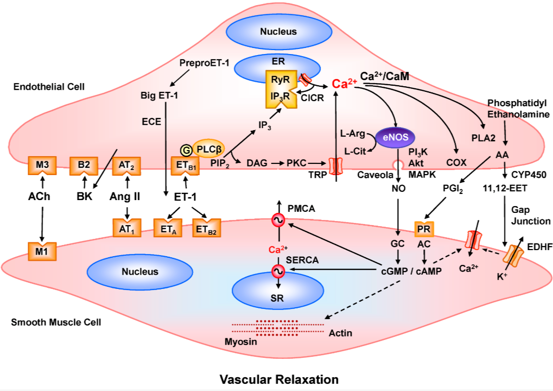
This figure explains how the endothelium, the inner lining of the vessel, relaxes the surrounding muscle by first raising its own calcium levels. When a relaxing signal reaches the lining cells, it releases calcium from their internal stores and allows more calcium to enter at the surface. That rise in calcium activates the enzyme that produces nitric oxide from the amino acid arginine. Nitric oxide then crosses into the smooth muscle and lowers the calcium there, partly by slowing calcium entry and partly by driving the pumps that clear it. It also makes the muscle less responsive to the remaining calcium, thereby relaxing the vessel. The same calcium rise in the lining sends two further relaxing signals to the muscle: one mediated by prostacyclin and one that hyperpolarizes the surface membrane. Both lower the muscle's calcium, so all three routes push the vessel toward relaxation.
Clinical Tips
1. An exaggerated, slow-to-settle increase in blood pressure during cold exposure or acute stress indicates reverse-mode sodium–calcium exchange, with continued calcium influx after the trigger has passed. The key feature is the slow recovery phase, not the peak value.
2. Pulse pressure widening before a rise in resting diastolic pressure suggests calcium sensitization stabilizing smooth muscle tone, leading to reduced arterial recoil in conduit vessels, while resistance vessels still appear normal.
3. Blood pressure that responds more to sustained mental stress than to physical exertion reflects store-operated calcium entry, which maintains smooth muscle in a primed state as intracellular stores undergo cycling.
4. Loss of the normal nocturnal blood pressure dip indicates delayed mitochondrial calcium handling, with oxidative stress maintaining redox-gated calcium channels in an open state when they should close.
5. AIC therapy acts in conditions where surface calcium entry, intracellular stores, and mitochondrial calcium handling are out of synchrony, with the characteristic feature being vascular tone failing to stabilize between signals rather than a consistently low reading.
Clinical Snapshot 1
The reading that should stand out is a systolic value that gradually drifts upward while diastolic pressure remains stable or trends lower. These patients often appear only borderline in the overall risk profile. Weight is reasonable, glucose and lipids remain near reference limits, and there is no formal diagnosis of hypertension at this stage. The key change is in the systolic component, with an increasing gap between systolic and diastolic values over time.
The primary mechanism is located in the wall of large arteries rather than in blood volume or renal regulation. The vessel loses full elastic recoil between beats. Vascular smooth muscle, which should fully relax between cardiac cycles, maintains a persistent low-level tone due to incomplete resetting of intracellular calcium handling. This sustained tone progressively remodels the vessel wall. Calcium-dependent enzymatic activity alters the architecture of elastin and collagen, shifting the artery toward a stiffer, more deposition-prone state. The normal elastic “cushion” that buffers pulsatile flow gradually diminishes, so each cardiac cycle transmits greater force forward. As a result, systolic pressure increases while diastolic pressure drifts downward or remains unchanged. The widening systolic–diastolic gap should be interpreted primarily as a structural arterial wall problem rather than a simple pressure phenomenon. A single measurement is insufficient; the longitudinal trend is more informative. The characteristic pattern is a progressive rise in systolic pressure over the years, with stable or slightly decreasing diastolic values, in individuals without other metabolic drivers that would explain the change. This pattern suggests progressive conduit artery stiffening driven by sustained calcium tone and calcium-mediated remodeling that predates overt hypertension.
The clinical implication is to target calcium handling within the vascular wall rather than focusing only on surface blood pressure values. AIC addresses abnormal calcium loading within the arterial wall and supports restoration of calcium-sensing regulation in vascular smooth muscle. As calcium load decreases, signaling across membrane channels, intracellular stores, and mitochondria re-equilibrates, and the persistent vascular tone diminishes. Over time, the systolic–diastolic gap narrows as the underlying mechanism improves rather than as a simple numerical effect.
Conclusion
Hypertension in this issue reads as a calcium-handling failure in the vessel wall that raises and then maintains vascular tone. Within the smooth muscle, calcium entry at the surface outpaces the pumps and stores that should clear it, leaving the mitochondria to buffer the excess. The endothelium should offset this by lifting its own calcium to release nitric oxide and relax the muscle, but that relaxing signal fades as its calcium handling slips. The same drift surfaces in three familiar shapes: a pressor spike that is slow to settle, a night pressure that no longer dips, and a top number that climbs while the bottom holds as the wall stiffens. Lowering pressure at its source is best read as a question of calcium direction in the vessel wall.
Wall tone depends on three coordinated steps: calcium entry across the cell membrane, release from internal stores, and uptake by mitochondria. When these fall out of phase, tone stays high between beats, and the wall remodels toward stiffness. AIC restores coordinated calcium handling across these three sites, so smooth muscle releases its standing tone, and the endothelium regains its capacity to relax the vessel. AIC also improves calcium transfer between internal stores and mitochondria, reducing the oxidative stress that keeps the cycle running, so the wall holds pressure in check rather than driving it upward.
Bring calcium signaling into your practice.
Practitioners can request a consultation to discuss AIC therapy and how these mechanisms apply to specific cases.
Newsletter content is educational and contributed by participating healthcare practitioners. These statements have not been evaluated by the FDA and are not intended to diagnose, treat, cure, or prevent any disease.