Executive Summary
Refractory fatigue is no longer a mystery of so-called 'non-responders.' A breakdown in intracellular calcium signaling, particularly across the endoplasmic reticulum (ER) –mitochondria axis, may underlie stalled energy recovery in biochemically optimized patients.
The Cellular Disconnect Behind Fatigue That Doesn’t Resolve
Many of us have witnessed the same frustrating pattern: patients presenting with chronic fatigue who, despite careful adrenal, mitochondrial, and thyroid support, show only partial or fleeting improvements. Recent findings in molecular medicine reveal a previously overlooked link—a progressive decline in intracellular calcium signaling. Mitochondrial energy production is intimately dependent on dynamic calcium flux; however, chronic inflammation and oxidative injury often impair calcium transport mechanisms, effectively starving mitochondria even when nutrient cofactors are present. Meanwhile, endoplasmic reticulum (ER) calcium depletion impairs both repair signaling and metabolic resilience, leaving cells locked in an energy-conservation state despite external support.
Adding further complexity, dysfunction in store-operated calcium entry (SOCE) prevents proper recharging of ER calcium stores, amplifying the block in mitochondrial activation. These patients are not resistant due to a lack of intervention—they are resistant because a fundamental cellular communication system remains impaired. Compounding this, unchecked intracellular calcium accumulation can open the mitochondrial permeability transition pore (mPTP), collapsing ATP synthesis entirely and deepening refractory fatigue states.
At a systemic level, long-term calcium misallocation contributes to soft tissue calcification and skeletal demineralization, further disrupting calcium-dependent bioenergetic cycles. Even the best-designed protocols risk producing only marginal gains without addressing the core dysfunction in calcium signaling and redistribution. Emerging approaches that support cellular decalcification and recalibrate intracellular calcium dynamics offer new leverage points for resolving these persistent cases. Rather than layering additional stimulants or mitochondrial cofactors, restoring the fundamental calcium signaling architecture holds the potential for a deeper, more durable recovery.
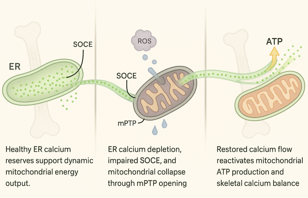
Figure concept and design by Dr. Server Bozdogan, MD, PhD. Cellular visualization illustrates calcium dynamics between the endoplasmic reticulum (ER) and mitochondria across functional, impaired, and recovered states. Created for clinical education purposes.
The three phases show a visual map of calcium signaling between the endoplasmic reticulum (ER) and mitochondria across three physiological states: normal, signaling collapse, and post-restoration. Disrupted calcium dynamics impair ATP production and underlie the patterns of refractory fatigue observed in resistant clinical cases.
Achieving Clinical Turnaround Through Intracellular Decalcification
Restoring bioenergetic flow in fatigue-resistant patients requires more than mitochondrial support—it demands clearing the traffic jam within calcium signaling itself. AIC therapy works by promoting intracellular decalcification and directing free calcium away from soft tissues and back toward skeletal storage, thereby relieving the molecular blockade at the ER-mitochondria interface. This shift helps re-establish dynamic calcium oscillations, unlocking mitochondrial responsiveness and resetting ATP production cycles that had stalled. Even without direct visualization, we can track its effects. AIC therapy doesn’t push the system; it retrains it. Patients regain physical tolerance, post-exertional resilience, and cellular rhythm—not through stimulation, but through restoration.
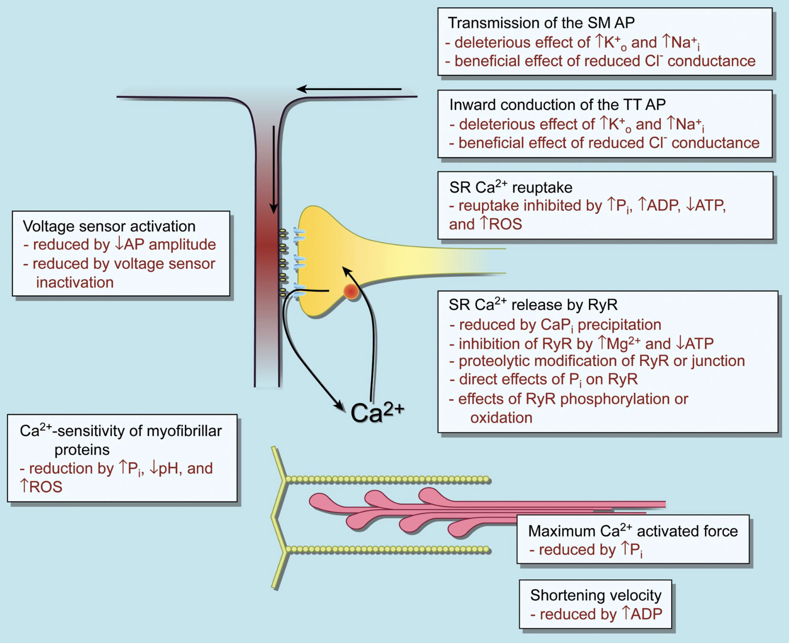
Allen DG, Lamb GD, Westerblad H. Skeletal muscle fatigue: cellular mechanisms. Physiol Rev. 2008;88(1):287-332. doi:10.1152/physrev.00015.2007
Each box highlights a distinct subcellular domain, with the heading indicating its functional role. The bullet points within each box summarize fatigue-associated cellular alterations that impair that function. Abbreviations: SM – surface membrane; TT – transverse tubule; SR – sarcoplasmic reticulum; AP – action potential.
Blocked Mitochondrial Recovery
Many patients plateau despite aggressive mitochondrial protocols, not because of supplement failure, but because the intracellular ignition switch—mitochondrial calcium influx—remains jammed. Even with CoQ10, NAD+, and B vitamin optimization, dysfunctional mitochondrial calcium uniporter (MCU) channels prevent the full activation of the TCA cycle, leaving energy output stuck at half throttle. Clinically, this explains why patients feel an initial surge after mitochondrial IVs or adaptogenic programs, only to crash weeks later when their underlying energy ignition never stabilizes.
Complicating this further, chronic inflammation does more than generate oxidative stress; it also rigidifies calcium channel structures, such as STIM1 and IP3R, thereby silencing critical intracellular communication. As a result, even high-dose antioxidant therapies often fail, because the root failure is not oxidative damage alone—it’s the collapse of the calcium signaling architecture that drives cellular recovery. Patients present with lingering fatigue, brain fog, and post-exertional slumps not because they lack antioxidants, but because their intracellular highways for energy signaling have collapsed in silence.
Collapse of Calcium Recovery
Energy restoration requires more than mitochondrial cofactors—it demands the reactivation of calcium signaling pathways that enable cells to recharge their internal repair systems. One of the most overlooked barriers is the failure of store-operated calcium entry (SOCE), a mechanism essential for refilling endoplasmic reticulum calcium stores after stress. When SOCE remains disrupted, even well-designed protocols cannot revive cellular energy cycles, and patients remain metabolically frozen.
Adding to this hidden dysfunction, unchecked intracellular calcium floods can force open the mitochondrial permeability transition pore (mPTP), collapsing membrane potential and halting ATP synthesis altogether. Clinically, this presents not as gradual fatigue, but as deep, non-restorative exhaustion where even minimal exertion triggers overwhelming crashes. Without correcting both SOCE dysfunction and intracellular calcium overload, mitochondrial recovery remains structurally impossible, regardless of the number of nutrients or hormones provided.
Decoding the Clinical Pattern
Patients with refractory fatigue often present with a mismatch between their clinical stability and persistent exhaustion—a pattern that rarely responds to mitochondrial, hormonal, or immune-focused therapies alone. Subtle signs, such as poor exercise tolerance, unpredictable crashes, unexplained muscle stiffness, or progressive soft tissue calcification, hint at a deeper intracellular signaling collapse that standard protocols often fail to address. Despite regular laboratory markers, these patients operate in a hidden state of energetic rigidity, where both mitochondrial and endoplasmic reticulum systems remain mechanically blocked. Actual recovery patterns only begin to emerge when cellular calcium distribution is corrected, freeing energy signaling pathways that have been silenced at a structural level. Without addressing the calcium misallocation at the heart of these patterns, interventions tend to stabilize symptoms temporarily but rarely achieve lasting restoration.
Conclusion
Refractory fatigue continues to challenge even the most comprehensive protocols, often because the disruption lies deeper within the silent breakdown of calcium communication between organelles. As shown in this issue’s visual framework, when calcium flow is impaired, cells lose access to their repair intelligence, and energy systems stall, regardless of external support. The recurring clinical pattern isn’t always due to biochemical imbalance—it’s a structural rigidity in the intracellular environment that blocks dynamic recovery. AIC therapy provides a strategic entry point by directly addressing this rigidity, restoring the flow between calcium compartments, and enabling the cell to resume its natural energy rhythm.
Bring calcium signaling into your practice.
Practitioners can request a consultation to discuss AIC therapy and how these mechanisms apply to specific cases.
Newsletter content is educational and contributed by participating healthcare practitioners. These statements have not been evaluated by the FDA and are not intended to diagnose, treat, cure, or prevent any disease.