
How Early Hair Loss Begins with Fine Hair
This newsletter reveals that early hair loss often begins with fine, weak regrowth, not because follicles are gone, but because they are still alive and already underperforming.
Executive Summary
Peach-fuzz hairs with preserved follicular openings usually indicate follicle miniaturization rather than follicle loss, suggesting that signaling reserve remains in living follicles. Calcium-dependent regulation of cell adhesion, stem-cell quiescence, extracellular matrix organization, and actin structure helps explain why some follicles remain present yet produce weak, very fine hair. In this setting, the biology points to the need to restore ionic calcium signaling and intracellular calcium handling, especially when follicle timing, structural integrity, and stress stability begin to fail.
Why Fine Hair Still Matters
Peach-fuzz scalp hair indicates that the follicle is still present but has shifted from terminal hair production toward weaker vellus-like output. This pattern is common in nonscarring thinning, especially androgenetic or patterned hair loss, which remains primarily androgen-driven, even though calcium-dependent signaling, cell adhesion, and stress biology can modify how preserved follicles behave.
Hair follicles depend on ionic calcium signals to control keratinocyte adhesion, orderly differentiation, and hair shaft assembly. In the skin, the calcium gradient and the calcium-sensing receptor pathway help initiate these differentiation programs, and stronger calcium-sensing receptor signaling in mice has been linked to faster early hair growth and altered follicle morphogenesis. The key clinical point is that a visible vellus-like hair means the follicle still has signaling activity, even when that activity is weak or misdirected.
Hair follicle miniaturization reflects structural and calcium signaling disturbances that reduce dermal papilla support and destabilize the growth cycle. At the tissue level, miniaturized follicles show a smaller dermal papilla, lower matrix output, and thinner shafts with shorter growth cycles. Disturbed calcium entry can damage this system in both directions: excess channel activity in transient receptor potential vanilloid 3 models drives premature follicular keratinocyte differentiation and apoptosis, whereas ORAI1 loss impairs keratinocyte behavior and is linked to narrower follicles and sporadic hair loss in mice.
When membrane influx, endoplasmic reticulum release, and mitochondrial uptake no longer coordinate well, the follicle handles energy less efficiently, and stress signaling increases. This state favors oxidative stress, endoplasmic reticulum strain, unstable stem-cell quiescence, and an easier shift into regression. Clinically, this appears as shaft diameter diversity, persistent vellus-like regrowth, shedding on top of thinning, and follicular openings that remain visible because the units are weak rather than absent.
Restoring coordinated ionic calcium dynamics offers a rational strategy to stabilize signaling and improve follicle cycling in miniaturized units. This requires better coordination among follicle stem cells, keratinocytes, and dermal papilla cells so that growth signals are read with more appropriate timing. It also depends on stronger coupling among membrane influx, ER release, and mitochondrial uptake, allowing stress output to fall and cycling to become more coherent.
AIC therapy restores calcium signaling across these compartments and supports more stable communication inside living miniaturized follicles. It also regulates cyclooxygenase-2, triggers endogenous calcitonin release, and supports cellular decalcification with a lower tendency for ectopic deposition around the follicle unit. Improving calcium regulation in the mitochondria and endoplasmic reticulum, and reducing oxidative stress, fits a scalp pattern in which vellus-like hairs show biologic reserve rather than complete follicle loss.
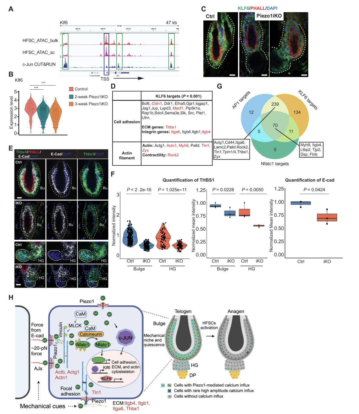
Wang J, Fu C, Chang S, Stephens C, Li H, Wang D, Fu YC, Green KJ, Yan J, Yi R. PIEZO1-mediated calcium signaling reinforces mechanical properties of hair follicle stem cells to promote quiescence. Sci Adv. 2025 May 30;11(22):eadt2771. doi: 10.1126/sciadv.adt2771. Epub 2025 May 28. PMID: 40435254; PMCID: PMC12118625.https://creativecommons.org/licenses/by/4.0/
As shown in Figure 1, PIEZO1-mediated PIEZO1-mediated calcium influx controlled by mechanical force transmitted through E-cadherin, reinforcing quiescence and the mechanical properties of hair follicle stem cells through the regulation of the extracellular matrix, cell adhesion, and the actin cytoskeleton by the transcription factors activator protein 1 (AP1), nuclear factor of activated T cells 1 (NFATC1), and Krüppel-like factor 6 (KLF6). The sequence begins with force applied through the adhesion molecule named in the caption. That force is linked to calcium influx through the mechanosensitive channel named in the caption. The calcium signal is then connected to quiescence and to the mechanical properties of hair follicle stem cells. The next level of control involves regulation of the extracellular matrix, cell adhesion, and the actin cytoskeleton. The final step places three transcription factors as the gene-level mediators of that structural program.
What to Notice Clinically
Small scalp details often separate weak follicles from absent follicles.
1. Wide shaft-diameter variability often means that neighboring follicles are reading calcium-dependent differentiation cues unevenly.
2. A scalp that sheds and thins at the same time often reflects mixed biology, with a telogen shift layered on top of a miniaturized follicle.
3. Vellus-like regrowth is not proof of normal recovery, because premature differentiation can still produce visible hair without restoring shaft strength.
4. In patterned hair loss, androgen signaling remains the primary driver, but calcium channel balance, cell adhesion, and stress routing still influence how surviving follicles cycle and differentiate.
Clinical Practice Snapshot
Peach-fuzz regrowth with preserved follicular openings marks a scalp pattern of retained follicular architecture with diminished output. Fine regrowth, shaft-caliber variability, and visible ostia can coexist across thinning areas, placing weak production beside follicles that remain structurally in place. Within that pattern, AIC Therapy is mechanistically aligned with living miniaturized units because it addresses calcium handling and stress signaling in the pathways that shape follicle cycling, differentiation, and hair shaft formation.
Conclusion
Peach-fuzz hair usually indicates that the follicle is still present but producing weak, incomplete growth. Visible follicular openings, shaft-diameter variability, and persistent vellus-like regrowth therefore point more strongly to follicle dysfunction than to total follicle loss.
In patterned hair loss, this pattern is best understood as a preserved but miniaturized follicle population. Androgens drive the overall process, while calcium-dependent signaling, adhesion biology, and cellular stress influence the quality and stability of the remaining growth. Calcium influx helps connect stem-cell quiescence, structural organization, and the mechanical stability of the follicle unit. Physiopathologically, this is most consistent with a living but unstable follicle population that retains biologic reserve while failing to restore normal shaft production.
This biologic profile makes AIC Therapy a coherent fit for hair loss defined by preserved, miniaturized, and functionally unstable follicles. In that setting, the therapeutic logic aligns with a follicle population that remains viable but no longer sustains stable terminal hair production.
Bring calcium signaling into your practice.
Practitioners can request a consultation to discuss AIC therapy and how these mechanisms apply to specific cases.
Newsletter content is educational and contributed by participating healthcare practitioners. These statements have not been evaluated by the FDA and are not intended to diagnose, treat, cure, or prevent any disease.