
When Calcium Signaling Quietly Sabotages Your Patients' Weight Loss
This newsletter reveals how misdirected calcium signaling can quietly keep weight loss stuck in patients who look fully compliant.
Executive Summary
Many patients stay at the same weight because their fat cells are loaded with calcium and low-grade inflammation, not because they ignore your plan. When intracellular calcium stays high, adipocytes hold on to triglycerides, hormones lose their impact, and lifestyle tools look weaker than they really are. This opens the door for gentle ionic calcium strategies that lower the abnormal calcium load in fat cells so the very same diet,finally starts to work.
How Calcium Signaling Turns “Compliant” Patients Into Weight-Resistant Cases
Many of your patients do everything right on paper and still stay stuck at the same weight. This stubborn weight is not only about calories but also about how fat cells handle calcium and respond to hormones and inflammation. High intracellular calcium inside fat cells stimulates lipogenesis, blocks lipolysis, and pushes triglycerides to stay stored instead of being burned.
Low dietary calcium, together with secondary changes in vitamin D and PTH, can increase intracellular calcium in adipocytes and support a higher defended fat mass, while cortisol mainly modulates energy balance through broader stress pathways. In this setting, insulin and leptin signals lose some of their effect, and your patient looks non-compliant even when they follow the plan.
Inflamed adipose tissue then becomes an active organ, releasing cytokines that maintain weight loss resistance and feed back on hormones and appetite. One more detail is that adipose tissue also expresses the calcium-sensing receptor, and its activation can increase proinflammatory cytokine production in adipose cells, which may help maintain this inflamed fat state. This means calcium is not just about bone density; it is an active intracellular signal that helps shift fat metabolism toward storage or mobilization depending on the hormonal and inflammatory context. If we can gently correct calcium handling inside fat cells while we work on diet, movement, sleep, and stress, the same lifestyle plan can start to give different results in resistant cases.
This is the point where the AIC (Anti Orbital Ionic Calcium) therapy comes into the picture as a possible clinical tool. AIC lowers the abnormal calcium load in fat cells, so they shift out of chronic storage mode and actually start releasing triglycerides. When COX-2-driven inflammation in adipose tissue is reduced and calcium signaling is normalized, insulin signaling becomes cleaner, and patients can access their fat stores more effectively on the same diet and exercise plan.
If you also support buffering of exercise-related acidosis and maintain stable mitochondrial calcium handling, skeletal muscle can rely more on oxidative metabolism, including fat oxidation, and patients are less likely to experience a severe post-exercise energy crash. At the same time, AIC triggers endogenous calcitonin, which provides built-in satiety and a glucose-smoothing effect, so portion sizes come down, and carb swings flatten without stimulants. Overall, the patient is taking calcium out of the wrong places, putting the signaling back in order, and lifting several of the metabolic brakes that keep inflamed, weight-loss-resistant patients stuck in your clinic. That is exactly why AIC can make weight loss actually happen.
Goudarzi F, Mohammadalipour A, Khodadadi I, Karimi S, Mostoli R, Bahabadi M, Goodarzi MT. The Role of Calcium in Differentiation of Human Adipose-Derived Stem Cells to Adipocytes. Mol Biotechnol. 2018 Apr;60(4):279-289. doi: 10.1007/s12033-018-0071-x. PMID: 29488128.
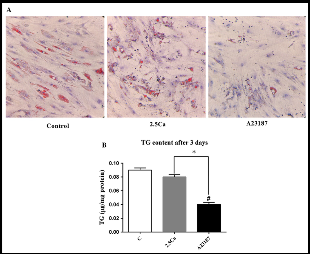
This figure compares fat droplets and triglyceride levels under different calcium settings.
Panel a uses Oil Red O staining and shows fewer fat droplets in cells given 2.5 millimolar calcium, and in cells given 1.8 millimolar calcium plus the ionophore A23187, compared with the control.
Panel b shows that after three days of differentiation, triglycerides are lowest in the 1.8 millimolar calcium plus A23187 group.
The 2.5 millimolar calcium group stays close to the control for triglycerides in this analysis.
Guney E, Arruda AP, Parlakgul G, Cagampan E, Min N, Lee GY, Greene L, Tsaousidou E, Inouye K, Han MS, Davis RJ, Hotamisligil GS. Aberrant Ca2+ signaling by IP3Rs in adipocytes links inflammation to metabolic dysregulation in obesity. Sci Signal. 2021 Dec 14;14(713):eabf2059. doi: 10.1126/scisignal.abf2059. Epub 2021 Dec 14. PMID: 34905386; PMCID: PMC10130146.
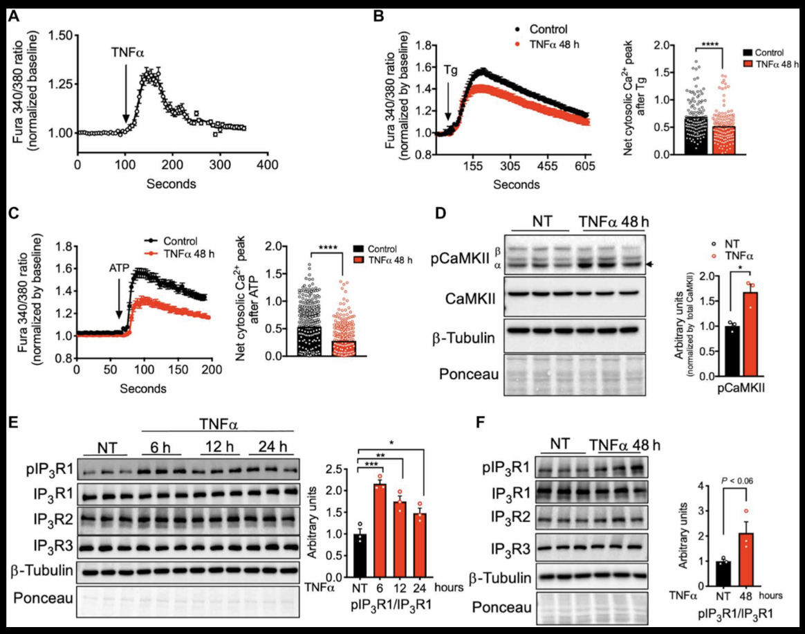
This figure shows how the inflammatory cytokine tumor necrosis factor alpha changes calcium handling inside a standard laboratory fat cell model. When tumor necrosis factor alpha is added, calcium in the watery part of these mature fat cells rises quickly. After forty-eight hours of continuous exposure, the main internal calcium store inside the cell holds less calcium, and when the researchers add thapsigargin or adenosine triphosphate, which generally cause a strong release of calcium from this store, much less calcium comes out in the inflamed cells than in control cells.
The figure also shows that a key calcium-dependent enzyme inside the cell becomes more activated under tumor necrosis factor alpha, which fits with stronger calcium signaling in the cell fluid. In the last panels, a specific channel that releases calcium from the internal store is more activated at several different time points after tumor necrosis factor alpha treatment. Taken together, the data show that inflammatory stress through tumor necrosis factor alpha shifts fat cells into a state where calcium is released differently from their internal stores and calcium balance inside the cell is rewired.
Misconceptions
Misconception 1
Many doctors still think high calcium intake burns body fat almost automatically.
Reality
In practice, calcium just tunes adipocyte signaling; diet and movement still decide.
Misconception 2
Some believe that normal serum calcium excludes a calcium-related mechanism in obesity.
Reality
Yet intracellular calcium in adipocytes rises, driving lipogenesis and blocking proper lipolysis.
Misconception 3
Many colleagues reduce obesity to calories alone, completely ignoring calcium-driven adipogenesis.
Reality
Calcium influences mitochondrial thermogenesis, gut hormones, low-grade inflammation, and activity tolerance.
Misconception 4
Soft tissue calcification is often dismissed as cosmetic, unrelated to weight issues.
Reality
Microcalcified vessels impair perfusion, limit exercise capacity, and quietly lower energy expenditure.
Calcium-Driven Weight Resistance
Some patients will tell you their body feels heavy, tight, and inflamed even though their food diary looks clean. They may also experience puffiness, fatigue, brain fog, cold hands and feet, night cramps, and morning joint stiffness, making them feel older than their actual age. Unstable teeth, fragile nails, or a surprisingly weak DEXA suggest that calcium is drifting out of bone and quietly settling in the wrong tissues. These patients may not see a small bump on the scale even after a significant dietary change. At this point, the question should be not just how many calories they consumed but also what signals are communicated to and within adipocytes. At this point, misdirected calcium signaling may be the main culprit preventing weight loss. AIC, as a targeted ionic calcium intervention, can help pull excess calcium out of fat and other soft tissues, trigger endogenous calcitonin, calm COX-2-driven inflammation, and finally let the same lifestyle plan lower the defended weight set point.
Conclusion
What we see in practice is a pattern where adipocytes are overloaded with calcium, bathed in inflammatory signals, and simply refuse to release fat, even when the food diary looks clean. When that intracellular calcium load is lowered and handled more dynamically, fat droplets shrink, triglycerides fall, and the same calorie intake starts to have a different effect on the scale. At the same time, chronic cytokine stress keeps pushing calcium out of its normal stores, so hormones like insulin and leptin send weaker messages, and weight loss feels blocked from the inside out.
Daily drivers such as poor sleep, sympathetic overdrive, and heavy caffeine use can sit on top of this biology, tightening vascular tone, worsening microcalcification, and leaving patients tired, cold, and less willing to move. Once we stop treating calcium as only a bone density nutrient and start reading it as a signal that shapes adipogenesis, thermogenesis, gut hormones, and exercise tolerance, the clinical picture of weight resistance becomes far less mysterious.
In this context, AIC therapy, used as a targeted ionic calcium intervention, can pull excess calcium out of fat and other soft tissues and release several of the internal brakes that keep weight locked in place. By calming calcium-driven inflammation, smoothing post-meal glucose swings, engaging endogenous calcitonin, and restoring access to fat oxidation, AIC therapy lets a familiar lifestyle plan finally translate into visible, more durable weight loss.
Bring calcium signaling into your practice.
Practitioners can request a consultation to discuss AIC therapy and how these mechanisms apply to specific cases.
Newsletter content is educational and contributed by participating healthcare practitioners. These statements have not been evaluated by the FDA and are not intended to diagnose, treat, cure, or prevent any disease.