This newsletter reveals how disrupted calcium homeostasis, microcalcifications, and microvascular stiffness jointly slow cognition, and it outlines practical ways to restore mineral balance for clearer thinking.
Executive Summary
Calcium stress in the aging brain alters endothelial signaling, loosens barrier control, and seeds microcalcifications that dampen neurovascular coupling, sleep-linked glymphatic flow, and synaptic timing. Correcting systemic mineral handling and gently restoring extracellular ionized calcium tone can recalibrate CaSR activity across endothelium, astrocytes, and neurons, quietly pointing toward an ionic calcium–forward strategy without overreliance on symptomatic aids.
Calcium Stress, Microcalcifications, and the Neurovascular Unit
Cognitive aging increasingly reflects calcium dyshomeostasis alongside synaptic wear. Microcalcifications in cortical tissue and small penetrating arterioles distort local field signaling and delay neurotransmission. The blood-brain barrier senses calcium stress through pericytes and astrocytic end feet, adjusting junctional proteins and transporters in both directions. These permeability shifts alter glucose and amino acid flux and slow the removal of metabolic acids, which feeds back to worsen intracellular calcium handling.
Neurons and glia under oxidative load accumulate mitochondrial Ca²⁺, depressing ATP production and priming apoptotic signaling. Endothelial Ca²⁺ imbalance activates calpain and NFAT programs that favor vesicle-mediated mineral deposition on the capillary wall. Microglia attempt to clear crystals, yet a pro-inflammatory milieu and stiff extracellular matrix reduce efficient phagocytosis. As mentioned earlier, persistent small vessel calcification then dampens neurovascular coupling and impairs glymphatic clearance during sleep.
Clinically, first-line tactics target the upstream drivers that raise the cerebral mineral burden, namely blood pressure, atherogenic lipids, and insulin resistance. Improving cardiorespiratory fitness and sleep quality restores pulsatile perfusion, which enhances endothelial nitric oxide signaling and calcium extrusion. Nutritional care that limits phosphate additives, ensures magnesium sufficiency, and supports vitamin K-dependent carboxylation of matrix Gla protein stabilizes endogenous anti-calcification shields. Hydration and gentle postprandial walks assist lactate washout, reducing intracellular acidification that otherwise opens calcium entry channels.
Within this framework, AIC Therapy can help normalize the extracellular ionized pool, supporting CaSR tone on endothelium, astrocytes, and neurons for steadier calcium influx and efflux dynamics. Given away from meals, immediately bioavailable Ca²⁺ may blunt stress-driven parathyroid surges and permit transient C cell-mediated calcitonin responses, which together can reduce procalcific signaling.

This figure shows how calcium deposits can appear in the brain, especially in aging and Alzheimer’s disease. The von Kossa stain (brown/black spots) and SEM images (white/gray areas) both reveal not only calcium in blood vessels but also tiny microcalcifications scattered through brain regions like the caudate nucleus and temporal cortex. These deposits are seen in older and Alzheimer’s brains but never in young ones, suggesting that calcium buildup increases with age. Interestingly, the vessel calcification and the small tissue deposits are not directly linked, meaning they likely form through different biological processes. This finding points to a deeper disruption of calcium balance within brain cells, which may open new directions for restoring healthy mineral homeostasis.
Tsolaki E, Csincsik L, Xue J, Lengyel I, Bertazzo S. Nuclear and cellular, micro and nano calcification in Alzheimer's disease patients and correlation to phosphorylated Tau. Acta Biomater . 2022;143:138-144. doi:10.1016/j.actbio.2022.03.003
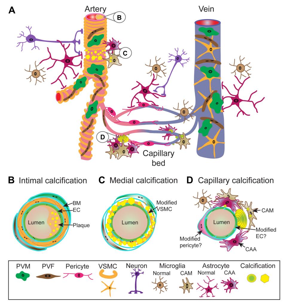
This figure illustrates the structure of blood vessels in the central nervous system and where calcifications can appear. In arteries, endothelial cells line the inner surface, while smooth muscle cells surround them, and calcium may accumulate either in the inner (intimal) layer or in the muscular (medial) layer when these cells change their function. Capillaries, which connect arteries and veins, are wrapped by pericytes and can show small mineral “beads” that extend into brain tissue. These calcified spots often trigger strong reactions in surrounding glial cells, while the exact changes in pericytes and endothelial cells remain unclear. Overall, the figure shows that vascular calcification in the brain involves many cell types, including fibroblasts, macrophages, and astrocytes, forming a complex and dynamic process along the entire vascular network.
Maheshwari U, Huang SF, Sridhar S, Keller A. The Interplay Between Brain Vascular Calcification and Microglia. Front Aging Neurosci . 2022;14:848495. Published 2022 Mar 2. doi:10.3389/fnagi.2022.848495
Five Common Clinical Misconceptions
Misconception 1: Cognitive decline is mainly a neuron problem.
Clinical angle 1: Many older adults show cognitive slowing not because of neuron loss, but because microcalcifications and vascular stiffness reduce local blood flow and calcium exchange. Clinically, improving microcirculation through regular movement, hydration, and magnesium repletion often proves more beneficial than focusing solely on memory enhancers.
Misconception 2: Brain microcalcifications are untreatable or irreversible.
Clinical angle 2: These deposits can be reduced by improving calcium handling in the body. Supporting vitamin K2 pathways, balancing magnesium and phosphate intake, and ensuring good sleep quality help the brain clear excess minerals naturally through the glymphatic system.
Misconception 3: Blood-brain barrier changes are always due to inflammation.
Clinical angle 3: Subtle calcium stress alone can loosen barrier junctions and alter nutrient flow. Clinically, combining antioxidant-rich nutrition with gentle cardiovascular conditioning can stabilize this barrier and protect metabolic exchange across brain vessels.
Misconception 4: Vascular calcification is a static aging process.
Clinical angle 4: Calcification is dynamic and influenced by daily habits. Interventions that improve endothelial flexibility—like nitric oxide support, controlled breathing, and avoiding phosphate additives—can slow or even reverse early vascular mineral buildup.
Misconception 5: Supporting brain health means focusing on neurotransmitters.
Clinical angle 5: Neurotransmitters depend on stable calcium signaling. Clinically, restoring ionic balance through proper mineral intake, regular movement, and stress management often enhances focus, mood, and sleep far better than stimulant-based approaches.
Case Reference: Restoring Calcium Balance in Neurovascular Function
Two patients showed how improving calcium dynamics can lead to fast, visible recovery.
A 53-year-old man had long struggled to swallow because of neck muscle weakness and fatigue. Before treatment, poor muscle coordination suggested disturbed calcium flow in motor nerves and local microcalcification. After starting Active Ionic Calcium (AIC), he regained normal swallowing within days without any other change in therapy. His improvement points to better neuromuscular calcium exchange and smoother signaling between nerves and muscles.
A 65-year-old woman with multiple sclerosis had chronic cold hands and weak circulation. She was on high-dose aspirin (320 mg daily) to support blood flow. After one week of AIC, her circulation improved, and her doctor safely reduced aspirin to 80 mg. The improvement reflected restored vessel tone, likely from more stable calcium receptor activity and nitric oxide signaling.
Both cases demonstrate that re-establishing ionic calcium equilibrium can improve neurovascular coordination and reduce functional calcific stress. These clinical responses align with the broader concept of brain decalcification, where supporting dynamic calcium exchange rather than blocking calcium pathways enhances cellular resilience, microcirculatory flow, and overall neurological function.
Conclusion
Age-related cognitive slowing tracks with disturbed calcium signaling that accumulates in tiny deposits across the cortex and small vessels, weakening local field transmission and delaying synaptic responses. Subtle barrier looseness shifts nutrient and acid clearance, while oxidative load drives mitochondrial Ca²⁺ uptake that suppresses ATP and nudges apoptotic programs.
Improving cardiorespiratory fitness, sleep quality, hydration, and postprandial movement restores pulsatile flow and lactate washout, which steadies endothelial nitric oxide and calcium extrusion. Daily nutrition that limits phosphate additives, ensures magnesium sufficiency, and supports vitamin K-dependent matrix Gla activation strengthens the body’s anti-calcification shield. Clinical experience shows that when calcium dynamics are rebalanced, neuromuscular coordination and peripheral perfusion can improve within days in selected patients.
AIC is well-positioned here because normalizing the extracellular ionized pool supports steadier CaSR tone and can blunt stress-driven parathyroid spikes. Brief C-cell calcitonin responses that follow may counter pro-calcific signaling and help nudge microvascular and parenchymal minerals back toward safe trafficking.
Bring calcium signaling into your practice.
Practitioners can request a consultation to discuss AIC therapy and how these mechanisms apply to specific cases.
Newsletter content is educational and contributed by participating healthcare practitioners. These statements have not been evaluated by the FDA and are not intended to diagnose, treat, cure, or prevent any disease.