
The Calcium-Mitochondria Loop Behind Neuroinflammation
This newsletter shows how disordered calcium handling drives mitochondrial stress and sustains neuroinflammation.
Executive Summary
In neurodegenerative decline, function is worse than the scan predicts, and recovery runs slowly. Rapid, repeated tasks fade first and decline after each infection or surgery. Underneath, calcium overloads the mitochondria, and the brain's immune cells switch on. They stay inflamed, and that inflammation drives the decline forward. This calcium problem occurs at three sites: the cell surface, internal stores, and mitochondria. Bringing calcium handling back to these sites quiets the inflammation, and the decline eases with it.
How disordered calcium handling drives mitochondrial stress and sustains neuroinflammation in neurodegenerative disease
Chronic neuroinflammation now drives neurodegenerative disease. It is not a secondary byproduct. The starting point sits inside the neuron. It lies in how the cell handles calcium. Neurons run on brief, well-timed calcium signals. Mitochondria are the primary intracellular calcium buffer. They take up the surplus during activity. This buffering keeps each signal short. It also protects the cell. When calcium handling slips, mitochondria take up too much. They can no longer clear the excess. Overloaded mitochondria lose efficiency and leak reactive oxygen species. Eventually, they open the pore that commits the cell to injury. This calcium-driven mitochondrial stress is an early event. It appears before neurons die. That early stress sets the pace of decline.
The injury does not stay inside the neuron. It recruits the brain's immune cells. The process then becomes self-reinforcing. Calcium-overloaded mitochondria release danger signals. These include fragments of their own damaged DNA. The signals spill into the cell and the surrounding space. Microglia are the brain's resident immune cells. They read these signals and become active. Active microglia run an inflammatory program. They release cytokines, as evidenced by elevated CRP and IL-6 levels in lab results. These cytokines feed back onto the mitochondria. They lower the membrane charge and cut cellular energy. They also increase oxidative stress. Microglia carry their own mitochondria, too. Once those are calcium-stressed, the cells stay inflammatory. Now the loop runs on its own. It sustains progressive neuronal injury and the functional decline of these diseases.
Slowing this process means restoring calcium control early. The fix belongs inside the cell. Disordered calcium handling at three sites holds the loop in place. These are the cell surface, the endoplasmic reticulum, and the mitochondria. The endoplasmic reticulum and mitochondria meet at a contact point. When calcium transfer goes wrong, oxidative stress climbs. When calcium handling regains coordination, mitochondrial overload stops. Oxidative stress falls, and the danger signals subside. Microglia then leave the chronic inflammatory state. Anti-orbital Ionic Calcium Therapy (AIC) restores coordinated calcium signaling across the surface, stores, and mitochondria. It reduces mitochondrial calcium overload and the oxidative stress that feeds the loop. It also eases the calcium-driven inflammatory signaling behind neuroinflammation. The clinical reading is a calmer neuroinflammatory baseline. Cellular energy holds up better, and functional decline slows across these diseases.
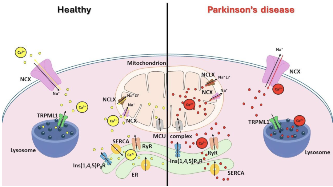
This figure explains how cells balance calcium and how Parkinson's disease disrupts that balance. The endoplasmic reticulum is the main internal calcium store. A pump fills it, and release channels let calcium back out. Nearby mitochondria take up that calcium for metabolism, and lysosomes hold and release more. The sodium-calcium exchanger (NCX) clears calcium at the surface and in mitochondria. A further mitochondrial exchanger helps maintain balance between influx and efflux. In Parkinson's disease, surface calcium entry increases, and these internal stores lose control.
Bastioli, G.; Piccirillo, S.; Graciotti, L.; Carone, M.; Sprega, G.; Taoussi, O.; Preziuso, A.; Castaldo, P. Calcium Deregulation in Neurodegeneration and Neuroinflammation in Parkinson’s Disease: Role of Calcium-Storing Organelles and Sodium–Calcium Exchanger. Cells 2024, 13, 1301. https://doi.org/10.3390/cells13151301. https://creativecommons.org/licenses/by/4.0/
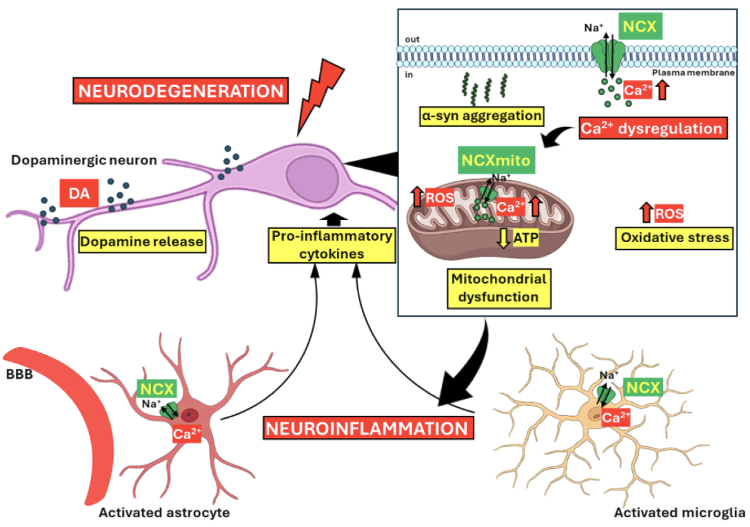
This figure illustrates how dysregulated calcium handling drives damage in Parkinson's disease. It groups five linked features: calcium disruption, mitochondrial dysfunction, oxidative stress, protein aggregation, and neuroinflammation. At the center is the sodium-calcium exchanger (NCX), which becomes dysregulated in the disease. The dysregulated exchanger contributes to intracellular calcium overload. That overload worsens mitochondrial damage and drives neuronal cell death. Misfolded alpha-synuclein protein builds up, and oxidative stress rises as well. Together, they further amplify both the neurodegeneration and the neuroinflammation.
Bastioli, G.; Piccirillo, S.; Graciotti, L.; Carone, M.; Sprega, G.; Taoussi, O.; Preziuso, A.; Castaldo, P. Calcium Deregulation in Neurodegeneration and Neuroinflammation in Parkinson’s Disease: Role of Calcium-Storing Organelles and Sodium–Calcium Exchanger. Cells 2024, 13, 1301. https://doi.org/10.3390/cells13151301 https://creativecommons.org/licenses/by/4.0/
Clinical Tips
1. A stepwise, non-recovering drop after infection or surgery marks the calcium-driven loop deepening under inflammatory load.
2. Rapid, repeated functions fade first because the calcium signal resets too slowly between pulses.
3. The protein that aggregates in these diseases binds the mitochondrial energy machinery directly. It forces calcium overload there, making the aggregate an active driver of the energy failure.
4. Microglia rely on mitochondrial energy to clear debris and damaged mitochondria. Once their own mitochondria are overloaded with calcium, cleanup fails, and inflammatory output takes over.
5. AIC restores safe mitochondrial calcium uptake at the core of this loop. With the overload eased, ordinary metabolic and inflammatory demands no longer produce a lasting decline.
Clinical Snapshot
The puzzling visits are the ones where the scan looks better than the person does. The functional loss is greater than the imaging predicts and harder to explain away. The fatigue, the slowing, the lost sharpness all run ahead of the structural loss. Much of that gap is not dead tissue. It is active neuroinflammation. Microglia, the brain's resident immune cells, have switched into an inflammatory state. In that state, they keep nearby neurons handling calcium poorly. The calcium signals these cells rely on stay disordered, and their energy stays low. So the neurons underperform without being gone. That is the part the scan cannot show. It is also the part still open to change.
Read the gap between the scan and daily function as active calcium-driven inflammation, not fixed damage. Look for the person whose function sits below what the imaging predicts. Look for symptoms with a live, modifiable quality rather than a fixed deficit. Much of the burden sits in living cells that signal poorly, not in tissue already gone. That reframing moves the work toward the active process. You treat the inflammatory calcium signaling that holds those neurons down. That is where the gains remain. AIC lowers the calcium-driven inflammatory signaling that keeps microglia switched on. It then re-coordinates neuronal calcium handling, allowing the suppressed function to return.
Conclusion
This issue reads neurodegeneration as a calcium-handling failure that fuels its own inflammation. In health, calcium is balanced among intracellular stores, mitochondria, and membrane exchangers. In disease, a calcium exchanger fails, overloading the mitochondria with calcium. Oxidative stress increases, and damaged mitochondria shift microglia toward lasting inflammation. Misfolded protein and inflammation then amplify the neuronal damage. In practice, this surfaces in a few familiar shapes. Function sits below what a scan predicts, and rapid, repeated tasks fade first. Decline steps down after an infection or surgery, while routine labs show inflammatory markers rising.
Slowing the progression of this disease comes down to the direction of calcium inside the cell. The loop runs while calcium overloads the mitochondria, and microglia stay inflamed. AIC re-coordinates calcium across stores, so mitochondrial overload eases. AIC also quiets the calcium-driven inflammation, so microglia stand down, and function is preserved.
Bring calcium signaling into your practice.
Practitioners can request a consultation to discuss AIC therapy and how these mechanisms apply to specific cases.
Newsletter content is educational and contributed by participating healthcare practitioners. These statements have not been evaluated by the FDA and are not intended to diagnose, treat, cure, or prevent any disease.