
The Calcium Bridge from Depression to Alzheimer’s
This newsletter reveals why unstable calcium signaling and impaired calcium clearance can push depressive circuits toward Alzheimer vulnerability and how restoring calcium homeostasis and compartmental handling may protect synaptic plasticity and cognitive resilience.
Executive Summary
Depression phenotypes and Alzheimer vulnerability can converge on a shared driver, intracellular calcium dysregulation that destabilizes network firing and stress biology. Slow recovery after mild stress, reward without pleasure, sleep fragmentation, psychomotor slowing, and brain fog with autonomic shifts can align with impaired calcium clearance and weak SERCA-mediated endoplasmic reticulum refilling. Correcting calcium dysregulation can stabilize endoplasmic reticulum to mitochondria calcium handling, support synaptic plasticity and energy matching, and reduce oxidative stress signals that erode cognition.
Calcium Dysregulation as the Depression–Alzheimer Bridge
Depression is linked to altered brain network firing and stress biology. Neurons translate electrical activity into intracellular calcium transients that drive synaptic plasticity and gene regulation. When calcium entry, buffering, and clearance are not coordinated, signaling becomes noisy and energy costly. That instability can bias circuits toward threat processing, anhedonia, and impaired cognitive control. Clinically, this can align with sleep fragmentation, psychomotor slowing, and reduced stress resilience.
A central clearance step in brain cells is the refilling of endoplasmic reticulum stores by SERCA. It sets the amplitude and timing of cytosolic calcium signals by controlling how quickly calcium is sequestered after receptor activation. Endoplasmic reticulum calcium content also shapes mitochondrial calcium uptake at contact sites, thereby coupling calcium signaling to adenosine triphosphate production and reactive oxygen species burden. Under chronic stress, inflammatory signaling and oxidative load can impair this routing, pushing calcium toward sustained cytosolic elevation or depleted stores. Such patterns can reduce synaptic adaptability and alter neuroendocrine feedback loops relevant to depressive phenotypes.
When this routing remains unstable, calcium dysregulation can extend beyond mood circuits into neurodegenerative vulnerability. In depression, prolonged cytosolic calcium transients and weak endoplasmic reticulum refilling can blunt plasticity in prefrontal and limbic networks.
In Alzheimer's disease, amyloid and tau pathology disrupt calcium control at synapses and in astrocytes, promoting persistent calcium imbalance. Across both conditions, altered SERCA function destabilizes endoplasmic reticulum store content and modifies calcium transfer to mitochondria, amplifying oxidative stress signals. Restoring calcium signaling benefits both Alzheimer's disease and depression because precise compartmental flux supports energy matching, limits stress-driven calcium spillover, and preserves plasticity programs. This shared calcium biology supports a single recovery logic framed around ionic calcium dynamics. Recovery logic starts with restoring ionic calcium dynamics across membranes and organelles.
A second aim is to improve systemic routing so that calcium signals remain sharp, time-limited, and energetically supported. Anti-orbital Ionic Calcium (AIC) Therapy restores calcium signaling at membrane calcium channels, endoplasmic reticulum calcium release, and mitochondrial calcium uniporter regulation, stabilizing calcium-dependent neuronal firing patterns relevant to depression and cognitive decline. AIC Therapy also modulates cyclooxygenase-2 and triggers endogenous calcitonin release from thyroidal C cells and neuroendocrine sources, thereby shifting inflammatory and stress signaling that intersects with mood and neurodegeneration. Moreover, AIC Therapy promotes cellular decalcification and reduces mitochondrial dysfunction and oxidative stress by improving calcium handling between mitochondria and the endoplasmic reticulum, including effects at mitochondria-associated membranes.
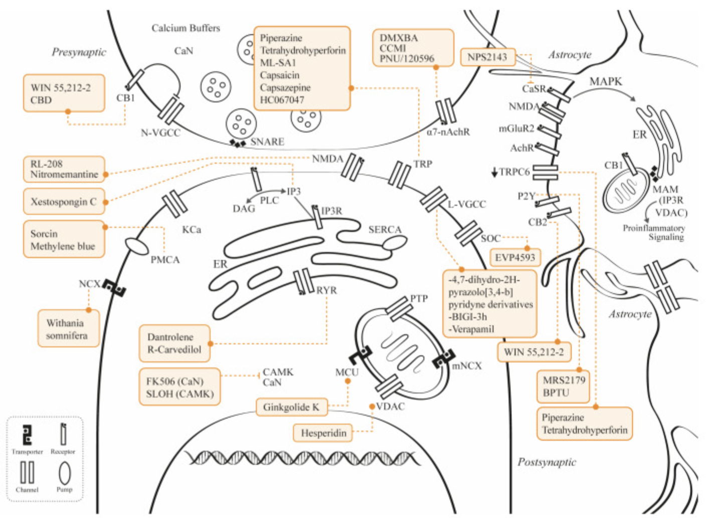
This figure illustrates potential therapies and the calcium-regulation proteins they primarily target. Many of these molecules also have anti-inflammatory, anti-amyloid, and antioxidative effects, but the figure focuses on their main interaction with proteins involved in calcium regulation. Black arrows show activation, and bars show inhibition. The down arrow (↓) next to transient receptor potential canonical channel 6 (TRPC6) means reduced expression of that channel. The legend defines many calcium-related abbreviations, including calcium-dependent phosphatase calcineurin (CaN), calcium-sensing receptor (CaSR), voltage-gated calcium channels (VGCC), mitochondrial calcium uniporter (MCU), inositol trisphosphate receptor (IP3R), ryanodine receptors (RyR), plasma-membrane calcium-ATPases (PMCA), sodium–calcium exchanger (NCX), and mitochondria-associated membranes (MAMs). These labels show which calcium-control proteins each therapy activates or inhibits.
Baracaldo-Santamaría, D.; Avendaño-Lopez, S.S.; Ariza-Salamanca, D.F.; Rodriguez-Giraldo, M.; Calderon-Ospina, C.A.; González-Reyes, R.E.; Nava-Mesa, M.O. Role of Calcium Modulation in the Pathophysiology and Treatment of Alzheimer’s Disease. Int. J. Mol. Sci. 2023, 24, 9067. https://doi.org/10.3390/ijms24109067.https://creativecommons.org/licenses/by/4.0/
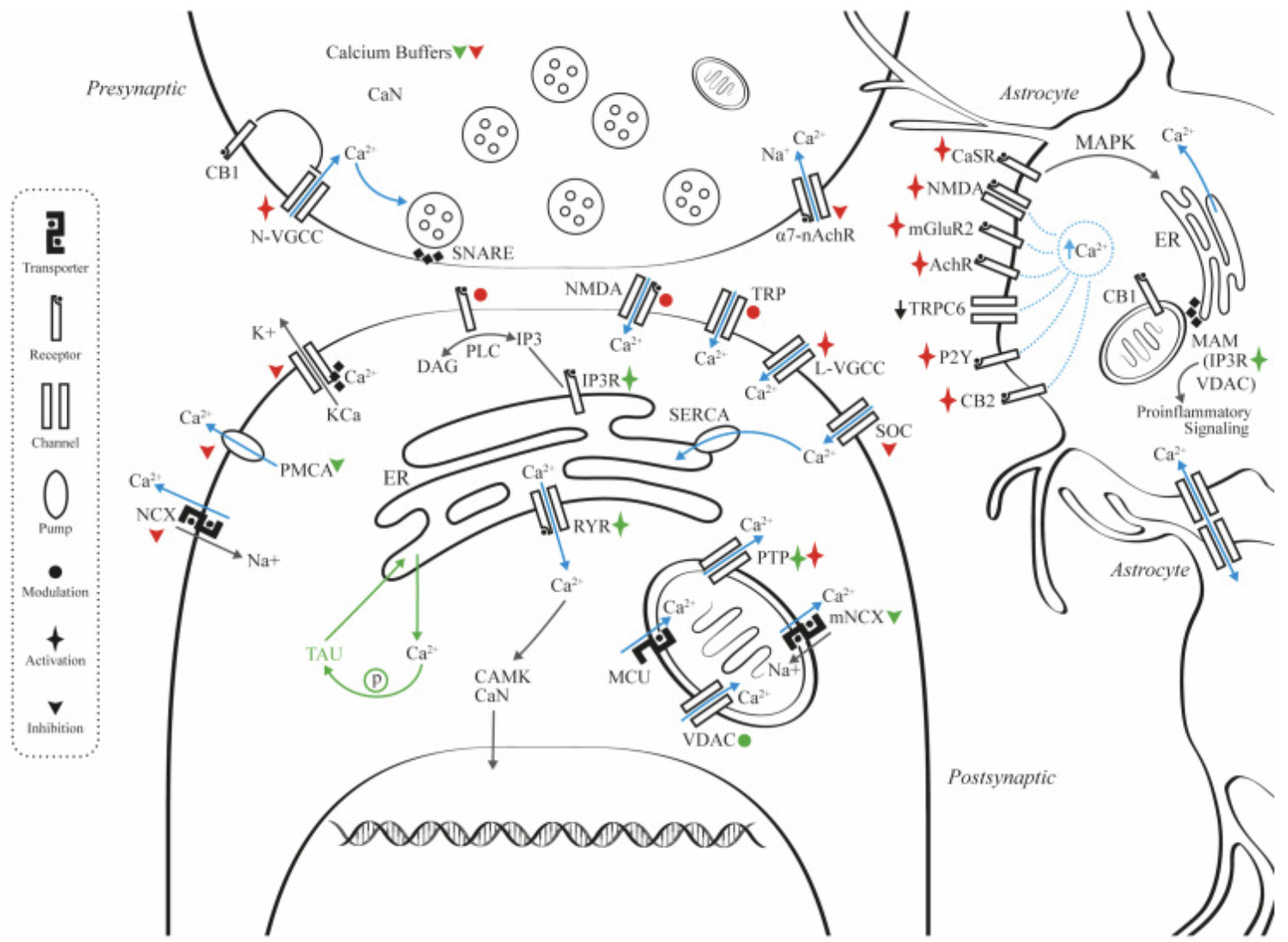
This figure explains cellular systems that affect calcium levels at presynaptic and postsynaptic sites and in astrocytes. It highlights key surface-membrane proteins, such as calcium transporters and receptors, and shows how their interactions with organelles and calcium buffers form a coordinated system that balances intracellular calcium concentration. The figure also illustrates how amyloidosis and tauopathy contribute to calcium dysregulation during Alzheimer's disease progression. Red symbols show the effects of amyloid on several membrane proteins, green symbols show the effects of tau protein and its interactions with intracellular calcium systems, and blue arrows show the direction of calcium flow between compartments. The letter P inside a circle indicates phosphorylation. Black arrows indicate activation, bars indicate inhibition, and the down arrow (↓) next to TRPC6 indicates reduced expression. Two key calcium terms shown are the calcium-sensing receptor (CaSR) and store-operated Ca2+ entry (SOCE).
Baracaldo-Santamaría, D.; Avendaño-Lopez, S.S.; Ariza-Salamanca, D.F.; Rodriguez-Giraldo, M.; Calderon-Ospina, C.A.; González-Reyes, R.E.; Nava-Mesa, M.O. Role of Calcium Modulation in the Pathophysiology and Treatment of Alzheimer’s Disease. Int. J. Mol. Sci. 2023, 24, 9067. https://doi.org/10.3390/ijms24109067.https://creativecommons.org/licenses/by/4.0/
Clinical Tips
1. Depression “slow-off” signal: Low mood or irritability lasting hours after a mild stressor may align with prolonged intracellular calcium spikes, not only context.
2. Depression “reward-without-pleasure” pattern: Goal-directed behavior persists while pleasure stays flat, consistent with reduced calcium-dependent plasticity in prefrontal–limbic circuits.
3. Depression “autonomic plus cognition” pairing: Brain fog with orthostatic symptoms, palpitations, or temperature swings can reflect calcium-linked coupling between neuroendocrine output and excitability.
4. Alzheimer “habituation gap”: Noise, crowds, or multitasking that does not settle during the same encounter can fit delayed calcium clearance and weak ER store refilling.
5. Alzheimer “variability fingerprint”: Day-to-day swings in attention, navigation, or stamina may reflect unstable ER–mitochondria coupling; brief testing can still look normal.
Clinical Practice Snapshot 1 :
A recurring presentation is reactive collapse: day-to-day function seems acceptable until a minor demand triggers a disproportionate shutdown. Another is sleep without recovery: sleep duration looks adequate, yet mornings bring sensory overload and a slow cognitive start. A third is switch-cost slowing: shifting between tasks feels unusually difficult while simple recall can remain relatively intact.
When these patterns cluster with a slow return to baseline after stress, a calcium-regulation phenotype becomes plausible. The proposed biology is prolonged cytosolic calcium spikes with inefficient endoplasmic reticulum refilling, which can make signaling noisy and energetically costly. Following time-to-settle after a defined trigger and the within-day drift of cognitive speed often captures this pattern more clearly than symptom checklists alone.
In this symptom cluster, AIC Therapy is described as supporting endogenous calcitonin release and cellular decalcification, thereby stabilizing calcium balance under stress. This framing treats prolonged post-stressor aftereffects as a calcium-homeostasis disturbance that remains “stuck on,” rather than purely situational reactivity. A shorter, slower return-to-baseline window and less persistence of the reactive-collapse pattern follow from that mechanism.
Clinical Practice Snapshot 2 :
An early clinic-usable pattern is cognitive stamina loss: performance can look near-normal early in the day, then drops sharply later the same day. Another is crowded-scene failure: attention and navigation degrade more in visually busy settings than in quiet rooms. A third is sensory intolerance with fatigue: background noise becomes increasingly aversive as energy wanes, often accompanied by irritability or withdrawal.
When good days and bad days alternate without a clear external driver, a calcium-control instability frame becomes workable. The proposed physiology is a compartmental mismatch between synapses and astrocytes, in which membrane calcium flux, endoplasmic reticulum stores, and mitochondrial energy coupling do not remain coordinated. Recording the time-of-day inflection point and the specific sensory context that precipitates errors can detect progression earlier than isolated memory questions.
In the early cognitive-variability patterns described above, AIC Therapy is positioned as tightening calcium control at membrane channels, endoplasmic reticulum release, and mitochondrial uptake alongside COX-2 inhibitory modulation, while improving mitochondria–endoplasmic reticulum calcium handling. This framing links crowded-scene failure and within-day cognitive stamina drop to periods when calcium flux and energy support drift out of sync, with oxidative stress signals amplifying errors under sensory load. The clinical expectation is not a single memory effect, but improved stability in real-world cognitive performance across settings and across the day.
Conclusion
Depression can be framed as a disorder of calcium signal kinetics and compartmental routing, with SERCA setting endoplasmic reticulum refilling that shapes network stability. A related Alzheimer's disease links amyloid and tau to disrupted calcium control at synapses and in astrocytes, converging on the same clearance and energy-coupling nodes. Clinically, the “slow-off” depression pattern and the “habituation gap” Alzheimer pattern both point toward delayed calcium clearance and weak store refilling rather than a purely symptom-based classification. The clinically relevant misconception is that intracellular calcium is only a concentration variable, while signal duration and directionality between compartments determine whether signaling supports plasticity or drives oxidative and inflammatory load.
A mapping approach that aligns candidate interventions to primary calcium-regulation proteins clarifies which step is being targeted, such as calcium entry channels, endoplasmic reticulum release receptors, extrusion pumps, exchangers, mitochondrial uptake systems, and mitochondria-associated membranes. The snapshot patterns of reactive collapse with slow return to baseline in depression, and cognitive stamina loss with crowded-scene failure in Alzheimer's disease, fit a shared routing phenotype that can be followed longitudinally with time-to-settle and time-of-day inflection points. When flux is biased toward sustained cytosolic elevation, both cognitive decline and mood symptoms can emerge from impaired synaptic adaptability and stressed glial support functions.
By design, AIC Therapy targets the same calcium-routing bottlenecks highlighted above, linking the mechanism to the clinical patterns. By restoring compartmental calcium control across membrane entry, endoplasmic reticulum release, and mitochondrial uptake, it addresses the slow-return-to-baseline and reactive-collapse signatures where stress responses fail to settle. AIC Therapy reduces the inflammatory gain that sustains calcium instability and magnifies fatigue, sensory intolerance, and within-day cognitive drop. By triggering endogenous calcitonin release with support for cellular decalcification, it reinforces calcium-setpoint stability when good days and bad days alternate without an external driver, stabilizing the bridge where depression phenotypes and Alzheimer vulnerability intersect.
Bring calcium signaling into your practice.
Practitioners can request a consultation to discuss AIC therapy and how these mechanisms apply to specific cases.
Newsletter content is educational and contributed by participating healthcare practitioners. These statements have not been evaluated by the FDA and are not intended to diagnose, treat, cure, or prevent any disease.