Why Is No One Connecting Calcium Breakdown to Sleep Dysfunction?
This newsletter reveals how sleep disorders may stem not only from neurohormonal imbalance but also from subtle disruptions in ionic calcium signaling—and introduces AIC therapy as a clinically promising mineral-based intervention.
Overlooked Calcium Clock: A New Lens on Sleep Dysfunction
We often assess melatonin levels, cortisol rhythms, or sleep hygiene when addressing persistent insomnia—but how often do we truly evaluate calcium homeostasis? Ionic calcium, as many clinicians overlook, is far more than a skeletal nutrient. It modulates neuronal excitability, stabilizes membrane thresholds, and serves as a second messenger in neurotransmitter release. Within sleep architecture—particularly the transition into slow-wave NREM sleep—calcium-dependent hyperpolarization plays a decisive role.
What’s less commonly discussed in clinical settings is the role of T-type calcium channels, especially Cav3.1 and Cav3.3 in thalamic relay neurons. These channels drive the rhythmic burst firing needed for sleep spindle generation. When calcium flux through them is disrupted, thalamocortical synchrony breaks down. This can fragment both stage 2 and slow-wave sleep—even in patients with normal melatonin output.
Clinically, we’ve observed that patients with “normal labs” but unrefreshing sleep often show signs of glial and synaptic dysregulation. Astrocytic calcium waves are involved in glutamate clearance and glymphatic fluid exchange, which is critical for overnight detoxification and memory consolidation. If glial calcium signaling is impaired, whether due to ionic depletion, mitochondrial stress, or dysfunctional TRP channels, this restorative phase of sleep may be lost.
Subclinical calcium deficiency or faulty calcium signaling may also distort thalamocortical oscillations, making it difficult to enter or maintain deep sleep. Patients often describe this as “light sleep,” frequent waking, or chaotic REM states that resist melatonin, GABA, or sleep hygiene interventions.
On a molecular level, calcium-calmodulin complexes directly regulate AANAT—the enzyme that controls melatonin biosynthesis. Inadequate calcium doesn't destroy the pineal gland; it simply disrupts its signal transduction. Likewise, intracellular calcium is required for GAD67 activity, the enzyme that drives GABA production in key brain regions like the hypothalamus and basal forebrain. If ionic calcium is missing, GABAergic inhibition weakens—and excitatory messengers like orexin, glutamate, or histamine dominate the night.
In practice, we’ve also seen signs like nocturnal leg cramps, paresthesia, or jaw clenching that don’t respond to magnesium or electrolyte repletion. These may actually reflect voltage-gated calcium channel dysregulation or intracellular calcium trapping that destabilizes neuromuscular inhibition.
In more chronic presentations, it’s worth considering that calcium may be mislocalized—leaving the extracellular space and depositing into soft tissues or organelles like mitochondria and the ER. This impairs ATP production, interferes with calcium-buffering proteins like calreticulin and parvalbumin, and compromises memory-linked synaptic pruning. It’s a form of cellular “sleep debt” that we’re not measuring.
Importantly, even patients with normal or elevated serum calcium may have a functional ionic deficiency at the synaptic level. Especially in individuals with high PTH or features of metabolic syndrome, calcium is often sequestered into the wrong compartments—leading to local deficits despite normocalcemia on labs.
All of this raises a clinically relevant question: Are we overlooking a mineral-driven mechanism in unresolved insomnia cases? And perhaps more urgently—could ionic calcium status be a predictive marker for sleep quality, independent of behavioral and hormonal factors?
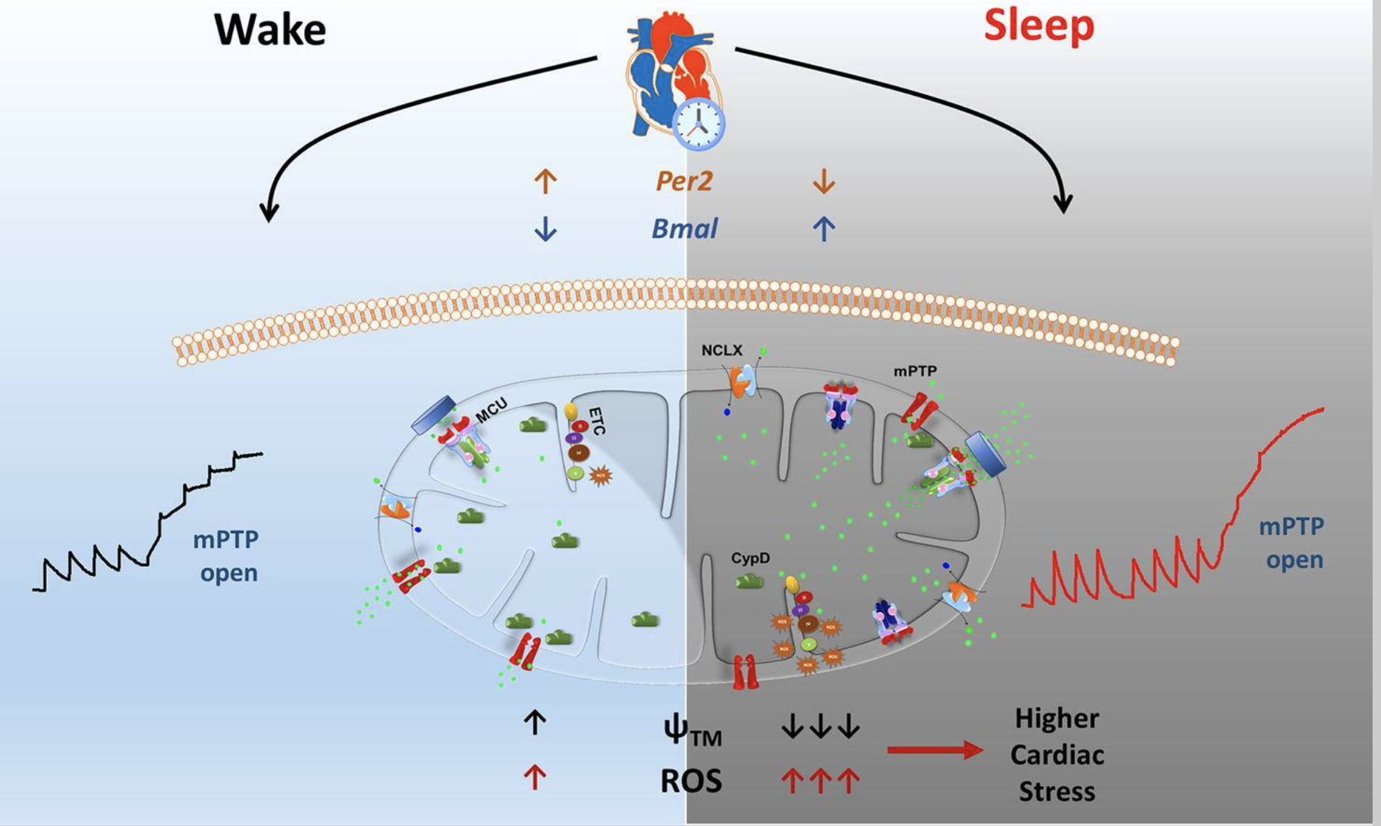
This figure illustrates how calcium enters mitochondria through a specific channel called the mitochondrial calcium uniporter (MCU), particularly during the sleep phase. During sleep, cardiac mitochondria increase their calcium uptake and retention, which leads to higher mitochondrial calcium levels. This calcium buildup alters the mitochondrial membrane potential and slows down ATP production by reducing the efficiency of the electron transport chain (ETC). At the same time, the elevated calcium induces higher production of reactive oxygen species (ROS), potentially contributing to oxidative stress. This rhythmic calcium handling is regulated by circadian genes like Bmal1 and Per2, which show sleep-specific expression patterns affecting mitochondrial calcium transport proteins. Therefore, the image illustrates how calcium flow into mitochondria during sleep connects to slower energy production and increased oxidative stress, which may raise vulnerability to cardiac events during the sleep-wake transition.
https://pmc.ncbi.nlm.nih.gov/articles/PMC8240107/
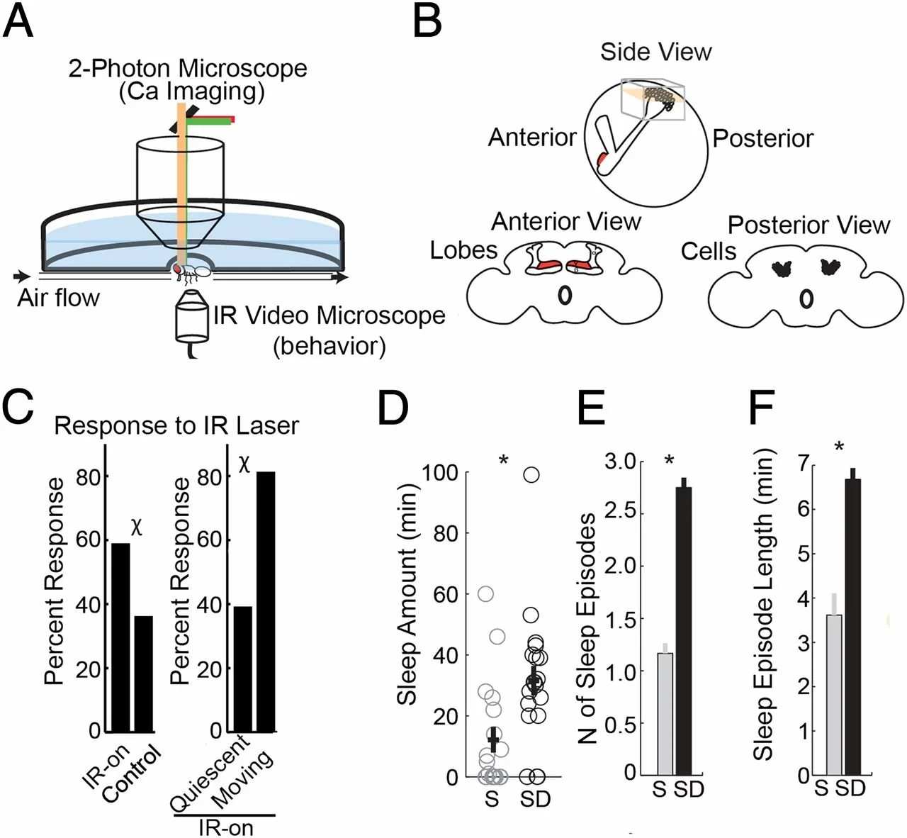
This figure illustrates how calcium imaging in the Drosophila brain can be used to identify sleep and wake states by combining two-photon microscopy with behavioral tracking. The mushroom body region, a key center for sleep regulation, shows distinct calcium activity patterns depending on behavioral state. Flies that were quiescent responded more strongly to laser stimulation than actively moving ones, indicating a shift in sensory responsiveness linked to sleep. Sleep-deprived flies exhibited significantly reduced total sleep time, fewer sleep episodes, and shorter episode durations. Together, the data suggest that sleep is associated with altered calcium dynamics, supporting the idea that calcium signaling plays a critical role in sleep regulation and homeostasis.
A New Direction for Sleep Modulation
Enter AIC Therapy, a modality uniquely positioned to recalibrate this imbalance. Unlike standard calcium supplements, AIC promotes rapid intracellular decalcification and restores ionic calcium gradients—without contributing to soft tissue calcification. It also stimulates endogenous calcitonin, which plays a role in modulating both neuronal excitability and circadian rhythm stability.
Preliminary clinical insights suggest that AIC may enhance not only total sleep time but also the quality of restorative sleep, particularly in patients resistant to hormonal or behavioral interventions. In light of this, it may be time to reframe sleep disorders—not just as neurochemical or behavioral in origin, but also as mineral dysregulation syndromes.
Advanced Clinical Tips
1. Patients waking consistently around 2 to 4 a.m., without clear cortisol or glycemic triggers, often have subtle disruptions in thalamic calcium signaling—especially postmenopausal women with early signs of bone turnover. It’s worth considering ionic calcium imbalance as a sleep-stage disruptor.
2. When someone sleeps a full night yet wakes up depleted, the issue may lie in mitochondrial calcium uptake. Energy isn’t being restored at the cellular level, even though the sleep duration looks normal. These cases often parallel blunted morning cortisol and poor tissue recovery.
3. Nocturnal bruxism or leg cramps that persist despite adequate magnesium may reflect intracellular calcium trapping or early soft tissue calcification. It’s not always about deficiency—it’s often about where the calcium is stuck.
4. Non-responders to GABAergic agents might not have a receptor problem—it could be a release issue. Calcium dysregulation at the synapse can block GABA from even reaching its targets, which changes the clinical approach entirely.
5. Shallow, irregular breathing during sleep—without full-blown apnea—may point toward ER calcium leak disrupting central rhythm generators in the brainstem. This pattern often slips past standard respiratory screening but shows up in fatigue and anxiety clusters.
6. Those with fragmented REM sleep, intense dreaming, or emotional exhaustion upon waking may be experiencing REM-phase calcium overload. Targeted decalcification using Anti-Orbital Ionic Calcium (AIC) has shown promise in restoring neurochemical boundaries during sleep.
Case Reference
A 60-year-old male with lifelong insomnia reported difficulty falling asleep since early childhood, often staying awake throughout the night and disrupting his family’s sleep. Despite decades of trying both conventional and alternative therapies, he remained unable to experience even one full hour of uninterrupted sleep. Over the years, he developed numbness from his shoulders to his arms, persistent mental fog, and increased alcohol use to manage his symptoms.
After initiating AIC Therapy, he described a dramatic change after the first dose, stating he felt “a big ice block melting” in his head, followed by deep, restorative sleep. The next morning, he reported waking up refreshed for the first time in over 50 years. Within weeks, he experienced full nights of sleep, emotional clarity, and resolution of arm numbness. He also noted reduced depressive symptoms and renewed enthusiasm for daily life. Thirty days into therapy, he continues to sleep soundly and shared, “This is the first time I truly feel rested—my arms, my thoughts, even my poems have changed.”
Conclusion
As demonstrated across neurobiological pathways, mitochondrial function, and clinical observation, calcium is not simply a co-factor—it plays a central regulatory role in sleep physiology. From synaptic inhibition and circadian enzyme activity to mitochondrial energy cycling and even the neuroarchitecture of REM states, calcium’s reach is deep and multifaceted. In our clinical experience, AIC Therapy has emerged as a reliable tool to correct hidden ionic imbalances—especially in cases where conventional interventions fall short. By restoring calcium gradients without adding to calcification burden, AIC supports restorative sleep from the inside out. Sleep dysfunction, increasingly, appears to be not just a neurohormonal disorder but a calcium signaling imbalance in disguise.
Bring calcium signaling into your practice.
Practitioners can request a consultation to discuss AIC therapy and how these mechanisms apply to specific cases.
Newsletter content is educational and contributed by participating healthcare practitioners. These statements have not been evaluated by the FDA and are not intended to diagnose, treat, cure, or prevent any disease.