This newsletter reveals vitiligo as a Ca²⁺-driven stress-immune loop where restoring ER-mitochondrial coherence calms active edges and supports repigmentation.
Executive Summary
Vitiligo reflects melanocyte stress plus immune attack, with disrupted intracellular Ca²⁺ signaling across melanocytes and keratinocytes as a central amplifier of oxidative injury and antigen presentation. Clinically, activity often declares itself through friction-line spread and an “active edge” (itch or burn, subtle erythema, confetti lesions, rapid margin change) even when total serum calcium is normal; therefore, tracking new-lesion count and margin velocity is the most useful readout. A calcium-axis support strategy that stabilizes Ca²⁺ transients via improved ER mitochondrial handling and a calcitonin-linked endocrine shift may quiet the edge first and then enable perifollicular “peppering” and progressive repigmentation.
Vitiligo Mechanisms: Ca²⁺ Signaling Instability, Barrier Breakdown, and Calcitonin-Linked Control
Vitiligo reflects melanocyte loss within a stressed epidermal unit. Melanocytes and keratinocytes rely on tightly regulated ionic calcium transients to coordinate adhesion, differentiation, melanosome transfer, and rapid stress sensing. When this signaling becomes unstable, melanocytes shift toward endoplasmic reticulum calcium dysregulation, mitochondrial calcium dysregulation, and increased oxidative stress, which in turn increase antigen presentation and immune recognition.
A second mechanism involves the keratinocyte compartment. The epidermal calcium gradient that supports barrier function can diminish, shifting keratinocyte maturation and weakening junction architecture. Barrier weakening increases exposure to irritants and external microbial products at the surface, thereby raising the local oxidative burden and further perturbing calcium flux. In vitiligo, oxidative stress can open calcium-permeable channels, drive repetitive cytosolic calcium spikes or sustained overload, and trigger melanocyte apoptosis. Microtrauma and friction can synchronize these calcium signals with innate immune activation and DAMP-driven signaling, recruiting and sustaining cytotoxic responses, thereby making depigmentation self-sustaining.
Clinically relevant therapeutic framing is not “more calcium” or “less calcium,” but reducing instability in calcium signaling and normalizing tighter regulation of calcium distribution and flux between the extracellular space, cytosol, ER stores, and mitochondrial calcium buffering. Endocrine calcium control systems influence how tissues buffer ionic calcium during inflammation and stress, which matters when epidermal homeostasis is already unstable.
Anti-orbital Ionic Calcium Therapy (AIC) targets this axis by supporting calcium signaling stability in the melanocyte niche, improving ER mitochondrial calcium handling, and reducing oxidative load. During vitiligo stress, calcium homeostasis can become disrupted, leading to calcium becoming trapped in the ER and mitochondria rather than moving normally between cellular compartments. Consequently, the cell loses normal cytosolic calcium pulses, which are needed for clear signaling. This imbalance increases organelle stress signals and can push melanocytes toward apoptosis, further triggering the innate immune response, which can repeatedly activate, contributing to depigmentation. AIC aims to stimulate endogenous calcitonin and support correct calcium handling, reducing ER and mitochondrial trapping, restoring cytosolic Ca²⁺ transients, lowering organelle stress, decreasing melanocyte death signals, and weakening the self-sustaining innate immune loop .
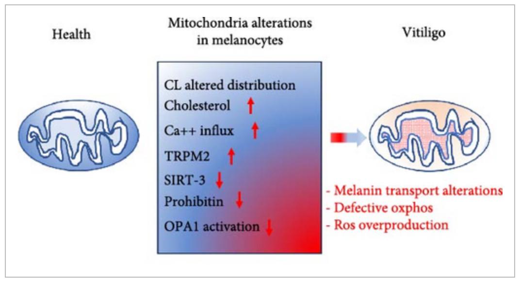
Oxidative phosphorylation changes have been reported in vitiligo melanocytes. Specifically, altered expression of selected electron transport chain proteins and altered complex I activity have been found. These functional deficits have been hypothesized to relate to changes in the amount and distribution of cardiolipin within the mitochondrial membrane, which may disrupt the proper organization of respiratory chain proteins. Mitochondrial dysfunction reduces adenosine triphosphate production, only partially offset by glucose metabolism. Although still to be confirmed, these findings support a possible crucial role for mitochondria in vitiligo pathogenesis
Matarrese P, Puglisi R, Mattia G, Samela T, Abeni D, Malorni W. An Overview of the Biological Complexity of Vitiligo. Oxid Med Cell Longev. 2024 Dec 19;2024:3193670. doi: 10.1155/omcl/3193670. PMID: 39735711; PMCID: PMC11671640. https://creativecommons.org/licenses/by/4.0/
Mechanism-to-Clinic Trigger
Friction-site spread usually suggests a Koebner response plus barrier stress. Do a quick friction check at the beltline, cuffs, pressure points from masks, and shaving areas.
The timing matters. New lesions after a new or intensified friction exposure, or brief recurrent flares at the same high-friction sites, often suggest a modifiable stress-driven pattern.
“Normal calcium” can mislead. Total serum calcium may be normal while intracellular calcium signaling is unstable, so lesion behavior remains the primary clinical indicator.
Activity is most evident at the edge. Symptoms and signs include itch or burning, subtle erythema, confetti-type lesions, and rapid edge expansion. Track new-lesion count and the rate of edge expansion.
Interpret calcitonin as a trend during and after flares, even when values remain within the reference range. In friction-driven, rapid-edge activity, AIC Therapy can serve as calcium-axis support; endpoints are simple: fewer new lesions, reduced edge activity, and slower edge expansion.
Clinical Practice Snapshot: AIC Therapy in Vitiligo Care
In the daily clinic, vitiligo often reflects melanocyte stress plus immune attack, and both can disrupt intracellular calcium control. When calcium signals become unstable, mitochondria and the endoplasmic reticulum can fail to buffer oxidative stress effectively. Melanocytes may then detach, reduce melanogenesis, or undergo apoptosis. For that reason, a therapy plan that aims to normalize calcium signaling is a practical priority when lesions show active edges or ongoing expansion. AIC therapy is presented here as a mechanism-focused option intended to stabilize calcium homeostasis and support melanocyte survival and functional recovery. In practice, we follow lesion spread, edge activity, symptoms such as itch or burning, and early perifollicular repigmentation as the main clinical endpoints under AIC therapy, alongside standard skin care. The more common sequence is progression control first, then perifollicular “peppering,” then repigmentation extending inward from the margins with improved color match.
If response occurs, it often starts at hair follicles and lesion margins, consistent with preserved melanocyte reservoirs and migration capacity. By stimulating endogenous calcitonin and associated endocrine signaling, AIC therapy reduces maladaptive calcium overload signaling, dampens stress-amplifying pathways, and promotes conditions that favor melanocyte adhesion and melanogenesis over apoptosis. This endocrine shift may also modulate local immune activation through calcium-dependent signaling in immune cells, potentially reducing persistent cytotoxic activity directed at melanocytes.
Conclusion
Vitiligo is not only pigment loss; it is a stress- and immune-mediated process within an unstable epidermal unit. Across melanocytes and keratinocytes, disrupted intracellular Ca²⁺ signaling amplifies oxidative injury and supports antigen presentation. This framework shifts our target from “more or less calcium” to better Ca²⁺ stability and better ER–mitochondrial handling under oxidative stress.
Barrier stress and friction often act as triggers, so Koebner-type spread and site-specific flares are practical clues in routine visits. Do not be reassured by “normal calcium,” because total serum calcium can be normal while intracellular Ca²⁺ control remains unstable. Clinical activity is best assessed at the edge, using itch or burning, subtle erythema, confetti lesions, edge expansion rate, and new-lesion counts.
Re-pigmentation, when it starts, often begins perifollicularly and at margins, which fits preserved follicular reservoirs and migration. For active vitiligo with edge progression, AIC Therapy is needed because it directly addresses this Ca²⁺ instability, reduces ER and mitochondrial trapping, and lowers the apoptotic drive in melanocytes. AIC Therapy is also needed because it supports calcitonin-mediated endocrine control of tissue Ca²⁺ handling, which can calm edge activity and help make repigmentation durable.
Bring calcium signaling into your practice.
Practitioners can request a consultation to discuss AIC therapy and how these mechanisms apply to specific cases.
Newsletter content is educational and contributed by participating healthcare practitioners. These statements have not been evaluated by the FDA and are not intended to diagnose, treat, cure, or prevent any disease.