This newsletter reveals how quiet, chronic inflammation gradually reshapes calcium handling, stiffens tissues from the inside out, and opens the door for gentler mineral-based strategies.
Executive Summary
Chronic low-grade inflammation does not just raise CRP; over the years, it trains vascular, fascial, and glandular tissues to behave like bone-friendly landing zones for calcium phosphate. When we pair classic natural tools for calming the immune system with a thoughtful ionic calcium support strategy, also paying attention to vitamin K, magnesium, phosphate load, bile, and renal clearance, we create a realistic approach to slowing and softening the calcification trajectory, rather than simply accepting it as normal aging.
How quiet inflammation teaches tissues to harden and misplace calcium
Most of us were trained to think of inflammation as something visible on the surface, such as a red joint, a hot rash, or a painful flare. In practice, much of the trouble stems from a quieter pattern, where immune cells continually send low-level danger signals for years without ever triggering a dramatic episode. Inside the tissue, this silent inflammation prompts endothelial cells, smooth muscle cells, and fibroblasts to alter their behavior, not just their laboratory values.
It does not stop there. Cells begin to express more bone-related proteins, increase alkaline phosphatase activity, and gradually transform the local matrix into a site where calcium phosphate crystals are very welcome. At the same time, protective factors such as matrix Gla protein, fetuin A, and pyrophosphate can be consumed, undercarboxylated, or simply overwhelmed, allowing the tissue to slowly lose its ability to maintain calcium in a flexible, soluble form.
What the patient feels is usually not “calcification” but stiffness, loss of elasticity, vague tightness, slower healing, or a sense that the body does not adapt as easily as before. In vessels, this manifests as higher pulse pressure, reduced microcirculation, and eventually, plaque that is harder, more brittle, and less forgiving during daily stress. In fascia, cartilage, and glandular tissue, it appears as small areas that fail to move and regenerate properly, even though routine imaging may still be reported as “age-related changes.”
For us as clinicians, the key idea is that silent inflammation and calcification are partners in the same long game; one keeps telling the story while the other writes it into the structure. AIC therapy fits into this picture as a way to stabilize ionic calcium handling and support decalcification , so that when we calm the inflammation, the tissue also has a realistic chance to clear the calcification and restore the normal cellular function.

In blood vessels, calcium phosphate mineral is not just a passive deposit; it is present inside secondary calciprotein particles, within calcifying extracellular vesicles, and in the extracellular matrix. Each of these calcium phosphate-containing structures can trigger pro-inflammatory signaling in immune cells and vascular smooth muscle cells, while also pushing smooth muscle cells toward an osteogenic, bone-like phenotype. Once activated, macrophages then amplify vascular calcification by releasing more calcifying extracellular vesicles and by further promoting osteogenic differentiation in nearby vascular smooth muscle cells. These osteogenic smooth muscle cells, in turn, drive additional matrix calcification through their own vesicle release and by strengthening macrophage-mediated pro-inflammatory responses. Taken together, this creates a vicious cycle in which inflammation and calcification continually reinforce each other within the vessel wall.
Viegas C, Araújo N, Marreiros C, Simes D. The interplay between mineral metabolism, vascular calcification and inflammation in Chronic Kidney Disease (CKD): challenging old concepts with new facts. Aging (Albany NY) . 2019;11(12):4274-4299. doi:10.18632/aging.102046
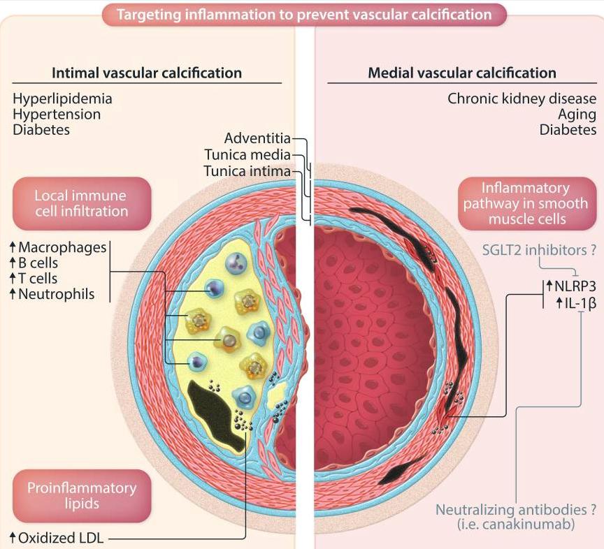
This figure contrasts the inflammatory patterns seen in intimal versus medial vascular calcification. Intimal calcification, which typically accompanies atherosclerosis, is linked to risk factors such as hyperlipidemia, hypertension, and diabetes. In this setting, local immune cell infiltration and pro-inflammatory lipid deposition in the tunica intima contribute to driving the calcification process. Medial calcification is also associated with chronic kidney disease, advancing age, and diabetes; however, it is usually not as closely linked to strong local inflammation. The article updates our understanding by showing that dampening smooth muscle cell inflammatory pathways, specifically NLRP3 and IL-1β, with the SGLT2 inhibitor canagliflozin can lessen calcification in the tunica media.
Turner ME, Aikawa E. Updating the paradigm: inflammation as a targetable modulator of medial vascular calcification. Cardiovasc Res . 2023;119(13):2259-2261. doi:10.1093/cvr/cvad139
Five Common Clinical Misconceptions
Misconception 1: Silent inflammation is only a lab pattern, not a structural problem.
Clinical angle: We often chase CRP, ESR, or cytokine panels and feel satisfied when they improve, but those signals have already been talking to the matrix for years. Even “mild” low-grade inflammation can push vascular and connective tissue cells to behave more like bone-forming cells, which means calcification is already being planned long before it is visible on a scan.
Misconception 2: Once calcification appears, there is nothing meaningful to do.
Clinical angle: Patients are frequently told that “calcium deposits are permanent,” which makes both them and the clinician feel powerless. In reality, the balance between deposition and removal is influenced by vitamin K status, magnesium, phosphate load, local pH, and blood flow, so a well-designed program can at least slow progression, sometimes soften the pattern, and often improve function even if the image does not completely normalize.
Misconception 3: Normal serum calcium means calcium distribution is healthy.
Clinical angle: Serum calcium is tightly regulated and will stay in range even while calcium quietly moves from where it should be dynamic, like bone and extracellular fluid, into places where it becomes rigid, like vessel walls, valves, or scar tissue. Looking only at total calcium can therefore be misleading; it is more helpful to consider ionized calcium, cofactor status, and signs of ectopic calcification as part of the same story.
Misconception 4: Treating silent inflammation mainly means adding more antioxidants and fish oil.
Clinical angle: These tools are useful, but they do not automatically reset how cells handle minerals in the matrix. If blood sugar swings, chronic infections, mechanical stress, or low vitamin K keep the tissue in a “repair, then harden” mode, antioxidants alone will not stop calcification; therefore, we need to consider cell signaling, matrix biology, and mineral regulators rather than just “more scavengers.”
Misconception 5: Decalcification needs aggressive chelation or strict calcium restriction.
Clinical angle: Strong chelation or severe calcium restriction can destabilize plaques, disrupt rhythm in ion handling, and overload excretory organs if the rest of the system is not prepared. A gentler and often safer approach is to stabilize ionic pools, support bile and renal clearance, ensure magnesium sufficiency, restore vitamin K-dependent proteins, and then use targeted ionic calcium support such as AIC within that framework to encourage calcium to move out of the wrong tissues in a controlled way.
Case Reference
Suzan L. is a 62-year-old woman from Langley with a 10-year history of multiple sclerosis who experienced a gradual decline despite numerous treatments. After turning to Eastern therapies, including six months of acupuncture, her acupuncturist recommended AIC therapy, Antiorbital Ionic Calcium Therapy, at a standard dose. Soon after starting AIC, she reported waking more refreshed and lighter, with strength returning to her legs, and over time, she was able to walk without a walker, which her son from Hope witnessed with clear surprise.
A patient like this has lived for years with chronic neuroinflammation that can impair microcirculation and connective tissue, promoting stiffness and an internal milieu that favors disturbed calcium handling and microcalcification. Her improvement in energy, leg strength, and gait is compatible with the idea that when silent inflammation is reduced and ionic calcium is stabilized, tissues may regain some flexibility and perfusion even if the primary diagnosis remains multiple sclerosis.
AIC can be viewed as one tool to help redirect calcium away from stiff, overloaded regions, contributing to better gait and daily function.
Conclusion
Stepping back from the details, the story running through this discussion is that low-grade inflammation does not remain confined to the blood; over time, it instructs vessels, fascia, cartilage, and glands to remodel their matrix in a more bone-like direction.
In arteries, different clinical pictures of calcification share a similar biology, where smooth muscle cell inflammatory pathways, such as NLRP3 and IL-1β, together with metabolic stressors, help transform the intima or media into a surface that welcomes calcium phosphate. Additionally, calcium travels in secondary calciprotein particles and calcifying extracellular vesicles, which both carry minerals and trigger further inflammatory signaling, creating a loop in which immune cells and smooth muscle cells continue to harden the wall they are trying to repair. Clinically, our patients experience that loop not as “calcification” but as rising pulse pressure, reduced microcirculation, stiff tissue, slower healing, and the familiar explanation that these are simply age-related changes.
Against that background, it is noteworthy that a woman with longstanding multiple sclerosis and clear functional decline reports improved morning energy, lighter legs, and the ability to walk again without a walker after completing a mineral-focused, integrative program that includes ionic calcium support.
For clinical practice, the key shift is to view calcium not only as a lab value or a supplement to restrict, but also as a mobile signal that can be gently redirected by calming the drivers of silent inflammation, restoring vitamin K-dependent proteins, supporting magnesium and phosphate balance, and maintaining bile and renal routes.
Within that kind of terrain-focused work, a targeted ionic calcium approach, such as AIC, makes sense as one of the tools that helps stabilize calcium pools and encourages excess minerals to leave stiff, overloaded tissues in a controlled manner, giving vessels, fascia, and even brain microcirculation a more realistic chance to regain flexibility.
Bring calcium signaling into your practice.
Practitioners can request a consultation to discuss AIC therapy and how these mechanisms apply to specific cases.
Newsletter content is educational and contributed by participating healthcare practitioners. These statements have not been evaluated by the FDA and are not intended to diagnose, treat, cure, or prevent any disease.