Cancer Immunity, Timed by Calcium
This newsletter shows how cytotoxic immunity in cancer depends on short, controlled free ionic Ca2+ pulses at the immune synapse, and how a low, steady calcium window can support cleaner killing and recovery.
Executive Summary
Cytotoxic killing is a timing event: NK cells and CD8 T cells require a brief, controlled rise in free ionic Ca2+ at the synapse to trigger degranulation, not a significant calcium surge. When Ca2+ entry is too weak, contact stalls; when it is excessive or prolonged, calcineurin–NFAT timing drifts, mitochondrial load rises, and cells slide toward strain and exhaustion-like behavior, which is why “more calcium equals more immunity” misleads in oncology care. A homeostasis-first ionic calcium approach, aimed at reducing calcium trapping in soft-tissue microdomains and restoring clean signaling pools, supports immune rhythm while avoiding peaks that the cell cannot buffer.
Short Calcium Pulses, Better Killing
Cancer immunity is often explained mainly through cytokines, but cytotoxic killing depends on calcium timing early on. NK cells and CD8 cytotoxic T cells do not need a big calcium surge. They need a short, controlled rise in free ionic calcium at the immune synapse (the contact zone). That brief timing step triggers degranulation, allowing perforin and granzymes to complete the kill.
Across patients, two common failure patterns show up. If ionic calcium entry is too weak, signaling at the immune synapse slows, contact becomes costly, and killing efficiency drops. If ionic calcium entry is too strong or lasts too long, calcineurin stays active too long, NFAT stays on too long, mitochondrial load rises, ATP demand rises, and recovery becomes harder. This is one reason why “more calcium equals more immunity” often fails in cancer care. The practical target is a low, yet sufficient ionic calcium range that remains stable throughout the day, without peaks that exceed the cell’s buffering capacity.
Two different calcium routes matter. Effector cells use ORAI1-driven calcium entry to execute cytotoxicity. In contrast, many tumors can use ER-to-mitochondria calcium transfer via InsP3R at MAM contacts to maintain ATP production. So, the aim is to support effector timing and mitochondrial resilience without pushing sustained activation in a setting already loaded with oxidative stress. AIC fits here as a restoration tool. In the AIC framework, reducing calcium trapping in soft-tissue microdomains helps return ionic calcium to signaling pools, supporting immune function and cellular energy handling.
Shao M, Teng X, Guo X, et al. Inhibition of Calcium Signaling Prevents Exhaustion and Enhances Anti-Leukemia Efficacy of CAR-T Cells via SOCE-Calcineurin-NFAT and Glycolysis Pathways. Adv Sci (Weinh). 2022;9(9):e2103508. doi:10.1002/advs.202103508
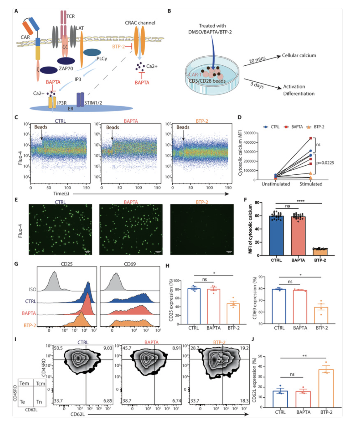
This figure compares two ways to control calcium after CAR-T activation: blocking calcium entry at the membrane versus buffering calcium inside the cell. CAR-T cells were activated with anti-CD3/CD28 beads and, at the same time, treated with two inhibitors: an intracellular calcium chelator and a calcium-entry blocker. Intracellular Ca2+ signals and cell-state markers were measured over time.
The intracellular chelator did not fully prevent the rise in the intracellular Ca2+ signal after activation, consistent with ongoing calcium entry exceeding its buffering capacity. In contrast, the calcium-entry blocker strongly reduced the activation-induced calcium influx.
After 48 hours, sorted 4-1BB CAR-T cells treated with the entry blocker, but not with the intracellular chelator, stayed more in naïve and central memory states and showed lower activation. Similar findings were reported in CD28 CAR-T cells under the same treatment window.
Calcium Signal Clinical Map
Some weeks look like poor follow-through during active cancer treatment. The patient does not make clear clinical progress. Infections linger. Recovery is slow, and there is no consistent daily pattern. Other weeks look different. Energy and basic function show up for a few hours, then fall apart over the next days. When the pattern is “good hours” followed by a restless evening, shallow sleep, and a next-day crash, this indicates that the signal stays on too long, not its absence. The immune system spends time in a high-cost energy state, and it does not return to baseline cleanly.
Common timing disturbers include stimulants taken late, late training, dehydration, irregular meals, and ongoing inflammatory load. When several factors stack up, it stops looking like a single-organ problem and becomes a 24-hour regulatory problem, where tissues do not return to baseline. What we look for clinically is simple. Sleep starts to come more easily. Post-stress crashes soften. Recovery feels steadier across an ordinary week. When these shifts happen together, calcium timing usually returns to a workable range instead of remaining prolonged noise.
Clinical Tips
Chronic antigen load often needs pacing, not stimulation. In a cancer patient, when viral flares keep coming back, recovery stays slow, and low-grade inflammation persists, the immune pattern can look like this: ATP is spent on basic survival, not on clean cytotoxic work. In these cases, reducing day-to-day physiologic noise often gives more usable immune output than adding another strong “booster.”
Look at the cost of the “up” period. If your plan gives a few good hours but then leads to irritability, insomnia, or a 48-hour slump, the system is being pushed into a state it cannot afford to maintain. A slower plan, repeated on a steady routine and adjusted one step at a time, usually protects week-to-week function better during active treatment.
If AIC Therapy is part of the plan, schedule matters more than intensity. Smaller, divided dosing on a stable rhythm is often more compatible with cytotoxic competence than intermittent peaks, which often disrupt sleep and trigger next-day depletion. In the AIC framework, even a 0.1 to 0.2 mg/dL shift in active ionic calcium can be enough to support tighter calcium control, so the aim is a steadier calcium signal: fewer spikes, smoother on-off control inside cells, and a stable baseline that supports calmer sleep and better next-day energy.
Keep effector support separate from tumor energetics when you choose supports. Tumor biology can benefit from sustained bioenergetic support, so strategies that push throughput and stimulation can backfire in the wrong setting. A homeostasis-first calcium frame, titrated conservatively and paired with hydration, regular meal windows, and sleep timing work, is less likely to move into that problem.
Case Reference
A 67-year-old postmenopausal woman with IgG lambda multiple myeloma presented with widespread bone disease, vertebral compression fractures, and hypercalcemia. Clinically, recovery was fragile, and infections were frequent, with weeks that often fell apart after minor stressors. Our working lens focused on calcium timing and immune execution, because cytotoxic function depends on signals that mitochondria can support.
With AIC therapy, the swings softened. Hypercalcemia moved into a more workable range and day-to-day resilience improved. Recurrent infections reportedly became less frequent and less intense, and the patient’s week looked less like repeated depletion after minor triggers. Kidney function improved to within the normal reported range. Anemia resolved. Inflammatory tone declined. Protein-related organ stress also appeared calmer, with free light chains trending toward a more balanced pattern and blood counts remaining stable. Overall, the clinical course shifted toward steadier mineral handling, with a more dependable immune rhythm, clearer day-to-day function, and a better quality-of-life pattern, alongside the objective clinical improvements noted above.
Conclusion
In cancer care, the key point is not the total calcium level. It is the signal's pattern. Free ionic Ca2+ should rise for a short time in the immune synapse, then go back to baseline. If Ca2+ entry is too weak, NK and CD8 T cells can still touch the target, but degranulation does not finish, so killing is less effective. If Ca2+ entry is too strong or lasts too long, the cell moves into an over-activated program that it cannot keep up.
Here is what “calcineurin to NFAT timing drift” means in practice. When the Ca2+ signal stays high for too long, calcineurin stays active too long, NFAT stays on too long, and gene programs shift toward high activation and lower staying power. Then mitochondria carry a bigger energy load, ATP needs go up, and the cell often loses clean killing and good recovery. Clinically, this can look like unstable weeks during treatment, with high-activation symptoms followed by a crash, worse sleep, slower recovery, and more infection-prone days. This is one reason “more calcium equals more immunity” is not reliable in everyday oncology follow-up.
In T-cell activation, it is usually better to control Ca2+ entry at the membrane than to try to buffer Ca2+ after a large influx has already happened. When entry is controlled, cells are less likely to move into an exhaustion-like state and are more likely to stay closer to naive and central memory behavior. This matters because these states are linked to better staying power and cleaner function over time.
AIC fits this model because the goal is not constant stimulation. The goal is to restore the calcium rhythm. In this framework, AIC aims to reduce calcium trapping in soft-tissue microdomains and return that calcium to signaling pools so that the system can use clean, low-amplitude pulses again. Kept away from meals and timed consistently, it is easier to support cytotoxic killing and recovery while avoiding peaks that can disturb sleep, increase ATP stress, and push immune bioenergetics in the wrong direction.
Bring calcium signaling into your practice.
Practitioners can request a consultation to discuss AIC therapy and how these mechanisms apply to specific cases.
Newsletter content is educational and contributed by participating healthcare practitioners. These statements have not been evaluated by the FDA and are not intended to diagnose, treat, cure, or prevent any disease.