Listen to this issue
This newsletter reveals how the loss of calcium control in liver cells drives fatty liver disease.
Executive Summary
Fatty liver resists the usual advice, holding on even as weight and diet improve. Blood sugar stays stubborn, liver enzymes drift up, and some livers move toward scarring. Underneath these signs, the liver cell has lost control of its calcium. This calcium problem sits in three places: the cell surface, the internal stores, and the mitochondria. When calcium accumulates in the mitochondria, oxidative stress increases and injury progresses. Bringing calcium handling back into order eases the liver, and these signs settle with it.
Calcium Homeostasis and the Development of Fatty Liver Disease
Fatty liver disease is common, and it starts with how liver cells handle calcium. Within the liver cell, calcium acts as a tightly controlled signal. The endoplasmic reticulum is the cell’s main calcium store. In fatty liver, fat overload drains calcium from this store. The pump that refills this store then works poorly in these cells. A poorly refilled store keeps the cell under endoplasmic reticulum stress, which drives the cell to store fat rather than clear it.
As fatty liver advances, calcium floods from the store into the mitochondria. The store and mitochondria meet at contact sites that allow calcium to pass. In obesity, these contacts multiply, and the calcium handoff grows excessive. A calcium-release gate at these contacts opens as the liver inflames. More calcium pours into the mitochondria than they can handle. Overloaded mitochondria lose efficiency and leak reactive oxygen species. This oxidative stress drives liver inflammation and promotes fat buildup, leading to injury. Calcium-stressed mitochondria also worsen insulin resistance, a core driver of fatty liver. Slowing fatty liver means restoring calcium control within liver cells.
The disorder sits in three places where calcium moved out of step. These are the cell surface, the internal store, and the mitochondria. When the store refills properly, that internal stress eases and fat clears. As the calcium handoff to mitochondria steadies, oxidative stress and inflammation settle. Anti-orbital Ionic Calcium Therapy (AIC) restores coordinated calcium control across the surface, stores, and mitochondria. The clinical reading is less liver fat, steadier blood sugar, and slower progression to inflammation.
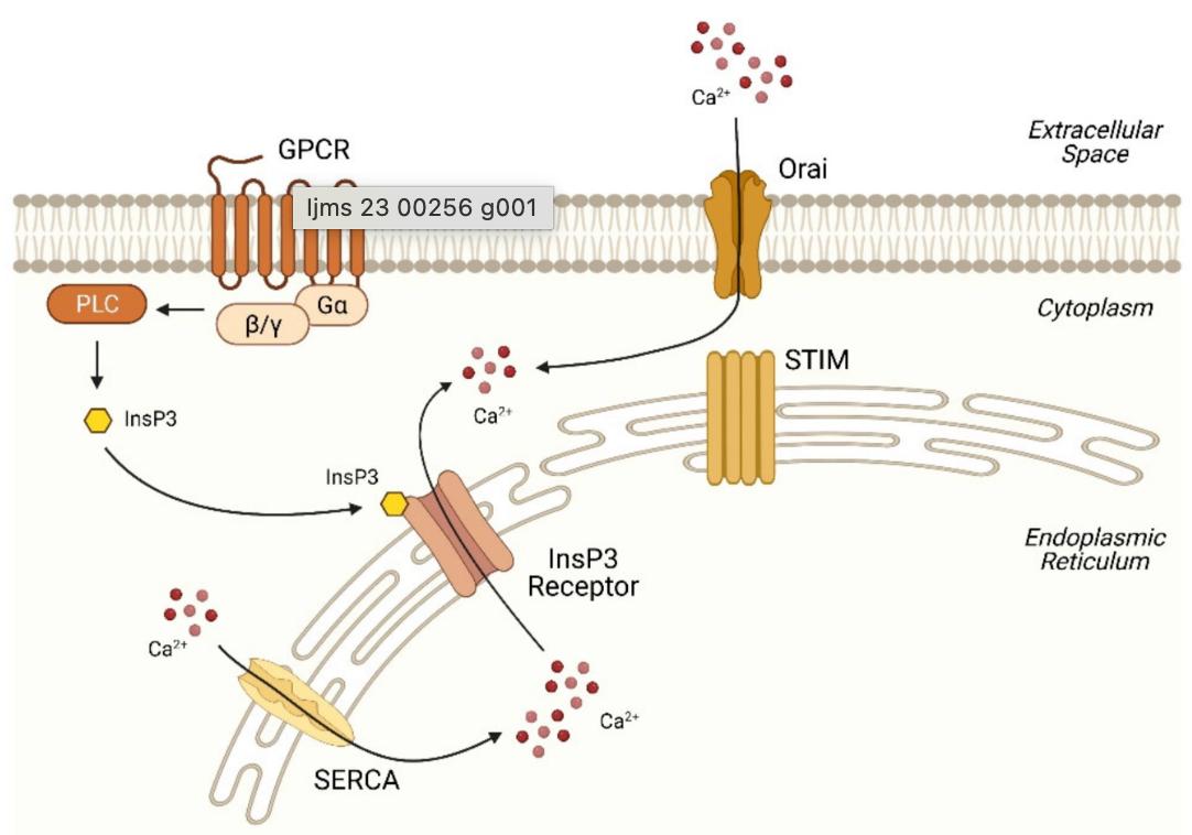
This figure explains how a liver cell releases calcium from its stores and then refills them. The store is the endoplasmic reticulum, the cell’s internal calcium reservoir. A signal at the cell surface starts the release. The surface receptor activates an enzyme that makes a messenger. That messenger opens a channel on the store, and calcium flows out into the cell. As the store empties, sensors on its open surface channels let calcium in from outside. A pump then returns calcium from the cell back into the store.
Chen, C.-C.; Hsu, L.-W.; Chen, K.-D.; Chiu, K.-W.; Chen, C.-L.; Huang, K.-T. Emerging Roles of Calcium Signaling in the Development of Non-Alcoholic Fatty Liver Disease. Int. J. Mol. Sci. 2022 , 23 , 256. https://doi.org/10.3390/ijms23010256 https://creativecommons.org/licenses/by/4.0/
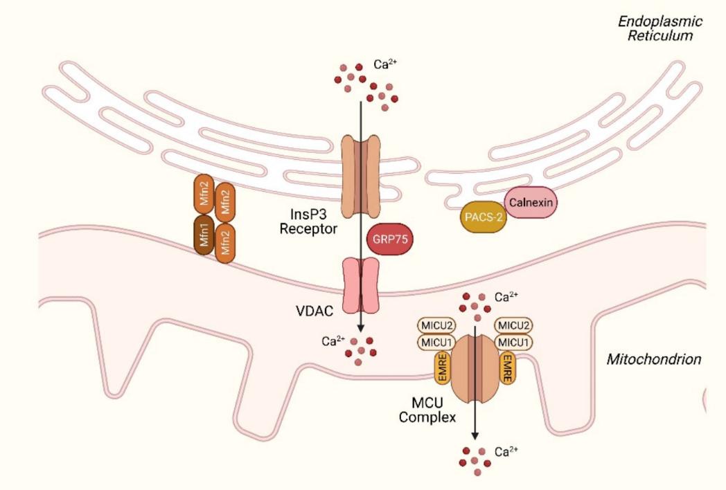
This figure illustrates how the liver cell releases calcium from its stores into the mitochondria. The store and the mitochondria meet at contact sites. These contact sites let calcium move directly from one to the other. The release channel on the store hands calcium to a channel on the mitochondrial surface. A connector protein links the two channels, and tethering proteins hold the contact in place. Calcium then crosses into the mitochondrial interior through an uptake channel . Gatekeeper proteins control when that channel opens, and a helper protein supports them.
Chen, C.-C.; Hsu, L.-W.; Chen, K.-D.; Chiu, K.-W.; Chen, C.-L.; Huang, K.-T. Emerging Roles of Calcium Signaling in the Development of Non-Alcoholic Fatty Liver Disease. Int. J. Mol. Sci. 2022 , 23 , 256. https://doi.org/10.3390/ijms23010256 https://creativecommons.org/licenses/by/4.0/
Clinical Tips
- In fatty liver, excess free fatty acids act as signals, not just fuel. The more fat the cell carries, the harder this calcium-release pathway is driven.
- As fatty liver advances, the calcium signal that drives liver-cell regeneration weakens. The liver then recovers poorly after injury, so each insult leaves lasting loss.
- The shift from simple fat to scarring is itself a calcium-driven event. Lipotoxic injury activates hepatic stellate cells via their own calcium signals.
- Bland-looking early steatosis already carries the calcium-handling lesion. The histology reads as low risk, while the disorder driving progression is already set.
- AIC rebalances the liver cell’s calcium release. The protective regenerative signal then holds, and the scar-driving activation loses its trigger.
Clinical Snapshot
The frustrating fatty livers are the ones that will not improve, no matter what. Weight comes off, the diet is clean, and still the liver stays bright. The problem is not always intake. It is disposable. Clearing stored fat is an active process that the cell runs, not a passive drain. That disposal program runs on calcium signals from the cell’s internal stores. In a fatty liver, those signals are disordered, so the program stalls. The cell holds its fat because the clearance program no longer switches on. So lower intake meets a liver that still will not empty its stores.
Read fatty liver that resists real lifestyle change as a failure of disposal, not a willpower failure. Look for steatosis that holds steady while weight and diet genuinely improve. Look for stalled progress that adherence alone does not explain. That pattern points to a clearance program inside the cell that has gone quiet. The work then turns toward waking that disposal program, not only trimming what comes in. AIC restores the calcium signaling that drives the cell’s fat-disposal machinery. With that signal back, the clearance program restarts, and stored fat finally moves.
Conclusion
This issue reads fatty liver as a calcium-handling failure inside the liver cell. In health, a surface signal releases calcium from the internal store. The store passes calcium to the mitochondria at their contact points, then a pump refills it. In fatty liver, fat overload throws this calcium cycle out of step. Steatosis then holds firm even when weight and diet genuinely improve. The liver heals poorly after an injury that should heal. Some livers slide from simple fat toward scarring. Liver enzymes drift up without a clear cause.
What turns this around is the order in which the cell moves calcium. In fatty liver, that sequence slips, so calcium pools where it should not. AIC restores the timed handoff of calcium from the store to the mitochondria. AIC also alleviates mitochondrial calcium overload, thereby subsiding the oxidative stress that drives injury.
Bring calcium signaling into your practice.
Practitioners can request a consultation to discuss AIC therapy and how these mechanisms apply to specific cases.
Newsletter content is educational and contributed by participating healthcare practitioners. These statements have not been evaluated by the FDA and are not intended to diagnose, treat, cure, or prevent any disease.